Download as PDF, PPTX
![ Organizer of First United States Lasik
Course
Organizer of Largest Lasik Wet lab course
in the world
First textbook chapter written on the lasik
technique
First textbook chapter written on hyperop-
ic lasik
First textbook chapter on Sutureless Cor-
neal Transplantation
Editor Advances in Refractive & Corneal
Surgery
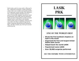
Helped teach the world LASIK
Popularized name LASIK
Over 30,000 surgeries performed
Largest Corneal Fellowship program in
USA
Reviewer for the Journal of the American
Medical Association [JAMA] and Middle
East African Council of Ophthalmology
worldwide firsts and notables: than 2 lines is about 0.5 to 2% for both LASIK and
PRK.
Inflammation (sterile white blood cell infiltrates)
in the treated area occur occasionally in surface
PRK/ usually while wearing the therapeutic band-
age soft contact lens. These usually respond well
to treatment with topical antibiotics and steroids.
Infiltrates are also occasionally seen with LASIK.
Steroid-induced glaucoma. Unlike LASIK, most
surface PRK/ patients require topical steroids for
one to four months to reduce scarring and prevent
regression. A small percentage of patients have a
genetic tendency to develop secondary glaucoma
when given steroids topically for this long. Most
of these cases will return to normal pressures when
steroids are stopped, but a small percentage will
require treatment for glaucoma from then on.
LASIK patients use steroid drops for only one
week and cannot develop steroid-induced glauco-
ma.
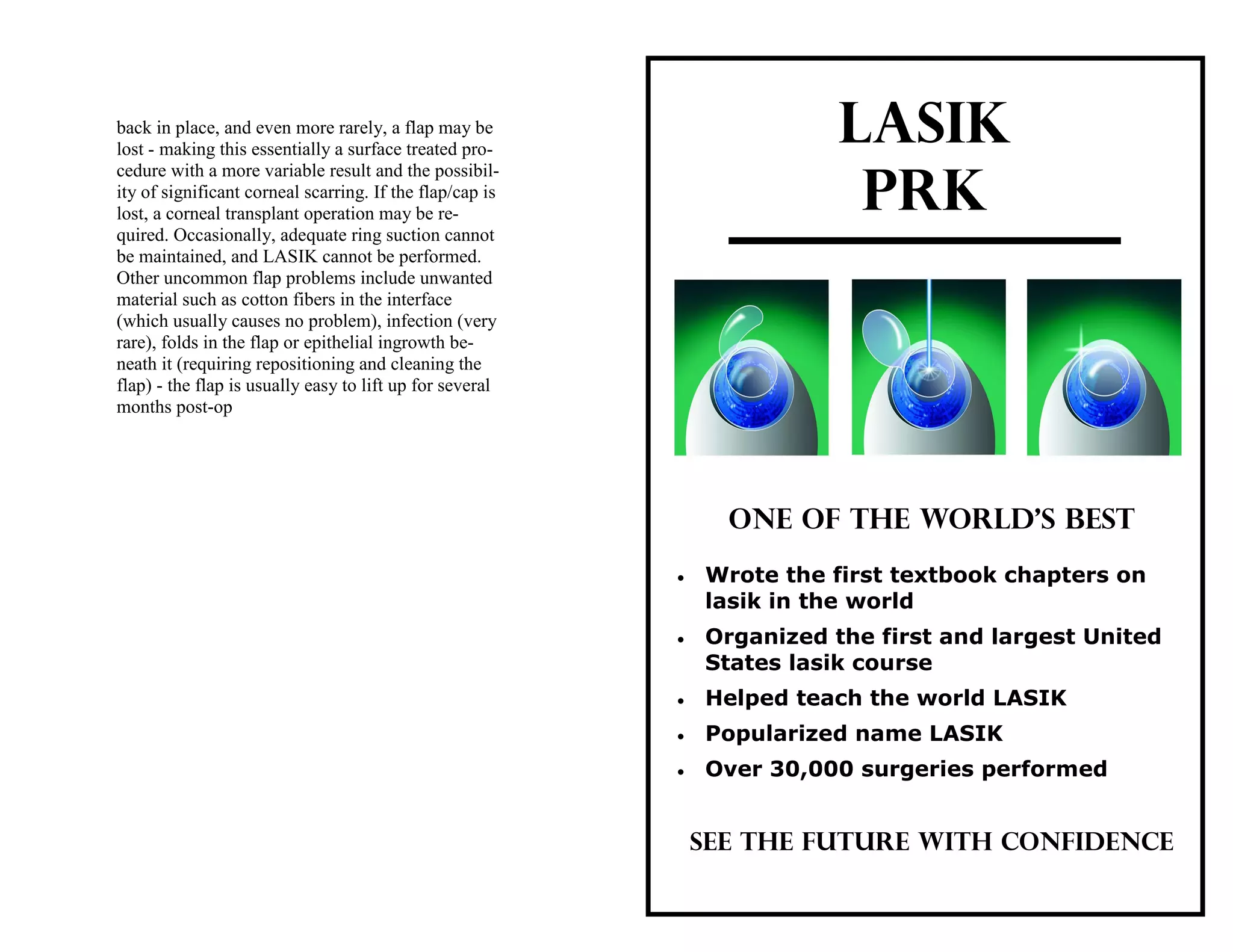
Complications Specific to LASIK
These include problems with the instru-
ment that makes the flap (microkeratome) which
result in incomplete flaps, irregular flaps, flaps
with thin spots or holes in it, or a flap that comes
totally off. These require the procedure to be abort-
ed, and then re-done in about three months. Occa-
sionally, if scarring occurs, the LASIK cannot be
repeated and the patient must wear a contact lens
for best vision. Rarely a flap may require suturing](https://image.slidesharecdn.com/lasikbrochure-170225172154/85/Lasik-brochure-by-Dr-Michael-Duplessie-2-320.jpg)


The document discusses LASIK and PRK, two surgical procedures for correcting refractive errors in the eye, detailing their processes, advantages, complications, and post-operative care. It highlights the experience and qualifications of Dr. Duplessie, who has performed over 30,000 procedures and is a pioneer in the field. While both procedures offer similar results, LASIK involves creating a corneal flap and has a slightly higher risk of complications compared to PRK, which involves surface treatment of the cornea.


![Mafe[1]](https://cdn.slidesharecdn.com/ss_thumbnails/mafe1-110519173429-phpapp01-thumbnail.jpg?width=640&height=640&fit=bounds)