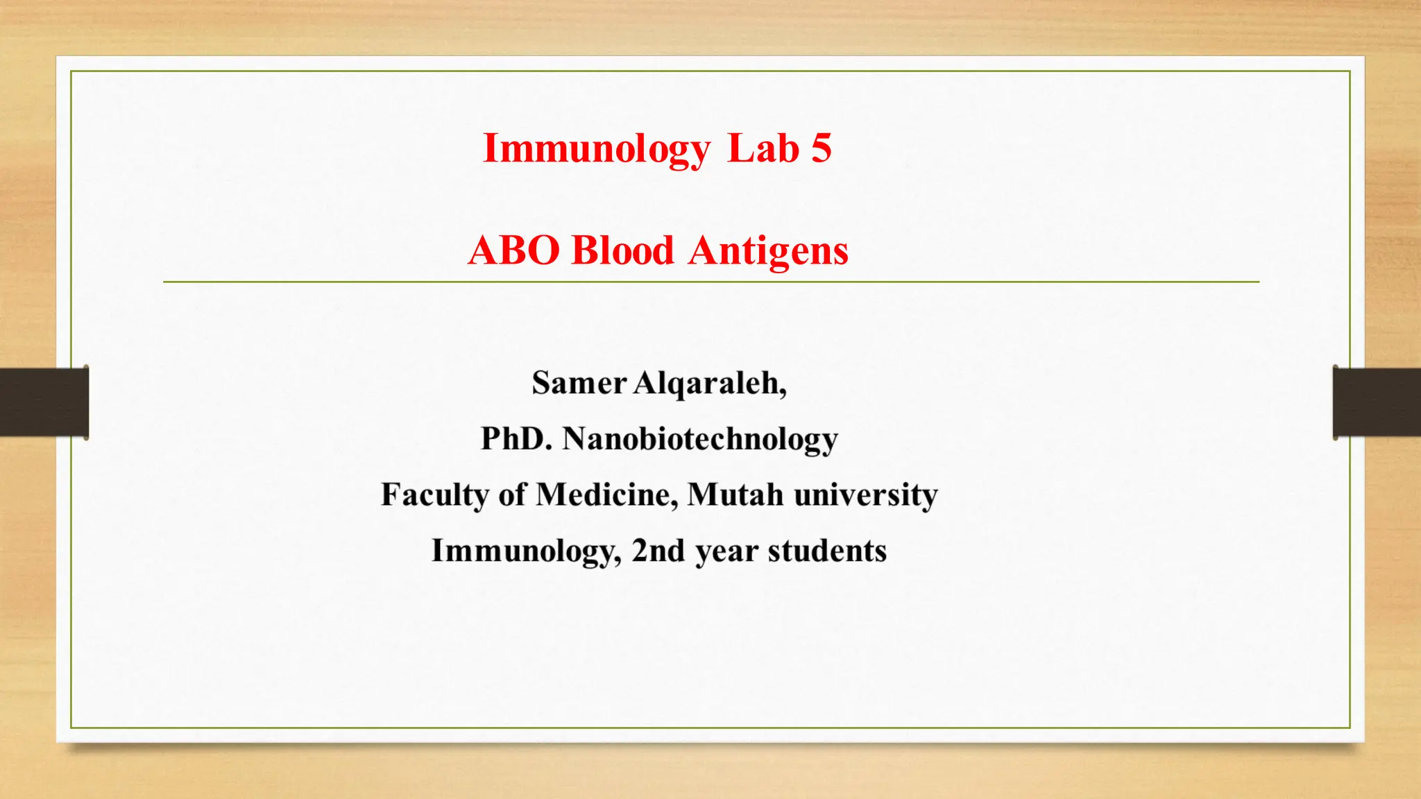
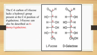
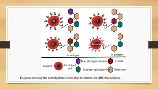
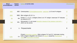
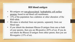
Immunology Lab 5
ABO Blood Antigens
ABO blood antigens
The ABO antigens are carbohydrates linked to cell surface proteins and lipids that are
synthesized by polymorphic glycosyltransferase enzymes.
Most individuals possess a fucosyltransferase that adds a fucose moiety to a nonterminal
sugar residue of the core glycan, and the resulted fucosylated glycan is called the H
antigen (O antigen).
A single gene on chromosome 9 encodes a glycosyltransferase enzyme that may further
modify the H antigen.
There are three allelic variants of this enzyme
1. O allele gene product: is devoid of enzymatic activity and cannot attach terminal
sugars to the H antigen and express only the H antigen.
2. A allele– encoded enzyme: transfers a terminal N-acetylgalactosamine moiety onto
the H antigen.
3. B allele gene product: transfers a terminal galactose moiety.
The C-6 carbon of l-fucose
lacks a hydroxyl group
present at the C-6 position of
d-galactose. l-Fucose can
also be described as 6-
deoxy-l-galactose.
Diagram showing the carbohydrate chains that determine the ABO blood group
➢ Mutations in the gene encoding the fucosyltransferase
that produces the H antigen without fucose are rare;
people who are homozygous for such a mutation are
said to have the Bombay blood group. And cannot
produce H, A, or B antigens. and cannot receive type
O, A, B, or AB blood.
Most Common Transplantation
-Blood Transfusion-
Percentages of the 8 blood groups
AB-negative (. 6 percent)
B-negative (1.5 percent)
AB-positive (3.4 percent)
A-negative (6.3 percent)
O-negative (6.6 percent)
B-positive (8.5 percent)
A-positive (35.7 percent)
O-positive (37.4 percent)
✓ O-negative is the universal blood type, meaning any other
blood type may receive it (see our blood type compatibility
chart here).
✓ This can quickly deplete the stores of O-negative that blood
centers have on the shelves.
✓ While 45% of the population is type O, less than 7% is O-
negative. So as you can see, the most needed type of blood is
also the hardest to collect.
✓ AB negative is the rarest of the eight main blood types - just
1% of our donors have it. Despite being rare, demand for AB
negative blood is low
Chromosome
Epitope or carrier,notes
System
symbol
Blood
groupingSystem
9
Carbohydrate( N-Acetylgalactosamine ,galactose .)A, B and H antigens
ABO
ABO
4
Main antigens M, N, S, s.
MNS
MNS
1
Protein. C, c, D, E, e antigens (there is no "d" antigen; lowercase "d" indicates
the absence of D
RH
Rh
7
Glycoprotein. K 1can cause hemolytic disease of the newborn (anti-Kell )
,
which can be severe.
KEL
Kell
6
Polysaccharide
Li
LI
1
Protein( chemokine receptor .)Main antigens Fya
and Fyb
.Individuals lacking
Duffy antigens altogether are immune to malaria caused by Plasmodium
vivax and Plasmodium knowlesi.
FY
Duffy
RH blood antigen
• Rh antigens are non-glycosylated, hydrophobic cell surface
proteins found in red blood cell membranes.
• 15% of the population has a deletion or other alteration of the
RhD allele.
• Rh status is inherited from our parents, separately from our
blood type.
• If you inherit the dominant Rhesus D antigen from one or both
of your parents, then you are Rh-positive (85% of us). If you do
not inherit the Rhesus D antigen from either parent, then you are
Rh-negative (15% of us).
Rh System
Rh Antigens and Encoding Genes
• Subsequentlyit was confirmed that the RH locus is on chromosome 1 and comprises two
highly homologous, very closely linked genes, RHD and RHCE.
• The Rh blood group system consists of 49 defined blood group antigens, among which the
five antigens (D, C, c, E, and e) are the most important.
• There is no d antigen. D antigen is the main that its presenceor absence mean RH+ or RH-
respectively.
• The main antigens are D, C, E, c and e, which are encoded by two adjacent gene loci, the
RHD gene which encodesthe RhD protein with the D antigen and the RHCE gene which
encodesthe RhCE protein with the C, E, c and e antigens
• The RHCE gene has four main alleles; CE, Ce, ce and cE.
• This concept of D and CcEe genes linked closely and transmitted together is consistent with
the Fisher nomenclature.
Examples on antigens in RH+ and -
D− C+ E+ c− e+ (RhD−)
D+ C+ E− c− e+ (RhD+)
✓ Each locus has its own set of alleles which are Dd , Cc , and Ee .
The D gene is dominant to the d gene, but Cc and Ee are co-
dominant (meaning that all of the inherited alleles lead to
expression of the coded antigens).
✓ Antibodies to Rh antigens can be involved in hemolytic transfusion
reactions and antibodies to the Rh(D) antigens confer significant
risk of hemolytic disease of the fetus and newborn.
Rh System
• Antibodies
✓ Antibodies directed against all Rh antigens, except d, have been
described: anti-D, anti-C, anti-c, anti-E and anti-e.
✓ Rh antigens are restricted to red cells and Rh antibodies result
from previous alloimmunization by previous pregnancy or
transfusion.
✓ Immune Rh antibodies are predominantly IgG.
✓ Anti-D is clinically the most important antibody.
✓ It may cause hemolytic transfusion reactions and was a common
cause of fetal death resulting from hemolytic disease of the
newborn before the introduction of anti-D prophylaxis.
…Rh Antibodies
hemolyticdisease of the newborn
• When the condition is caused by the Rh D antigen-antibody incompatibility,
it is called Rh D Hemolytic disease of the newborn
• The major clinical significance of anti-Rh antibodies is related to hemolytic
reactions associated with pregnancy that are similar to transfusion reactions.
• (Rh-negative mothers) carrying an Rh-positive fetus can be sensitized by
fetal red blood cells that enter the maternal circulation, usually during
childbirth. IgG antibodies are generated in Rh-negative mothers.
• Subsequent pregnancies in which the fetus is Rh positive are at risk because
the maternal anti-Rh D IgG antibodies can cross the placenta and mediate
the destruction of the fetal red blood cells. This causes anemia, dyspnea,
jaundice and erythroblastosis fetalis.

Lab 5.pdf

  • 1.
    Immunology Lab 5 ABOBlood Antigens
  • 2.
    ABO blood antigens TheABO antigens are carbohydrates linked to cell surface proteins and lipids that are synthesized by polymorphic glycosyltransferase enzymes. Most individuals possess a fucosyltransferase that adds a fucose moiety to a nonterminal sugar residue of the core glycan, and the resulted fucosylated glycan is called the H antigen (O antigen). A single gene on chromosome 9 encodes a glycosyltransferase enzyme that may further modify the H antigen. There are three allelic variants of this enzyme 1. O allele gene product: is devoid of enzymatic activity and cannot attach terminal sugars to the H antigen and express only the H antigen. 2. A allele– encoded enzyme: transfers a terminal N-acetylgalactosamine moiety onto the H antigen. 3. B allele gene product: transfers a terminal galactose moiety.
  • 3.
    The C-6 carbonof l-fucose lacks a hydroxyl group present at the C-6 position of d-galactose. l-Fucose can also be described as 6- deoxy-l-galactose.
  • 5.
    Diagram showing thecarbohydrate chains that determine the ABO blood group
  • 6.
    ➢ Mutations inthe gene encoding the fucosyltransferase that produces the H antigen without fucose are rare; people who are homozygous for such a mutation are said to have the Bombay blood group. And cannot produce H, A, or B antigens. and cannot receive type O, A, B, or AB blood.
  • 8.
  • 9.
    Percentages of the8 blood groups AB-negative (. 6 percent) B-negative (1.5 percent) AB-positive (3.4 percent) A-negative (6.3 percent) O-negative (6.6 percent) B-positive (8.5 percent) A-positive (35.7 percent) O-positive (37.4 percent)
  • 10.
    ✓ O-negative isthe universal blood type, meaning any other blood type may receive it (see our blood type compatibility chart here). ✓ This can quickly deplete the stores of O-negative that blood centers have on the shelves. ✓ While 45% of the population is type O, less than 7% is O- negative. So as you can see, the most needed type of blood is also the hardest to collect. ✓ AB negative is the rarest of the eight main blood types - just 1% of our donors have it. Despite being rare, demand for AB negative blood is low
  • 11.
    Chromosome Epitope or carrier,notes System symbol Blood groupingSystem 9 Carbohydrate(N-Acetylgalactosamine ,galactose .)A, B and H antigens ABO ABO 4 Main antigens M, N, S, s. MNS MNS 1 Protein. C, c, D, E, e antigens (there is no "d" antigen; lowercase "d" indicates the absence of D RH Rh 7 Glycoprotein. K 1can cause hemolytic disease of the newborn (anti-Kell ) , which can be severe. KEL Kell 6 Polysaccharide Li LI 1 Protein( chemokine receptor .)Main antigens Fya and Fyb .Individuals lacking Duffy antigens altogether are immune to malaria caused by Plasmodium vivax and Plasmodium knowlesi. FY Duffy
  • 12.
    RH blood antigen •Rh antigens are non-glycosylated, hydrophobic cell surface proteins found in red blood cell membranes. • 15% of the population has a deletion or other alteration of the RhD allele. • Rh status is inherited from our parents, separately from our blood type. • If you inherit the dominant Rhesus D antigen from one or both of your parents, then you are Rh-positive (85% of us). If you do not inherit the Rhesus D antigen from either parent, then you are Rh-negative (15% of us).
  • 13.
    Rh System Rh Antigensand Encoding Genes • Subsequentlyit was confirmed that the RH locus is on chromosome 1 and comprises two highly homologous, very closely linked genes, RHD and RHCE. • The Rh blood group system consists of 49 defined blood group antigens, among which the five antigens (D, C, c, E, and e) are the most important. • There is no d antigen. D antigen is the main that its presenceor absence mean RH+ or RH- respectively. • The main antigens are D, C, E, c and e, which are encoded by two adjacent gene loci, the RHD gene which encodesthe RhD protein with the D antigen and the RHCE gene which encodesthe RhCE protein with the C, E, c and e antigens • The RHCE gene has four main alleles; CE, Ce, ce and cE. • This concept of D and CcEe genes linked closely and transmitted together is consistent with the Fisher nomenclature. Examples on antigens in RH+ and - D− C+ E+ c− e+ (RhD−) D+ C+ E− c− e+ (RhD+)
  • 14.
    ✓ Each locushas its own set of alleles which are Dd , Cc , and Ee . The D gene is dominant to the d gene, but Cc and Ee are co- dominant (meaning that all of the inherited alleles lead to expression of the coded antigens). ✓ Antibodies to Rh antigens can be involved in hemolytic transfusion reactions and antibodies to the Rh(D) antigens confer significant risk of hemolytic disease of the fetus and newborn.
  • 16.
    Rh System • Antibodies ✓Antibodies directed against all Rh antigens, except d, have been described: anti-D, anti-C, anti-c, anti-E and anti-e. ✓ Rh antigens are restricted to red cells and Rh antibodies result from previous alloimmunization by previous pregnancy or transfusion. ✓ Immune Rh antibodies are predominantly IgG.
  • 17.
    ✓ Anti-D isclinically the most important antibody. ✓ It may cause hemolytic transfusion reactions and was a common cause of fetal death resulting from hemolytic disease of the newborn before the introduction of anti-D prophylaxis. …Rh Antibodies
  • 18.
    hemolyticdisease of thenewborn • When the condition is caused by the Rh D antigen-antibody incompatibility, it is called Rh D Hemolytic disease of the newborn • The major clinical significance of anti-Rh antibodies is related to hemolytic reactions associated with pregnancy that are similar to transfusion reactions. • (Rh-negative mothers) carrying an Rh-positive fetus can be sensitized by fetal red blood cells that enter the maternal circulation, usually during childbirth. IgG antibodies are generated in Rh-negative mothers. • Subsequent pregnancies in which the fetus is Rh positive are at risk because the maternal anti-Rh D IgG antibodies can cross the placenta and mediate the destruction of the fetal red blood cells. This causes anemia, dyspnea, jaundice and erythroblastosis fetalis.