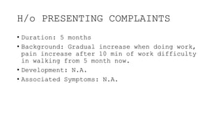
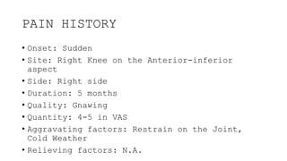
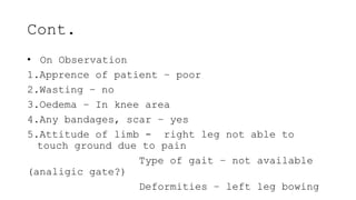
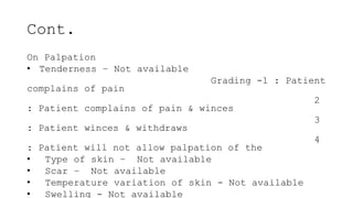
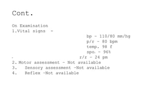
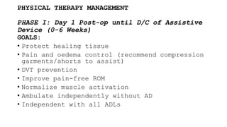
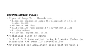
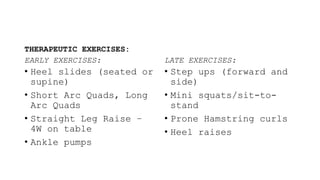
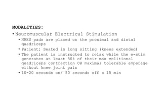
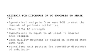
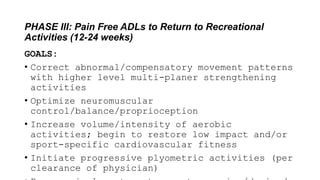
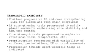
CASE PRESENTATION
DISCLAMIER
All the information provided in the presentation
is done with the verbal consent of the Patient
and his bystanders. All information provided are
done in accordance with
• Indian Medical Council Act 1956
• Information Technology Act
DEMOGRAPHIC DATA
• Name: Mr. ABC
• Age: 75
• Gender: Male
• Dominance: Right
• Occupation:
• C/o:
• Address: xxxxxxxxxxxx, Jalandhar Cant.
• Phone: xxxxxxxx84
CHIEF COMPLAINTS
• Difficulty in walking with pain in the right
knee
H/o PRESENTING COMPLAINTS
• Duration: 5 months
• Background: Gradual increase when doing work,
pain increase after 10 min of work difficulty
in walking from 5 month now.
• Development: N.A.
• Associated Symptoms: N.A.
PAIN HISTORY
• Onset: Sudden
• Site: Right Knee on the Anterior-inferior
aspect
• Side: Right side
• Duration: 5 months
• Quality: Gnawing
• Quantity: 4-5 in VAS
• Aggravating factors: Restrain on the Joint,
Cold Weather
• Relieving factors: N.A.
PAST HISTORY
• Known History of Tuberculosis – Not Applicable
• Bronchial Asthma – Not Applicable
• Blood Pressure – History from Past one month
(Pain related?)
• Diabetes – Not Applicable
• Cardiac Problems – Not Applicable (No Signs in
ECHO)
• Accidental History
OTHER PARTICULARS
FAMILY HISTORY: Not Applicable
PERSONAL HISTORY:
1. Diet – mixed
2. Appetite – adequate
3. Bowel/Bladder – clear
4. sleep – altered
5. Addiction – Not Available
SOCIO-ECONOMIC HISTORY
MEDICAL HISTORY
cont:
• SOCIO-ECONOMIC HISTORY – upper middle class
“according to 2023 annual report) (1)
•
Cont.
MEDICAL HISTORY –
Assessment
1.On Observation
2.On Palpation
3.On Examination
Cont.
• On Observation
1.Apprence of patient – poor
2.Wasting – no
3.Oedema – In knee area
4.Any bandages, scar – yes
5.Attitude of limb - right leg not able to
touch ground due to pain
Type of gait – not available
(analigic gate?)
Deformities – left leg bowing
Cont.
On Palpation
• Tenderness – Not available
Grading -1 : Patient
complains of pain
2
: Patient complains of pain & winces
3
: Patient winces & withdraws
4
: Patient will not allow palpation of the
• Type of skin – Not available
• Scar – Not available
• Temperature variation of skin - Not available
• Swelling - Not available
Cont.
On Examination
1.Vital signs -
bp – 110/80 mm/hg
p/r – 80 bpm
temp. 98 f
spo2 – 96%
• r/r – 24 pm
2. Motor assessment – Not available
3. Sensory assessment –Not available
4. Reflex –Not available
HEMATOLOGY REVIEW
• HEMOTOLOGY
• SERUM ANALYSIS
ANTHESI
PHYSICAL THERAPY MANAGEMENT
PHASE I: Day 1 Post-op until D/C of Assistive
Device (0-6 Weeks)
GOALS:
• Protect healing tissue
• Pain and oedema control (recommend compression
garments/shorts to assist)
• DVT prevention
• Improve pain-free ROM
• Normalize muscle activation
• Ambulate independently without AD
• Independent with all ADLs
PRECAUTION/RED FLAGS:
• Signs of Deep Vein Thromboses
• Localized tenderness along the distribution of deep
venous system
• Entire LE swelling
• Calf swelling >3cm compared to asymptomatic limb
• Pitting oedema
• Collateral superficial veins
• Mechanical block or clunk
• Lack of full knee extension by 4-6 weeks (Refer to
surgeon/or APP team for re-evaluation)
• AD required for ambulation after post-op week 6
THERAPEUTIC EXERCISES:
EARLY EXERCISES:
• Heel slides (seated or
supine)
• Short Arc Quads, Long
Arc Quads
• Straight Leg Raise –
4W on table
• Ankle pumps
LATE EXERCISES:
• Step ups (forward and
side)
• Mini squats/sit-to-
stand
• Prone Hamstring curls
• Heel raises
MODALITIES:
• Edema Control
• Cryotherapy at least 5x daily for the first week
• Cryotherapy at least 3x daily for week 1-6
• Neuromuscular Electrical Stimulation
• NMES pads are placed on the proximal and distal
quadriceps
• Patient: Seated in long sitting (knees extended)
• The patient is instructed to relax while the e-stim
generates at least 50% of their max volitional
quadriceps contraction OR maximal tolerable amperage
without knee joint pain
• 10-20 seconds on/ 50 seconds off x 15 min
CRITERIA TO PROGRESS TO PHASE II:
• Normalized gait pattern for community
ambulation (≥800 ft) without Assistive Device
• Knee extension normalized, knee flexion to 110
degrees
• SLR 2x10 without quad lag
• Minimal to no reactive pain and swelling with
ADLs and PT exercises
• Muscle activation and isolation is normalized
PHASE II: D/C of AD to Pain Free ADLs (6-12 weeks)
GOALS:
• Restore full PROM and AROM
• Progressively improve strength of the affected
LE musculature (core and LE muscles)
• Normalize postural/pelvic and LE control with
DL and SL activities
• Normalize gait at preferred walking speed for
community distances
• Tolerate ADLs without pain or limitation
PRECAUTIONS/RED FLAGS
• OK to progress strengthening exercises and
functional tasks as appropriate pending no
reactive pain or effusion
• Increase aerobic conditioning/endurance related
tasks monitoring reactive edema
THERAPEUTIC EXERCISES:
EARLY EXERCISES:
• Wall squats
• Mini lunges
• Step ups- progress to
single leg step ups
• Step downs
• 4 way hip
• Leg Press with light
resistance, higher reps
• Open Chain knee
extension
LATE EXERCISES:
• Full squat to 70 degrees
• Side steps with band
• Heel Taps
• Resisted walking
• Advanced bridges
• SLS and balance
progressions (like
wobble board)
MODALITIES:
• Neuromuscular Electrical Stimulation
• NMES pads are placed on the proximal and distal
quadriceps
• Patient: Seated in long sitting (knees extended)
• The patient is instructed to relax while the e-stim
generates at least 50% of their max volitional
quadriceps contraction OR maximal tolerable amperage
without knee joint pain
• 10-20 seconds on/ 50 seconds off x 15 min
CRITERIA FOR DISCHARGE OR TO PROGRESS TO PHASE
III:
• Symmetrical and pain free knee ROM to meet the
demands of patients activities
• Good (4/5) LE strength
• Symmetrical DL squat to at least 70 degrees
knee flexion
• Good quality movement as graded on forward step
down test
• Normalized gait pattern for community distances
of ambulation
PHASE III: Pain Free ADLs to Return to Recreational
Activities (12-24 weeks)
GOALS:
• Correct abnormal/compensatory movement patterns
with higher level multi-planer strengthening
activities
• Optimize neuromuscular
control/balance/proprioception
• Increase volume/intensity of aerobic
activities; begin to restore low impact and/or
sport-specific cardiovascular fitness
• Initiate progressive plyometric activities (per
clearance of physician)
PRECAUTIONS/RED FLAGS:
• Avoid sacrificing quality for quantity during
strengthening
• Ensure patient maintains full flexibility and
pain-free ROM as strength continues to increase
• Monitor/minimize reactive oedema when
increasing demand of task
• Closely monitor return to sport progression
THERAPEUTIC EXERCISES:
• Continue progressive LE and core strengthening
(DLSL for closed and open chain exercises)
• LE strengthening tasks progressed to multi-
planer movements emphasizing core stability and
hip/knee control
• Core strength tasks progressed to emphasize
rotational tasks (chops/lifts, etc)
• Proprioception progressed with variability of
surfaces, perturbations, UE or trunk movements
• Progression towards sport-specific tasks as
indicated
CARDIOVASCULAR EXERCISES:
• Dynamic Warm Up initiated
• Upright Bike/Elliptical progression (per PT and
patient preference)
• Swimming progression (per PT and patient
preference)
REFERENCES
• 1. Akram Z, Khairnar MR, Kusumakar A, Kumar JS,
Sabharwal H, Priyadarsini SS, et al. Updated B.
G. Prasad Socioeconomic Status Classification
for the year 2023. Journal of Indian
Association of Public Health Dentistry. 2023
Jun 30;21(2):204–5.
doi:10.4103/jiaphd.jiaphd_123_23

KNEE REPLACEMENT PHYSIOTHERAPY CASE.pptx

  • 1.
  • 2.
    DISCLAMIER All the informationprovided in the presentation is done with the verbal consent of the Patient and his bystanders. All information provided are done in accordance with • Indian Medical Council Act 1956 • Information Technology Act
  • 3.
    DEMOGRAPHIC DATA • Name:Mr. ABC • Age: 75 • Gender: Male • Dominance: Right • Occupation: • C/o: • Address: xxxxxxxxxxxx, Jalandhar Cant. • Phone: xxxxxxxx84
  • 4.
    CHIEF COMPLAINTS • Difficultyin walking with pain in the right knee
  • 5.
    H/o PRESENTING COMPLAINTS •Duration: 5 months • Background: Gradual increase when doing work, pain increase after 10 min of work difficulty in walking from 5 month now. • Development: N.A. • Associated Symptoms: N.A.
  • 6.
    PAIN HISTORY • Onset:Sudden • Site: Right Knee on the Anterior-inferior aspect • Side: Right side • Duration: 5 months • Quality: Gnawing • Quantity: 4-5 in VAS • Aggravating factors: Restrain on the Joint, Cold Weather • Relieving factors: N.A.
  • 7.
    PAST HISTORY • KnownHistory of Tuberculosis – Not Applicable • Bronchial Asthma – Not Applicable • Blood Pressure – History from Past one month (Pain related?) • Diabetes – Not Applicable • Cardiac Problems – Not Applicable (No Signs in ECHO) • Accidental History
  • 8.
    OTHER PARTICULARS FAMILY HISTORY:Not Applicable PERSONAL HISTORY: 1. Diet – mixed 2. Appetite – adequate 3. Bowel/Bladder – clear 4. sleep – altered 5. Addiction – Not Available SOCIO-ECONOMIC HISTORY MEDICAL HISTORY
  • 9.
    cont: • SOCIO-ECONOMIC HISTORY– upper middle class “according to 2023 annual report) (1) •
  • 10.
  • 12.
  • 13.
    Cont. • On Observation 1.Apprenceof patient – poor 2.Wasting – no 3.Oedema – In knee area 4.Any bandages, scar – yes 5.Attitude of limb - right leg not able to touch ground due to pain Type of gait – not available (analigic gate?) Deformities – left leg bowing
  • 14.
    Cont. On Palpation • Tenderness– Not available Grading -1 : Patient complains of pain 2 : Patient complains of pain & winces 3 : Patient winces & withdraws 4 : Patient will not allow palpation of the • Type of skin – Not available • Scar – Not available • Temperature variation of skin - Not available • Swelling - Not available
  • 16.
    Cont. On Examination 1.Vital signs- bp – 110/80 mm/hg p/r – 80 bpm temp. 98 f spo2 – 96% • r/r – 24 pm 2. Motor assessment – Not available 3. Sensory assessment –Not available 4. Reflex –Not available
  • 17.
  • 18.
  • 19.
    PHYSICAL THERAPY MANAGEMENT PHASEI: Day 1 Post-op until D/C of Assistive Device (0-6 Weeks) GOALS: • Protect healing tissue • Pain and oedema control (recommend compression garments/shorts to assist) • DVT prevention • Improve pain-free ROM • Normalize muscle activation • Ambulate independently without AD • Independent with all ADLs
  • 20.
    PRECAUTION/RED FLAGS: • Signsof Deep Vein Thromboses • Localized tenderness along the distribution of deep venous system • Entire LE swelling • Calf swelling >3cm compared to asymptomatic limb • Pitting oedema • Collateral superficial veins • Mechanical block or clunk • Lack of full knee extension by 4-6 weeks (Refer to surgeon/or APP team for re-evaluation) • AD required for ambulation after post-op week 6
  • 21.
    THERAPEUTIC EXERCISES: EARLY EXERCISES: •Heel slides (seated or supine) • Short Arc Quads, Long Arc Quads • Straight Leg Raise – 4W on table • Ankle pumps LATE EXERCISES: • Step ups (forward and side) • Mini squats/sit-to- stand • Prone Hamstring curls • Heel raises
  • 22.
    MODALITIES: • Edema Control •Cryotherapy at least 5x daily for the first week • Cryotherapy at least 3x daily for week 1-6 • Neuromuscular Electrical Stimulation • NMES pads are placed on the proximal and distal quadriceps • Patient: Seated in long sitting (knees extended) • The patient is instructed to relax while the e-stim generates at least 50% of their max volitional quadriceps contraction OR maximal tolerable amperage without knee joint pain • 10-20 seconds on/ 50 seconds off x 15 min
  • 23.
    CRITERIA TO PROGRESSTO PHASE II: • Normalized gait pattern for community ambulation (≥800 ft) without Assistive Device • Knee extension normalized, knee flexion to 110 degrees • SLR 2x10 without quad lag • Minimal to no reactive pain and swelling with ADLs and PT exercises • Muscle activation and isolation is normalized
  • 24.
    PHASE II: D/Cof AD to Pain Free ADLs (6-12 weeks) GOALS: • Restore full PROM and AROM • Progressively improve strength of the affected LE musculature (core and LE muscles) • Normalize postural/pelvic and LE control with DL and SL activities • Normalize gait at preferred walking speed for community distances • Tolerate ADLs without pain or limitation
  • 25.
    PRECAUTIONS/RED FLAGS • OKto progress strengthening exercises and functional tasks as appropriate pending no reactive pain or effusion • Increase aerobic conditioning/endurance related tasks monitoring reactive edema
  • 26.
    THERAPEUTIC EXERCISES: EARLY EXERCISES: •Wall squats • Mini lunges • Step ups- progress to single leg step ups • Step downs • 4 way hip • Leg Press with light resistance, higher reps • Open Chain knee extension LATE EXERCISES: • Full squat to 70 degrees • Side steps with band • Heel Taps • Resisted walking • Advanced bridges • SLS and balance progressions (like wobble board)
  • 27.
    MODALITIES: • Neuromuscular ElectricalStimulation • NMES pads are placed on the proximal and distal quadriceps • Patient: Seated in long sitting (knees extended) • The patient is instructed to relax while the e-stim generates at least 50% of their max volitional quadriceps contraction OR maximal tolerable amperage without knee joint pain • 10-20 seconds on/ 50 seconds off x 15 min
  • 28.
    CRITERIA FOR DISCHARGEOR TO PROGRESS TO PHASE III: • Symmetrical and pain free knee ROM to meet the demands of patients activities • Good (4/5) LE strength • Symmetrical DL squat to at least 70 degrees knee flexion • Good quality movement as graded on forward step down test • Normalized gait pattern for community distances of ambulation
  • 29.
    PHASE III: PainFree ADLs to Return to Recreational Activities (12-24 weeks) GOALS: • Correct abnormal/compensatory movement patterns with higher level multi-planer strengthening activities • Optimize neuromuscular control/balance/proprioception • Increase volume/intensity of aerobic activities; begin to restore low impact and/or sport-specific cardiovascular fitness • Initiate progressive plyometric activities (per clearance of physician)
  • 30.
    PRECAUTIONS/RED FLAGS: • Avoidsacrificing quality for quantity during strengthening • Ensure patient maintains full flexibility and pain-free ROM as strength continues to increase • Monitor/minimize reactive oedema when increasing demand of task • Closely monitor return to sport progression
  • 31.
    THERAPEUTIC EXERCISES: • Continueprogressive LE and core strengthening (DLSL for closed and open chain exercises) • LE strengthening tasks progressed to multi- planer movements emphasizing core stability and hip/knee control • Core strength tasks progressed to emphasize rotational tasks (chops/lifts, etc) • Proprioception progressed with variability of surfaces, perturbations, UE or trunk movements • Progression towards sport-specific tasks as indicated
  • 32.
    CARDIOVASCULAR EXERCISES: • DynamicWarm Up initiated • Upright Bike/Elliptical progression (per PT and patient preference) • Swimming progression (per PT and patient preference)
  • 33.
    REFERENCES • 1. AkramZ, Khairnar MR, Kusumakar A, Kumar JS, Sabharwal H, Priyadarsini SS, et al. Updated B. G. Prasad Socioeconomic Status Classification for the year 2023. Journal of Indian Association of Public Health Dentistry. 2023 Jun 30;21(2):204–5. doi:10.4103/jiaphd.jiaphd_123_23