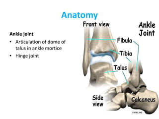
SlideShare a Scribd company logo
1 of 63
Download to read offline
Examination of foot and Ankle
Dr Manoj Das
Department of Orthopedics
Institute Of Medicine , TUTH, Nepal
objectives
• Assess
• Diagnose
• Treat
overview
• The ankle and foot is a complex structure comprised
of 28 bones (including 2 sesamoid bones) and 55
articulations (including 30 synovial joints),
interconnected by ligaments and muscles
• In addition to sustaining substantial forces, the foot
and ankle serve to convert the rotational movements
that occur with weight bearing activities into sagittal,
frontal, and transverse movements
Anatomy
• Anatomically and biomechanically, the foot is
often subdivided into:
• The rearfoot or hindfoot (the talus and
calcaneus)
• The midfoot (the navicular, cuboid and the 3
cuneiforms)
• The forefoot (the 14 bones of the toes, the 5
metatarsals, and the medial and lateral
sesamoids)
Anatomy
Ankle joint
• Articulation of dome of
talus in ankle mortice
• Hinge joint
Lateral ligament complex
• Lateral complex
– Ant. talofibular
– calcaneofibular
– Post. talofibular
• Syndesmosis
– Ant. Inf. tibiofibular
– Post.Inf. tibiofibular
Syndesmotic Structures
• Syndesmosis:
– Ant. Inf. Tibiofibular
ligament
– Post. Inf. Tibiofibular
ligament
– Transverse tibiofibular
ligament
– Interosseous membrane
Medial Ankle Structures
• Major Ligament
complex is called the
Deltoid Ligament.
• It is the strongest of the
ankle ligaments
Subtalar joint
– The subtalar joint is a
synovial, bicondylar
compound joint consisting of
two separate, modified ovoid
surfaces with their own joint
cavities (one male and one
female)
HISTORY TAKING
• Take a HISTORY
– What is the patient’s chief complaint?
– Pain?
• Where? When? How bad? What is it like?
• What makes it better?
• What makes it worse?
– Acute Injury vs. Chronic
– Progression of Symptoms?
HISTORY TAKING: Background
Information
• Any Previous Injuries
• Past Surgical History
• Past Medical History
• Medications
• Allergies
• Social History
– Work situation (laboring type job?)
– Home situation
Examination of the foot and ankle
STEPS in the PHYSICAL EXAMINATION
Consent
Privacy
Exposure
Gait analysis
Obsevation
Palpation
Range of motion
Neurovascular assessment
Special tests
Exposure
Both shoes and socks off. At least have
trousers rolled up to the knees,
preferably down to
underwear
Gait Analysis
OBJECTIVES
• Identify the phases of gait
and
perform a functional gait
analysis.
GAIT ANALYSIS
STRIDE LENGTH
• Symmetrical side-to-side?
• Shortened?
FOOT PROGRESSION
• Symmetrical?
• Neutral?
• Internal?
• External?
Observation
• Built
• Posture
• Weight bearing: equal on both sides
• Compare weight bearing and non wreight bearing
position of foot in
- Anterior View
- Posterior View
-Lateral View
• See for Contour of Foot
soft tissue swelling
Bony callosity
Observation- Deformities
• Forefoot Varus
mid tarsal joint- Inversion
Subtalar joint- Neutral
Forefoot Valgus
mid tarsal joint- Eversion
Subtalar joint- Neutral
Observation- Deformities…
Talipes Eqinus
• Plantar flexed foot
• Can cause plantar fascitis,
metatarsalgia
Observation- Deformities…
• Claw Toes
MTP joint- Hyper Extension
IP joint- Flexion
• Hammer Toes
MTP- Hyperextended
PIP- Flexed
DIP- Hyperextended
Observation- Deformities…
Hallux Rigidus
-Stiffness of Great toe at
MTP
- May be due to OA
Observation –Deformities…
• Splay foot
Spread of Metatarsal
• Rocker Bottom Foot
Forefoot in dorsiflexion
Arch may be absent
Standing and Weight bearing:
Anteropsterior view
• Weight Bearing:
Equal on both feet and forefoot/hindfoot
• Position of foot
Supination/pronation
• Ask the Patient to walk on heel and toes:
Gives the idea about muscle power or functional
range of motion
• Does the patient use Cane or stick?
Use of cane on opposite side decrease the load on
ankle by 1/3 of body weight
Standing and Weight bearing:
Anteropsterior view
• Check the toes
if parallel/ straight/
• Spurs/ exostosis/Swelling
• Check for tibia/ knee
Standing and Weight bearing:
Lateral view
• Observe longitudnal arch of
foot
• Medial longitudnal arch
should be higher than
lateral
Standing and Weight bearing:
Posterior view
• Bulk of calf : compare on both sides
• Achillis tendon : Vertical on both sides
• Observe calcaneum for
shape
position
callosity
• Position of malleolus
Foot Print Pattern
• Light film of baby’s oil on
patient foot and apply
powder
• Ask patient to step on piece
of colored paper
• Obsreve for pattern of foot
PALPATION
SURFACE ANATOMY IS THE KEY!!!
Palpate for
local rise of temperature
Local tenderness
Palpation of specific areas-
Palpation(Bony)…
Medial aspect
Palpation(Bony)…
Lateral Aspect
Palpation (soft tissue)…
Zone 1
• Head of 1st MT bone
• Patholology – gout, hallux
valgus
Palpation (soft tissue)…
Zone 2
• Navicular tubercle and talar
head
Palpation (soft tissue)…
Zone 3 - Medial malleolus
• Palpate
- Deltoid ligament
• palpate follwing structure in
depression between posterior
aspect of medial malleoli and
achillis tendon
-Tibialis posterior tendon
-Flexor digitorum longus tendon;
- Posterior tibial artery and tibial
nerve;
-Flexor hallucis longus tendon
Palpation (soft tissue)…
Zone 4 - Dorsum of foot
between malleoli
• 3 important tendons and one
vessel that pass between the
malleoli. From medial to
lateral they are:
- Tibialis anterior tendon
Extensor hallucis longus
tendon
- Dorsal pedal artery;
Extensor digitorum longus
tendon
-Peroneus Tertiu
Zone 4 - Dorsum of foot
between malleoli…
Palpation (soft tissue)…
Zone 5 – Lateral Malleoli
• 3 clinically important
ligaments, which comprise the
lateral collateral ligaments of
the ankle joint . From anterior
to posterior, they are:
-Anterior talofibular ligament
-Calcaneofibular ligament
-Posterior talofibular ligament
• Zone 6 sinus tarsi
commonly involved in
ankle sprain
• Zone 7 head of 5th MT
Tailors bunion
Palpation (soft tissue)…
• Zone 8 Calcaneum
Retrocalcaneal bursa/
calcaneal bursa
• Zone 9 plantar surface
Palpation (soft tissue)…
Zone 10 toes
Range of Motion
Range of Motion
Ankle motion
Check the range of motion
• Dorsiflexion- 10 to 30
-Reduce the talonavicular
joint
• Plantar flexion – 20 to 50
Range of Motion…
Hind foot – Inversion and
Eversion
• Patient sitting on stool with
knee flexed at 70 degree
• Hold ankle firmly from
dorsum to fix talus by
dosiflexion
• Hold body of calcaneum in
between thumb on one side
and index and middle finger
on other side with other
hand
• Turn in for inversion and
turn out for eversion
• I= 35 degree E= 25 degree
Range of Motion
Adduction and Abduction of Fore
foot
• Hold hind foot from dorsum
with one hand
• Hold forefoot with other
hand
• Passively deviate forefoot
inward for adduction and
outward for adduction
Range of Motion….
First MTP joint motion
• Principally involved in toe
off phase of gait
• Stabilize foot and move
great toe through flexion
and extension
NEUROVASCULAR ASSESSMENT
• Nerve Function
- motor
- Sensory
- Reflexes
• Vascular Status
– Distal pulses
– Capillary refill
Neurological examination(Motor)…
Dorsiflexers
• Tibialis Anterior
Deep Peroneal Nerve L4
• Extensor Hallucis Longus L5
• Extensor Digitorum Longus
L5
Neurological examination(Motor)…
Plantar Flexors
• Peroneus Longus and Brevis
-Suprficial peroneal Nerve,
S1
• Gastrnemius and Soleus
- Tibial Nerve, S1 S2
• Flexor Digitorum Longus
-Tibial Nerve L5
• Tibialis Posterior
- Tibial Nerve L5
Neurological examination(Reflexes)…
Ankle Reflex, S1
Neurological examination(Sensory)…
Special test
Stress test
For medial and lateral
collateral ligament
- Place the ankle in neutral
position
- Hold the lower leg firmly from
front by one hand
- Hold the foot at about level of
talus by opposite hand
- For testing the lateral
collateral ligament , invert the
foot and for testing of medial
collateral ligament stress has
to be given in opposite
direction
Evaluating for Syndesmotic injury
• 2 Tests for injury to the
syndesmosis
– The Squeeze test
– External rotation test
Anterior Drawer test
• For integrity of capsule and
anterior talofibular ligament
• Pulling the heel
anteromedially against
resistance applied by the
other hand over anterior
aspect of lower leg
• Anterior subluxation of 3
mm of talus is pathological
Test for rupture of tendo-Achilles
Thompson test
• Prone position with feet
projecting beyond
examining table
• Calf muscle squeezed
• Normal or partially torn-
planter flexion
• Complete rupture- No
movement of foot
Test for rupture of tendo-Achilles
Needle test
• For integrity of distal 10 cm of
tendo-achillles
• Prone position
• 25 G hypodermic needle
pierced through skin at 10 cm
above upper end of calcaneum
and just medial to midline of
calf
• Foot passively plantiflexed and
dorsiflexed
• Normal- needle swivel in
direction opposite to
movement of foot
Test for pre-achillles and post achilles
pathologies
• Pt asked to walk on toes
with heel off the ground-
pain in pre achilles
pathology
• Walk on heel- pain in post
achilles pathology
• Achilles tendinitis – pain in
both mode of walking, more
on walking on toes
Ankle Dorsiflexion Test
• To determine whether
gastronimius or soleus
causing limitation of ankle
dorsiflexion
• With flexion of the knee
joint, ankle dorsiflexion
achieved – Gastronemius
• Not affected by flexion of
knee- Soleus
Homan’s sign
• Test for deep vein
thrombhophlebitis
• Forcibly dorsiflex ankle
with leg in extension
• Pain in calf muscle
Measurement of equinus
deformity
• Position- lying on bed on
lateral position
• Passively dorsiflex as far as
possible
• Measure angle between
long axis of leg and long axis
of midfoot
• Substract 90 from angle
Tibial Torsion Test
• To determine whether
toeing in is due to internal
rotation of tibia
• Normally a line drawn
between malleoli is rotated
is rotated externally 15
degree from a
perpendicular line drawn
from the tibial tubercle to
ankle
• In tibial torsion the
malleolar line may face
directly anterioly close to
perpendicular line
Forefoot Adduction Correction Test
• Forefoot adduction is common
in children which may or may
not need correction
• If adduction can be corrected
manually and abduction can
be done beyond neutral
position – NO TREATMENT
• If only partially corrected to
neutral or less than neutral –
CAST CORRECTION
Colman Block test
- coleman block test evaluates hindfoot flexibility and
pronation of forefoot;
-
- initial deformity is in the forefoot followed by
subsequent changes in the hindfoot
- test is performed by placing the patient's foot on
wood block, 2.5 to 4 cm thick, with the heel and lateral
border of foot on the block and bearing full
weight while the first, second, & 3rd metatarsals are
allowed to hang freely into plantar flexion and
pronation;
- Interpretation:
- if heel varus corrects while the patient is standing
on the block, hindfoot is considered flexible;
- if subtalar joint is supple & correct w/ block test,
then surgical procedures may be directed to correcting
forefoot pronation, which is usually due to plantar
flexion of 1st metatarsal;
- if hindfoot is rigid, then surgical correction of both
forefoot & hindfoot are required
Examination of footwear
• Distortion of shape-
uderlying rigid defomity
• Wrinkling of footwear- in
persistent varus of heel,
deep wrinkles on inner
aspect of heel
• Bulging out thinning
• Deformity of sole
Last but not the least….
• DON’T FORGET
TO EXAMINE
SPINE , HIP AND
KNEE !!!!
Thank You

More Related Content

Similar to mypptfoot-160706125859.pdf

Basic introduction of human anatomy
Basic introduction of human anatomyBasic introduction of human anatomy
Basic introduction of human anatomyDr Neeraj Tiwari
 
Patellofemoral Pain Syndrome
Patellofemoral Pain SyndromePatellofemoral Pain Syndrome
Patellofemoral Pain SyndromeDrFarhaPT
 
Rotational deformities of lower extremity in children
Rotational deformities of lower extremity in childrenRotational deformities of lower extremity in children
Rotational deformities of lower extremity in childrenAhmed Ashour dr.
 
Manualmusletesting 361 390
Manualmusletesting 361 390Manualmusletesting 361 390
Manualmusletesting 361 390Anaum1990
 
Assessmentofthemusclo skletalsystem- post rn
Assessmentofthemusclo skletalsystem- post rnAssessmentofthemusclo skletalsystem- post rn
Assessmentofthemusclo skletalsystem- post rnshaheen khowaja
 
Ankle & Foot Xray & Surgical Approaches
Ankle & Foot Xray & Surgical ApproachesAnkle & Foot Xray & Surgical Approaches
Ankle & Foot Xray & Surgical ApproachesMirant Dave
 
gait analysis.pdf
gait analysis.pdfgait analysis.pdf
gait analysis.pdfzaiqar1
 
Gait analysis and.ppt by ramachandra
Gait analysis and.ppt by ramachandraGait analysis and.ppt by ramachandra
Gait analysis and.ppt by ramachandraramachandra reddy
 
Arches of the foot and plantar fascitiis
Arches of the foot and plantar fascitiisArches of the foot and plantar fascitiis
Arches of the foot and plantar fascitiisDr Nirav Mungalpara
 
Assessment of the musclo skletal system
Assessment of the musclo skletal systemAssessment of the musclo skletal system
Assessment of the musclo skletal systemMohamed Mansor
 
Assessment of the musclo skletal system
Assessment of the musclo skletal systemAssessment of the musclo skletal system
Assessment of the musclo skletal systemMohamed Mansor
 
CTEV DEFORMITY.pptx
CTEV DEFORMITY.pptxCTEV DEFORMITY.pptx
CTEV DEFORMITY.pptxEnejoJoseph
 

Similar to mypptfoot-160706125859.pdf (20)

History and examintion of knee joint slideshare
History and examintion of knee joint slideshareHistory and examintion of knee joint slideshare
History and examintion of knee joint slideshare
 
Basic introduction of human anatomy
Basic introduction of human anatomyBasic introduction of human anatomy
Basic introduction of human anatomy
 
Patellofemoral Pain Syndrome
Patellofemoral Pain SyndromePatellofemoral Pain Syndrome
Patellofemoral Pain Syndrome
 
Ctev
CtevCtev
Ctev
 
Rotational deformities of lower extremity in children
Rotational deformities of lower extremity in childrenRotational deformities of lower extremity in children
Rotational deformities of lower extremity in children
 
Manualmusletesting 361 390
Manualmusletesting 361 390Manualmusletesting 361 390
Manualmusletesting 361 390
 
Assessmentofthemusclo skletalsystem- post rn
Assessmentofthemusclo skletalsystem- post rnAssessmentofthemusclo skletalsystem- post rn
Assessmentofthemusclo skletalsystem- post rn
 
Joints and their movements
Joints and their movementsJoints and their movements
Joints and their movements
 
Ankle & Foot Xray & Surgical Approaches
Ankle & Foot Xray & Surgical ApproachesAnkle & Foot Xray & Surgical Approaches
Ankle & Foot Xray & Surgical Approaches
 
Lower Extremities.pptx
Lower Extremities.pptxLower Extremities.pptx
Lower Extremities.pptx
 
Flat foot and Cavus foot
 Flat foot and Cavus foot Flat foot and Cavus foot
Flat foot and Cavus foot
 
gait analysis.pdf
gait analysis.pdfgait analysis.pdf
gait analysis.pdf
 
Gait analysis and.ppt by ramachandra
Gait analysis and.ppt by ramachandraGait analysis and.ppt by ramachandra
Gait analysis and.ppt by ramachandra
 
Gait Cycle.pptx
Gait Cycle.pptxGait Cycle.pptx
Gait Cycle.pptx
 
Gait Cycle.pptx
Gait Cycle.pptxGait Cycle.pptx
Gait Cycle.pptx
 
Arches of the foot and plantar fascitiis
Arches of the foot and plantar fascitiisArches of the foot and plantar fascitiis
Arches of the foot and plantar fascitiis
 
Assessment of the musclo skletal system
Assessment of the musclo skletal systemAssessment of the musclo skletal system
Assessment of the musclo skletal system
 
Assessment of the musclo skletal system
Assessment of the musclo skletal systemAssessment of the musclo skletal system
Assessment of the musclo skletal system
 
CTEV PPT.pptx
CTEV PPT.pptxCTEV PPT.pptx
CTEV PPT.pptx
 
CTEV DEFORMITY.pptx
CTEV DEFORMITY.pptxCTEV DEFORMITY.pptx
CTEV DEFORMITY.pptx
 

Recently uploaded

Capitol Tech U Doctoral Presentation - April 2024.pptx
Capitol Tech U Doctoral Presentation - April 2024.pptxCapitol Tech U Doctoral Presentation - April 2024.pptx
Capitol Tech U Doctoral Presentation - April 2024.pptxCapitolTechU
 
Organic Name Reactions for the students and aspirants of Chemistry12th.pptx
Organic Name Reactions  for the students and aspirants of Chemistry12th.pptxOrganic Name Reactions  for the students and aspirants of Chemistry12th.pptx
Organic Name Reactions for the students and aspirants of Chemistry12th.pptxVS Mahajan Coaching Centre
 
Enzyme, Pharmaceutical Aids, Miscellaneous Last Part of Chapter no 5th.pdf
Enzyme, Pharmaceutical Aids, Miscellaneous Last Part of Chapter no 5th.pdfEnzyme, Pharmaceutical Aids, Miscellaneous Last Part of Chapter no 5th.pdf
Enzyme, Pharmaceutical Aids, Miscellaneous Last Part of Chapter no 5th.pdfSumit Tiwari
 
Computed Fields and api Depends in the Odoo 17
Computed Fields and api Depends in the Odoo 17Computed Fields and api Depends in the Odoo 17
Computed Fields and api Depends in the Odoo 17Celine George
 
Software Engineering Methodologies (overview)
Software Engineering Methodologies (overview)Software Engineering Methodologies (overview)
Software Engineering Methodologies (overview)eniolaolutunde
 
Biting mechanism of poisonous snakes.pdf
Biting mechanism of poisonous snakes.pdfBiting mechanism of poisonous snakes.pdf
Biting mechanism of poisonous snakes.pdfadityarao40181
 
Alper Gobel In Media Res Media Component
Alper Gobel In Media Res Media ComponentAlper Gobel In Media Res Media Component
Alper Gobel In Media Res Media ComponentInMediaRes1
 
Roles & Responsibilities in Pharmacovigilance
Roles & Responsibilities in PharmacovigilanceRoles & Responsibilities in Pharmacovigilance
Roles & Responsibilities in PharmacovigilanceSamikshaHamane
 
18-04-UA_REPORT_MEDIALITERAСY_INDEX-DM_23-1-final-eng.pdf
18-04-UA_REPORT_MEDIALITERAСY_INDEX-DM_23-1-final-eng.pdf18-04-UA_REPORT_MEDIALITERAСY_INDEX-DM_23-1-final-eng.pdf
18-04-UA_REPORT_MEDIALITERAСY_INDEX-DM_23-1-final-eng.pdfssuser54595a
 
Types of Journalistic Writing Grade 8.pptx
Types of Journalistic Writing Grade 8.pptxTypes of Journalistic Writing Grade 8.pptx
Types of Journalistic Writing Grade 8.pptxEyham Joco
 
Final demo Grade 9 for demo Plan dessert.pptx
Final demo Grade 9 for demo Plan dessert.pptxFinal demo Grade 9 for demo Plan dessert.pptx
Final demo Grade 9 for demo Plan dessert.pptxAvyJaneVismanos
 
Introduction to AI in Higher Education_draft.pptx
Introduction to AI in Higher Education_draft.pptxIntroduction to AI in Higher Education_draft.pptx
Introduction to AI in Higher Education_draft.pptxpboyjonauth
 
call girls in Kamla Market (DELHI) 🔝 >༒9953330565🔝 genuine Escort Service 🔝✔️✔️
call girls in Kamla Market (DELHI) 🔝 >༒9953330565🔝 genuine Escort Service 🔝✔️✔️call girls in Kamla Market (DELHI) 🔝 >༒9953330565🔝 genuine Escort Service 🔝✔️✔️
call girls in Kamla Market (DELHI) 🔝 >༒9953330565🔝 genuine Escort Service 🔝✔️✔️9953056974 Low Rate Call Girls In Saket, Delhi NCR
 
भारत-रोम व्यापार.pptx, Indo-Roman Trade,
भारत-रोम व्यापार.pptx, Indo-Roman Trade,भारत-रोम व्यापार.pptx, Indo-Roman Trade,
भारत-रोम व्यापार.pptx, Indo-Roman Trade,Virag Sontakke
 
Crayon Activity Handout For the Crayon A
Crayon Activity Handout For the Crayon ACrayon Activity Handout For the Crayon A
Crayon Activity Handout For the Crayon AUnboundStockton
 
ECONOMIC CONTEXT - LONG FORM TV DRAMA - PPT
ECONOMIC CONTEXT - LONG FORM TV DRAMA - PPTECONOMIC CONTEXT - LONG FORM TV DRAMA - PPT
ECONOMIC CONTEXT - LONG FORM TV DRAMA - PPTiammrhaywood
 
POINT- BIOCHEMISTRY SEM 2 ENZYMES UNIT 5.pptx
POINT- BIOCHEMISTRY SEM 2 ENZYMES UNIT 5.pptxPOINT- BIOCHEMISTRY SEM 2 ENZYMES UNIT 5.pptx
POINT- BIOCHEMISTRY SEM 2 ENZYMES UNIT 5.pptxSayali Powar
 
Meghan Sutherland In Media Res Media Component
Meghan Sutherland In Media Res Media ComponentMeghan Sutherland In Media Res Media Component
Meghan Sutherland In Media Res Media ComponentInMediaRes1
 

Recently uploaded (20)

Capitol Tech U Doctoral Presentation - April 2024.pptx
Capitol Tech U Doctoral Presentation - April 2024.pptxCapitol Tech U Doctoral Presentation - April 2024.pptx
Capitol Tech U Doctoral Presentation - April 2024.pptx
 
Organic Name Reactions for the students and aspirants of Chemistry12th.pptx
Organic Name Reactions  for the students and aspirants of Chemistry12th.pptxOrganic Name Reactions  for the students and aspirants of Chemistry12th.pptx
Organic Name Reactions for the students and aspirants of Chemistry12th.pptx
 
Enzyme, Pharmaceutical Aids, Miscellaneous Last Part of Chapter no 5th.pdf
Enzyme, Pharmaceutical Aids, Miscellaneous Last Part of Chapter no 5th.pdfEnzyme, Pharmaceutical Aids, Miscellaneous Last Part of Chapter no 5th.pdf
Enzyme, Pharmaceutical Aids, Miscellaneous Last Part of Chapter no 5th.pdf
 
Computed Fields and api Depends in the Odoo 17
Computed Fields and api Depends in the Odoo 17Computed Fields and api Depends in the Odoo 17
Computed Fields and api Depends in the Odoo 17
 
Software Engineering Methodologies (overview)
Software Engineering Methodologies (overview)Software Engineering Methodologies (overview)
Software Engineering Methodologies (overview)
 
Biting mechanism of poisonous snakes.pdf
Biting mechanism of poisonous snakes.pdfBiting mechanism of poisonous snakes.pdf
Biting mechanism of poisonous snakes.pdf
 
Alper Gobel In Media Res Media Component
Alper Gobel In Media Res Media ComponentAlper Gobel In Media Res Media Component
Alper Gobel In Media Res Media Component
 
Roles & Responsibilities in Pharmacovigilance
Roles & Responsibilities in PharmacovigilanceRoles & Responsibilities in Pharmacovigilance
Roles & Responsibilities in Pharmacovigilance
 
18-04-UA_REPORT_MEDIALITERAСY_INDEX-DM_23-1-final-eng.pdf
18-04-UA_REPORT_MEDIALITERAСY_INDEX-DM_23-1-final-eng.pdf18-04-UA_REPORT_MEDIALITERAСY_INDEX-DM_23-1-final-eng.pdf
18-04-UA_REPORT_MEDIALITERAСY_INDEX-DM_23-1-final-eng.pdf
 
Types of Journalistic Writing Grade 8.pptx
Types of Journalistic Writing Grade 8.pptxTypes of Journalistic Writing Grade 8.pptx
Types of Journalistic Writing Grade 8.pptx
 
ESSENTIAL of (CS/IT/IS) class 06 (database)
ESSENTIAL of (CS/IT/IS) class 06 (database)ESSENTIAL of (CS/IT/IS) class 06 (database)
ESSENTIAL of (CS/IT/IS) class 06 (database)
 
Final demo Grade 9 for demo Plan dessert.pptx
Final demo Grade 9 for demo Plan dessert.pptxFinal demo Grade 9 for demo Plan dessert.pptx
Final demo Grade 9 for demo Plan dessert.pptx
 
Introduction to AI in Higher Education_draft.pptx
Introduction to AI in Higher Education_draft.pptxIntroduction to AI in Higher Education_draft.pptx
Introduction to AI in Higher Education_draft.pptx
 
call girls in Kamla Market (DELHI) 🔝 >༒9953330565🔝 genuine Escort Service 🔝✔️✔️
call girls in Kamla Market (DELHI) 🔝 >༒9953330565🔝 genuine Escort Service 🔝✔️✔️call girls in Kamla Market (DELHI) 🔝 >༒9953330565🔝 genuine Escort Service 🔝✔️✔️
call girls in Kamla Market (DELHI) 🔝 >༒9953330565🔝 genuine Escort Service 🔝✔️✔️
 
भारत-रोम व्यापार.pptx, Indo-Roman Trade,
भारत-रोम व्यापार.pptx, Indo-Roman Trade,भारत-रोम व्यापार.pptx, Indo-Roman Trade,
भारत-रोम व्यापार.pptx, Indo-Roman Trade,
 
Crayon Activity Handout For the Crayon A
Crayon Activity Handout For the Crayon ACrayon Activity Handout For the Crayon A
Crayon Activity Handout For the Crayon A
 
ECONOMIC CONTEXT - LONG FORM TV DRAMA - PPT
ECONOMIC CONTEXT - LONG FORM TV DRAMA - PPTECONOMIC CONTEXT - LONG FORM TV DRAMA - PPT
ECONOMIC CONTEXT - LONG FORM TV DRAMA - PPT
 
POINT- BIOCHEMISTRY SEM 2 ENZYMES UNIT 5.pptx
POINT- BIOCHEMISTRY SEM 2 ENZYMES UNIT 5.pptxPOINT- BIOCHEMISTRY SEM 2 ENZYMES UNIT 5.pptx
POINT- BIOCHEMISTRY SEM 2 ENZYMES UNIT 5.pptx
 
Model Call Girl in Tilak Nagar Delhi reach out to us at 🔝9953056974🔝
Model Call Girl in Tilak Nagar Delhi reach out to us at 🔝9953056974🔝Model Call Girl in Tilak Nagar Delhi reach out to us at 🔝9953056974🔝
Model Call Girl in Tilak Nagar Delhi reach out to us at 🔝9953056974🔝
 
Meghan Sutherland In Media Res Media Component
Meghan Sutherland In Media Res Media ComponentMeghan Sutherland In Media Res Media Component
Meghan Sutherland In Media Res Media Component
 

mypptfoot-160706125859.pdf

  • 1. Examination of foot and Ankle Dr Manoj Das Department of Orthopedics Institute Of Medicine , TUTH, Nepal
  • 3. overview • The ankle and foot is a complex structure comprised of 28 bones (including 2 sesamoid bones) and 55 articulations (including 30 synovial joints), interconnected by ligaments and muscles • In addition to sustaining substantial forces, the foot and ankle serve to convert the rotational movements that occur with weight bearing activities into sagittal, frontal, and transverse movements
  • 4. Anatomy • Anatomically and biomechanically, the foot is often subdivided into: • The rearfoot or hindfoot (the talus and calcaneus) • The midfoot (the navicular, cuboid and the 3 cuneiforms) • The forefoot (the 14 bones of the toes, the 5 metatarsals, and the medial and lateral sesamoids)
  • 5. Anatomy Ankle joint • Articulation of dome of talus in ankle mortice • Hinge joint
  • 6. Lateral ligament complex • Lateral complex – Ant. talofibular – calcaneofibular – Post. talofibular • Syndesmosis – Ant. Inf. tibiofibular – Post.Inf. tibiofibular
  • 7. Syndesmotic Structures • Syndesmosis: – Ant. Inf. Tibiofibular ligament – Post. Inf. Tibiofibular ligament – Transverse tibiofibular ligament – Interosseous membrane
  • 8. Medial Ankle Structures • Major Ligament complex is called the Deltoid Ligament. • It is the strongest of the ankle ligaments
  • 9. Subtalar joint – The subtalar joint is a synovial, bicondylar compound joint consisting of two separate, modified ovoid surfaces with their own joint cavities (one male and one female)
  • 10. HISTORY TAKING • Take a HISTORY – What is the patient’s chief complaint? – Pain? • Where? When? How bad? What is it like? • What makes it better? • What makes it worse? – Acute Injury vs. Chronic – Progression of Symptoms? HISTORY TAKING: Background Information • Any Previous Injuries • Past Surgical History • Past Medical History • Medications • Allergies • Social History – Work situation (laboring type job?) – Home situation
  • 11. Examination of the foot and ankle STEPS in the PHYSICAL EXAMINATION Consent Privacy Exposure Gait analysis Obsevation Palpation Range of motion Neurovascular assessment Special tests
  • 12. Exposure Both shoes and socks off. At least have trousers rolled up to the knees, preferably down to underwear
  • 13. Gait Analysis OBJECTIVES • Identify the phases of gait and perform a functional gait analysis. GAIT ANALYSIS STRIDE LENGTH • Symmetrical side-to-side? • Shortened? FOOT PROGRESSION • Symmetrical? • Neutral? • Internal? • External?
  • 14. Observation • Built • Posture • Weight bearing: equal on both sides • Compare weight bearing and non wreight bearing position of foot in - Anterior View - Posterior View -Lateral View • See for Contour of Foot soft tissue swelling Bony callosity
  • 15. Observation- Deformities • Forefoot Varus mid tarsal joint- Inversion Subtalar joint- Neutral Forefoot Valgus mid tarsal joint- Eversion Subtalar joint- Neutral
  • 16. Observation- Deformities… Talipes Eqinus • Plantar flexed foot • Can cause plantar fascitis, metatarsalgia
  • 17. Observation- Deformities… • Claw Toes MTP joint- Hyper Extension IP joint- Flexion • Hammer Toes MTP- Hyperextended PIP- Flexed DIP- Hyperextended
  • 18. Observation- Deformities… Hallux Rigidus -Stiffness of Great toe at MTP - May be due to OA
  • 20. Observation –Deformities… • Splay foot Spread of Metatarsal • Rocker Bottom Foot Forefoot in dorsiflexion Arch may be absent
  • 21. Standing and Weight bearing: Anteropsterior view • Weight Bearing: Equal on both feet and forefoot/hindfoot • Position of foot Supination/pronation • Ask the Patient to walk on heel and toes: Gives the idea about muscle power or functional range of motion • Does the patient use Cane or stick? Use of cane on opposite side decrease the load on ankle by 1/3 of body weight
  • 22. Standing and Weight bearing: Anteropsterior view • Check the toes if parallel/ straight/ • Spurs/ exostosis/Swelling • Check for tibia/ knee
  • 23. Standing and Weight bearing: Lateral view • Observe longitudnal arch of foot • Medial longitudnal arch should be higher than lateral
  • 24. Standing and Weight bearing: Posterior view • Bulk of calf : compare on both sides • Achillis tendon : Vertical on both sides • Observe calcaneum for shape position callosity • Position of malleolus
  • 25. Foot Print Pattern • Light film of baby’s oil on patient foot and apply powder • Ask patient to step on piece of colored paper • Obsreve for pattern of foot
  • 26. PALPATION SURFACE ANATOMY IS THE KEY!!! Palpate for local rise of temperature Local tenderness Palpation of specific areas-
  • 29. Palpation (soft tissue)… Zone 1 • Head of 1st MT bone • Patholology – gout, hallux valgus
  • 30. Palpation (soft tissue)… Zone 2 • Navicular tubercle and talar head
  • 31. Palpation (soft tissue)… Zone 3 - Medial malleolus • Palpate - Deltoid ligament • palpate follwing structure in depression between posterior aspect of medial malleoli and achillis tendon -Tibialis posterior tendon -Flexor digitorum longus tendon; - Posterior tibial artery and tibial nerve; -Flexor hallucis longus tendon
  • 32. Palpation (soft tissue)… Zone 4 - Dorsum of foot between malleoli • 3 important tendons and one vessel that pass between the malleoli. From medial to lateral they are: - Tibialis anterior tendon Extensor hallucis longus tendon - Dorsal pedal artery; Extensor digitorum longus tendon -Peroneus Tertiu
  • 33. Zone 4 - Dorsum of foot between malleoli…
  • 34. Palpation (soft tissue)… Zone 5 – Lateral Malleoli • 3 clinically important ligaments, which comprise the lateral collateral ligaments of the ankle joint . From anterior to posterior, they are: -Anterior talofibular ligament -Calcaneofibular ligament -Posterior talofibular ligament • Zone 6 sinus tarsi commonly involved in ankle sprain • Zone 7 head of 5th MT Tailors bunion
  • 35. Palpation (soft tissue)… • Zone 8 Calcaneum Retrocalcaneal bursa/ calcaneal bursa • Zone 9 plantar surface
  • 38. Range of Motion Ankle motion Check the range of motion • Dorsiflexion- 10 to 30 -Reduce the talonavicular joint • Plantar flexion – 20 to 50
  • 39. Range of Motion… Hind foot – Inversion and Eversion • Patient sitting on stool with knee flexed at 70 degree • Hold ankle firmly from dorsum to fix talus by dosiflexion • Hold body of calcaneum in between thumb on one side and index and middle finger on other side with other hand • Turn in for inversion and turn out for eversion • I= 35 degree E= 25 degree
  • 40. Range of Motion Adduction and Abduction of Fore foot • Hold hind foot from dorsum with one hand • Hold forefoot with other hand • Passively deviate forefoot inward for adduction and outward for adduction
  • 41. Range of Motion…. First MTP joint motion • Principally involved in toe off phase of gait • Stabilize foot and move great toe through flexion and extension
  • 42. NEUROVASCULAR ASSESSMENT • Nerve Function - motor - Sensory - Reflexes • Vascular Status – Distal pulses – Capillary refill
  • 43. Neurological examination(Motor)… Dorsiflexers • Tibialis Anterior Deep Peroneal Nerve L4 • Extensor Hallucis Longus L5 • Extensor Digitorum Longus L5
  • 44. Neurological examination(Motor)… Plantar Flexors • Peroneus Longus and Brevis -Suprficial peroneal Nerve, S1 • Gastrnemius and Soleus - Tibial Nerve, S1 S2 • Flexor Digitorum Longus -Tibial Nerve L5 • Tibialis Posterior - Tibial Nerve L5
  • 48. Stress test For medial and lateral collateral ligament - Place the ankle in neutral position - Hold the lower leg firmly from front by one hand - Hold the foot at about level of talus by opposite hand - For testing the lateral collateral ligament , invert the foot and for testing of medial collateral ligament stress has to be given in opposite direction
  • 49. Evaluating for Syndesmotic injury • 2 Tests for injury to the syndesmosis – The Squeeze test – External rotation test
  • 50. Anterior Drawer test • For integrity of capsule and anterior talofibular ligament • Pulling the heel anteromedially against resistance applied by the other hand over anterior aspect of lower leg • Anterior subluxation of 3 mm of talus is pathological
  • 51.
  • 52. Test for rupture of tendo-Achilles Thompson test • Prone position with feet projecting beyond examining table • Calf muscle squeezed • Normal or partially torn- planter flexion • Complete rupture- No movement of foot
  • 53. Test for rupture of tendo-Achilles Needle test • For integrity of distal 10 cm of tendo-achillles • Prone position • 25 G hypodermic needle pierced through skin at 10 cm above upper end of calcaneum and just medial to midline of calf • Foot passively plantiflexed and dorsiflexed • Normal- needle swivel in direction opposite to movement of foot
  • 54. Test for pre-achillles and post achilles pathologies • Pt asked to walk on toes with heel off the ground- pain in pre achilles pathology • Walk on heel- pain in post achilles pathology • Achilles tendinitis – pain in both mode of walking, more on walking on toes
  • 55. Ankle Dorsiflexion Test • To determine whether gastronimius or soleus causing limitation of ankle dorsiflexion • With flexion of the knee joint, ankle dorsiflexion achieved – Gastronemius • Not affected by flexion of knee- Soleus
  • 56. Homan’s sign • Test for deep vein thrombhophlebitis • Forcibly dorsiflex ankle with leg in extension • Pain in calf muscle
  • 57. Measurement of equinus deformity • Position- lying on bed on lateral position • Passively dorsiflex as far as possible • Measure angle between long axis of leg and long axis of midfoot • Substract 90 from angle
  • 58. Tibial Torsion Test • To determine whether toeing in is due to internal rotation of tibia • Normally a line drawn between malleoli is rotated is rotated externally 15 degree from a perpendicular line drawn from the tibial tubercle to ankle • In tibial torsion the malleolar line may face directly anterioly close to perpendicular line
  • 59. Forefoot Adduction Correction Test • Forefoot adduction is common in children which may or may not need correction • If adduction can be corrected manually and abduction can be done beyond neutral position – NO TREATMENT • If only partially corrected to neutral or less than neutral – CAST CORRECTION
  • 60. Colman Block test - coleman block test evaluates hindfoot flexibility and pronation of forefoot; - - initial deformity is in the forefoot followed by subsequent changes in the hindfoot - test is performed by placing the patient's foot on wood block, 2.5 to 4 cm thick, with the heel and lateral border of foot on the block and bearing full weight while the first, second, & 3rd metatarsals are allowed to hang freely into plantar flexion and pronation; - Interpretation: - if heel varus corrects while the patient is standing on the block, hindfoot is considered flexible; - if subtalar joint is supple & correct w/ block test, then surgical procedures may be directed to correcting forefoot pronation, which is usually due to plantar flexion of 1st metatarsal; - if hindfoot is rigid, then surgical correction of both forefoot & hindfoot are required
  • 61. Examination of footwear • Distortion of shape- uderlying rigid defomity • Wrinkling of footwear- in persistent varus of heel, deep wrinkles on inner aspect of heel • Bulging out thinning • Deformity of sole
  • 62. Last but not the least…. • DON’T FORGET TO EXAMINE SPINE , HIP AND KNEE !!!!