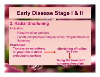
KIENBOCK DISEASE


   DR. MANOJ BHAMA
    SENIOR RESIDENT,
DEPT. OF ORTHOPAEDICS,
 S.P. MEDICAL COLLEGE,
      BIKANER, INDIA
   mbhama@gmail.com
KIENBOCK DISEASE
• Synonyms: Avascular Necrosis of Lunate
• First Described by Robert Kienbock in
  1910; as “traumatic softening” of Lunate
  bone
•    It is a painful disorder of wrist, due to
  avascular necrosis of carpal lunate, due to
  unknown cause
AETIOLOGY
• Aetiology = unknown, but several cause have
  been proposed
• vascular compromise from repetitive trauma
  causes microfracture & excessive stress on
  microscopic architecture (sports injury)
• Ulnar minus variant:- Individual having ulnar
  minus variance are at increased risk. Short Ulna
   increases shear force across the lunate 
  Causes vascular insufficiency
EPIDIMIOLOGY
• Age= 15-40 (young individuals)
• Sex= Men
• Location= Unilateral, Dominant wrist
PATHOLOGY
Pathological changes proceed in 4 stages:-
• Stage 1: Ischemia with naked eye or radiological
  examination
• Stage 2: Trabeculae Necrosis with reactive new
  bone formation & increased radiographic
  density, but little or no distortion of shape.
• Stage 3: Collapse of Bone
• Stage 4: Disruption of Radio-carpal congruence
  & secondary OA
PATHOLOGY
• The natural history of Kienbock’s disease
  is
  Progressive Sclerosis

                Fragmentation

                                Arthrosis
CLINICAL FEATURE
• There may be history of trauma with wrist in
  severe dorsiflexion
The lesion presents with
1. Dorsal wrist pain:- Pain may be produced in
  lunate region by axial strike/injury at distal end of
  middle finger
2. Swelling
3. Decreased Grip strength
4. Decreased range of motion; particularly in
  extension. In later stage movement may be
  painful
IMAGING
• X-ray at first show no abnormality but
  bone scan may reveals increased activity
• Later x-ray may show either mottled or
  diffuse density of bone – to – osteoarthritic
  changes in wrist
• MRI Most reliable way of detecting the
  early change
CLASSIFICATION
• Kienbock disease advances through 4
  radiological stages
• Stage I : Normal architecture; consistent with
             Microfracture
             (Lunate abnormal on bone scan)
• Stage II : Lunate sclerosis without collapse
• Stage III : Lunate collapse or Fragmentation &
  proximal migration of capitate
• Stage IV : Perilunate arthritis changes
TREATMENT
A Conservative
     Casting of wrist for several wks ; for
  early stages of disease ( St I or II, before
  sclerosis, fragmentation or collapse)
     But- Unacceptable & Ineffective
B Operative
• Surgery is only definitive treatment
Early Disease Stage I & II
• Unloading the Lunate*
• 1. Ulnar lengthening
• Transverse osteotomy at distal ulna 
  Distraction  Cortical iliac graft 
  Tightening of plate screw
Early Disease Stage I & II
2. Radial Shortening
Indication
   – Negative ulnar variance
   – Lunate compression fracture without fragmentation or
      flattening
Procedure
  Transverse osteotomy           shortening of radius
  3” proximal to distal                 by 2 cm
  articulating surface
                                 fixing the bone with
                                 compression plate
In stage III
1. Ulnar lengthening
2. Silicon prosthesis
      - silicon synovitis
      - foreign body cyst
3. Interacarpal fusion
4. Lunate excision
5. Arthrodesis Tri scaphe
                   Scaphocapitate
In stage IV
• Proximal carpal row resection
• Wrist arthrodesis

Kienbock Disease

  • 1.
    KIENBOCK DISEASE DR. MANOJ BHAMA SENIOR RESIDENT, DEPT. OF ORTHOPAEDICS, S.P. MEDICAL COLLEGE, BIKANER, INDIA mbhama@gmail.com
  • 2.
    KIENBOCK DISEASE • Synonyms:Avascular Necrosis of Lunate • First Described by Robert Kienbock in 1910; as “traumatic softening” of Lunate bone • It is a painful disorder of wrist, due to avascular necrosis of carpal lunate, due to unknown cause
  • 3.
    AETIOLOGY • Aetiology =unknown, but several cause have been proposed • vascular compromise from repetitive trauma causes microfracture & excessive stress on microscopic architecture (sports injury) • Ulnar minus variant:- Individual having ulnar minus variance are at increased risk. Short Ulna  increases shear force across the lunate  Causes vascular insufficiency
  • 4.
    EPIDIMIOLOGY • Age= 15-40(young individuals) • Sex= Men • Location= Unilateral, Dominant wrist
  • 5.
    PATHOLOGY Pathological changes proceedin 4 stages:- • Stage 1: Ischemia with naked eye or radiological examination • Stage 2: Trabeculae Necrosis with reactive new bone formation & increased radiographic density, but little or no distortion of shape. • Stage 3: Collapse of Bone • Stage 4: Disruption of Radio-carpal congruence & secondary OA
  • 6.
    PATHOLOGY • The naturalhistory of Kienbock’s disease is Progressive Sclerosis  Fragmentation  Arthrosis
  • 7.
    CLINICAL FEATURE • Theremay be history of trauma with wrist in severe dorsiflexion The lesion presents with 1. Dorsal wrist pain:- Pain may be produced in lunate region by axial strike/injury at distal end of middle finger 2. Swelling 3. Decreased Grip strength 4. Decreased range of motion; particularly in extension. In later stage movement may be painful
  • 8.
    IMAGING • X-ray atfirst show no abnormality but bone scan may reveals increased activity • Later x-ray may show either mottled or diffuse density of bone – to – osteoarthritic changes in wrist • MRI Most reliable way of detecting the early change
  • 9.
    CLASSIFICATION • Kienbock diseaseadvances through 4 radiological stages • Stage I : Normal architecture; consistent with Microfracture (Lunate abnormal on bone scan) • Stage II : Lunate sclerosis without collapse • Stage III : Lunate collapse or Fragmentation & proximal migration of capitate • Stage IV : Perilunate arthritis changes
  • 10.
    TREATMENT A Conservative Casting of wrist for several wks ; for early stages of disease ( St I or II, before sclerosis, fragmentation or collapse) But- Unacceptable & Ineffective B Operative • Surgery is only definitive treatment
  • 11.
    Early Disease StageI & II • Unloading the Lunate* • 1. Ulnar lengthening • Transverse osteotomy at distal ulna  Distraction  Cortical iliac graft  Tightening of plate screw
  • 12.
    Early Disease StageI & II 2. Radial Shortening Indication – Negative ulnar variance – Lunate compression fracture without fragmentation or flattening Procedure Transverse osteotomy shortening of radius 3” proximal to distal by 2 cm articulating surface fixing the bone with compression plate
  • 13.
    In stage III 1.Ulnar lengthening 2. Silicon prosthesis - silicon synovitis - foreign body cyst 3. Interacarpal fusion 4. Lunate excision 5. Arthrodesis Tri scaphe Scaphocapitate
  • 14.
    In stage IV •Proximal carpal row resection • Wrist arthrodesis