This guideline provides 25 recommendations for diagnosing, assessing prognosis, and treating peripheral artery disease (PAD) in patients with diabetes and foot ulcers or gangrene. It was developed by an international collaborative group and is based on systematic reviews of the literature. The recommendations cover diagnosing PAD, assessing likelihood of healing and amputation risk, deciding when revascularization is needed, choosing between endovascular and surgical procedures, post-procedure care, and general risk factor management. The goal is to help clinicians provide better care and reduce complications from diabetes-related foot disease.

![© 2023
The InternationalWorking Group on the Diabetic Foot
Intersocietal PAD Guideline
IWGDF
Guidelines
RECOMMENDATIONS
DIAGNOSIS
Clinical question: In a person with diabetes with or without a foot ulcer does medical history and clinical
examination (including pulse palpation) compared with a reference test (imaging- digital subtraction
angiography [DSA], magnetic resonance angiography [MRA], computed tomography angiography [CTA],
colour Duplex ultrasound [CDUS]) accurately identify PAD and reliably diagnose PAD?
Clinical question: In a person with diabetes with or without a foot ulcer, which non-invasive bedside
testing alone or in combination compared with reference tests (imaging- digital subtraction angiography
[DSA], magnetic resonance angiography [MRA], computed tomography angiography [CTA], colour
Duplex ultrasound [CDUS]) should be performed to accurately and reliably diagnose PAD?
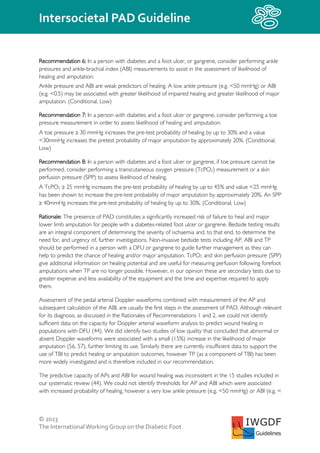
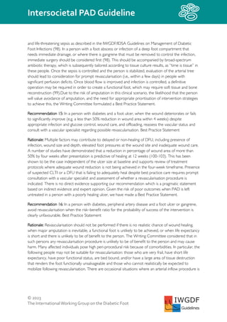
Recommendation 1: In a person with diabetes without a foot ulcer, take a relevant history for peripheral
artery disease, examine the foot for signs of ischaemia and palpate the foot pulses at least annually, or
with any change in clinical status of the feet. (Strong, Low)
Recommendation 2: In a person with diabetes without a foot ulcer, if peripheral artery disease (PAD) is
suspected, consider performing pedal Doppler waveforms in combination with ankle brachial index
(ABI) and toe-brachial index (TBI).
No single modality has been shown to be optimal for the diagnosis of PAD, and there is no value above
which PAD can be excluded. However, PAD is less likely in the presence of ABI 0.9-1.3; TBI ≥ 0.70; and
triphasic or biphasic pedal Doppler waveforms. (Conditional, Low)
Rationale: Diagnosis and treatment of PAD is critical due to the increased risk of developing DFU as
well as the increased rate of complications from co-existent cardiovascular disease including myocardial
infarction and stroke (32). Evidence for the diagnostic accuracy of pulse palpation for PAD in people
with diabetes without DFU is limited with two studies of low quality demonstrating that although
presence of pulses does not exclude disease , there is a small increase in ability to rule disease in where
a foot pulse is absent or weak (positive likelihood ratio [PLR]1.84 to 2.46) (35, 36);(The PLR gives the
change in odds of experiencing an outcome if the test is positive, whereas the negative likelihood ratio
[NLR] expresses a change in odds of experiencing an outcome if the test is negative. A PLR or NLR of
1.0 means that the test does not change the probability of the outcome over and above the pre-test
probability and therefore is not a useful diagnostic test). However, it is important to recognise that pulse
palpation should therefore be performed, and results considered in the context of other clinical
examinations that may be associated with PAD including hair loss, muscle atrophy and reduced
peripheral skin temperature. It should be noted that these clinical examinations are highly subjective and
such findings may also be associated with neuropathy. PAD may also be asymptomatic or have an
atypical presentation in people with diabetes as in other elderly or at-risk populations (24, 37, 38). For
example, peripheral neuropathy can mask pain symptoms and autonomic neuropathy can result in a
warm foot, meaning that the widely recognised signs and symptoms of PAD may not be present (39).
These recommendations are applicable to all people with diabetes. When DFU is absent, but there are
clinical signs and symptoms of PAD or PAD is suspected, for example due to long-standing diabetes,](https://image.slidesharecdn.com/iwgdf-2023-05-pad-guideline-230718145745-71f141fc/85/IWGDF-2023-05-PAD-Guideline-pdf-16-320.jpg)
![© 2023
The InternationalWorking Group on the Diabetic Foot
Intersocietal PAD Guideline
IWGDF
Guidelines
chronic hyperglycaemia, other diabetes complications such as peripheral neuropathy or presence of
atherosclerotic disease in other vascular beds, more frequent screening vascular assessment including
additional bedside testing is necessary. These recommendations are consistent with other (inter)national
guidelines on the management of diabetes, endorsing annual clinical assessment for PAD (and for other
foot complications) in people with diabetes (40-43).
Although based on low quality evidence, data demonstrating increased likelihood of PAD in those with
weak or absent pulses and elevated risk of cardiovascular morbidity and mortality support the
preference of a person with diabetes for clinical examination including pulse palpation to be performed
(32, 44). The non-invasive nature of clinical examination and pulse palpation suggest these assessments
would be valued by people with diabetes as initial diagnostic tests. As equipment is not required, the
Writing Committee considered pulse palpation and other forms of clinical examination having low
resource requirements, can be applied on a broad scale by a range of practitioners, and offer a method
to increase equity of health care access that is both feasible for health care providers and acceptable for
people with diabetes. We therefore made this a strong recommendation based on low certainty of
evidence and expert opinion.
Bedside testing techniques that provide objective measurement of peripheral blood flow in the lower
extremity (e.g., ankle-brachial index [ABI], toe-brachial index [TBI] and pedal Doppler waveforms) have
been shown to be useful as a means to diagnose and exclude PAD in people with diabetes. Our
systematic review demonstrates that multiple bedside testing techniques that offer objective
measurement of the peripheral circulation in the lower limb are useful as a means to rule disease in or
out for people with diabetes without a DFU but who are suspected of having PAD (44).
We identified forty studies investigating the diagnostic accuracy of non-invasive bedside tests in
populations with diabetes (44). Twenty-five of the studies were prospective, two cross sectional and the
remainder retrospective. Overall, the studies were of low quality and evidence was judged as being of
low certainty. . Although we could not identify the absolute threshold or ‘normal’ values of bedside
tests, we suggest that PAD is a more likely to be present in this population with an ABI <0.9 or >1.3, a
TBI <0.70, and presence of one or more monophasic Doppler waveforms from assessment of pedal
arteries with continuous wave Doppler (CWD) (44). In people without DFU, an ABI of <0.90 is
associated with a moderate to large increase in likelihood of PAD with PLRs ranging from 2.1 to 19.9,
however the ability to rule disease out is limited (NLR 0.29 to 0.84). A TBI <0.70 has a moderate ability
to diagnose and exclude PAD (PLRs 2.0 to 3.55, NLRs 0.25 to 0.44) and the presence of a visual
monophasic pedal Doppler waveform has a moderate ability to diagnose and exclude PAD (PLR 7.09,
NLR 0.19). Non-invasive tests are therefore likely to be beneficial for people without a DFU, however
high quality studies of diagnostic accuracy are required. A summary of results is provided in
Supplementary Table 1.
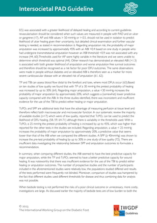
When calculating the ABI in the leg of a person with and without DFU for the purposes of diagnosing
PAD we advise to use the lower systolic blood pressure of either the dorsal pedis or posterior tibial
artery as this improves the diagnostic accuracy of the test (44). For PAD affecting arteries below the
knee this calculation method identifies the most severe disease while using the higher pressure identifies
the least affected artery. We also recommend using the three tests (ABI, TBI and pedal Doppler](https://image.slidesharecdn.com/iwgdf-2023-05-pad-guideline-230718145745-71f141fc/85/IWGDF-2023-05-PAD-Guideline-pdf-17-320.jpg)

![© 2023
The InternationalWorking Group on the Diabetic Foot
Intersocietal PAD Guideline
IWGDF
Guidelines
performed to improve the likelihood of healing of a below knee amputation, so as to avoid an above
knee amputation.
There is evidence from several observational studies of a 50% healing rate for ischaemic DFU in people
with diabetes unsuitable for revascularisation and this should also be considered in determining choice of
care (67, 94). The decision to proceed to primary amputation, or to adopt a palliative approach, should
be made in conjunction with the person and the multidisciplinary team (104) including a vascular
specialist unless an emergency procedure is indicated as discussed earlier. The Writing Committee
considered that in these circumstances where healing is improbable a person is unlikely to value the
outcomes from revascularisation over no revascularisation. Similarly in such circumstances the benefit of
revascularisation will not outweigh the potential harms.
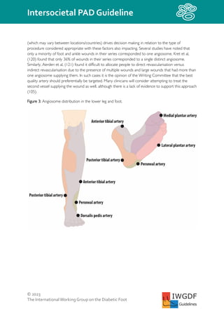
Clinical question: In people with diabetes, PAD and either a foot ulcer or gangrene how does
endovascular revascularisation compare to open or hybrid revascularisation?
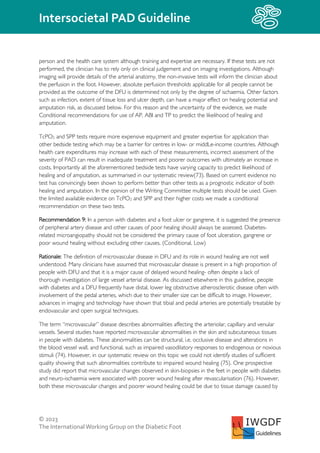
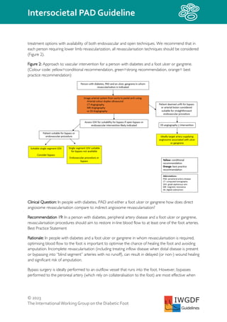
Recommendation 17: In a person with diabetes, peripheral artery disease and either a foot ulcer or
gangrene who have an adequate single segment saphenous vein in whom infrainguinal revascularisation
is indicated and who are suitable for either approach, we suggest bypass in preference to endovascular
therapy (Conditional, Moderate)
Recommendation 18: A person with diabetes, peripheral artery disease (PAD) and a foot ulcer or
gangrene, should be treated in centres with expertise in, or rapid access to, endovascular and surgical
bypass revascularisation. In this setting, consider making treatment decisions based on the risk to and
preference of the individual, limb threat severity, anatomic distribution of PAD, and the availability of
autogenous vein. Best Practice Statement
Rationale: Once the decision to revascularise has been made, the next decision is whether an
endovascular, an open (i.e., bypass or endarterectomy) procedure, or a combination of both (i.e. hybrid
procedure) should be performed. Recommendation 18 highlights the complementary role of open and
endovascular techniques in contemporary vascular practice. In particular, endovascular techniques have
largely replaced open surgery in the management of aorto-iliac disease and also allow treatment of foot
and pedal arch disease.
The majority of studies we identified in our systematic review on endovascular and bypass surgical
outcomes were observational and retrospective case series, with a high risk of bias (105). The BEST CLI
trial was a large randomised clinical trial with low risk of bias comparing an endovascular first with a
surgical first approach. People with CLTI who were deemed appropriate for revascularisation for
infrainguinal arterial occlusive disease were included (106). The primary outcome was above-ankle
amputation of the index limb or a major reintervention in the index limb (new bypass, vein graft
interposition revision, thrombectomy or thrombolysis) or death. It was designed in two parallel-cohort
trials: (Cohort 1) people who had adequate single segment great saphenous vein (GSV) available for use
as a bypass conduit, and (Cohort 2) people without adequate single segment GSV who required an
alternate conduit. Treatment with a GSV bypass first approach was superior to endovascular therapy
first for the primary outcome (hazard ratio [HR], 0.68; 95% confidence interval [CI] 0.59-0.79; P
<0.001). In Cohort 2 the primary outcomes were similar between the two groups. Subgroup analysis of](https://image.slidesharecdn.com/iwgdf-2023-05-pad-guideline-230718145745-71f141fc/85/IWGDF-2023-05-PAD-Guideline-pdf-31-320.jpg)
![© 2023
The InternationalWorking Group on the Diabetic Foot
Intersocietal PAD Guideline
IWGDF
Guidelines
Rationale: The Writing Committee decided to not write their own guidelines on pharmacological
interventions in people with diabetes, PAD and a foot ulcer or gangrene in order to reduce
cardiovascular risk or to prevent major limb events as defined above. There are already a number of
guidelines on cardiovascular risk prevention in people with diabetes and cardiovascular disease, and thus
another guideline would have little added value. We decided to base our Best Practice Statements on
the Global Vascular Guidelines for CLTI produced by the ESVS, SVS and World Federation of Vascular
Societies (WFVS) (17), as these address the specific population of people with CLTI. The advice on
antiplatelet therapy is in line with the recent ESVS antithrombotic guidelines (124). When we felt it was
applicable, we used the guidelines of the American Diabetes Association (ADA), the European
Association for the Study of Diabetes (EASD) and other guidelines on peripheral artery disease
(European Society of Cardiology [ESC]-ESVS, European Society of Vascular Medicine [ESVM], ESC-
EASD, ESC- European Atherosclerosis Society [EAS]) (13-16, 18-20).
PAD runs a more aggressive course in those with diabetes mellitus compared with those without
diabetes, with an elevated risk of lower leg amputation. In addition, the combination of diabetes and
PAD is associated with a high risk of developing complications in other vascular beds. As discussed
previously, persons with an ischaemic diabetes-related foot ulcer have an overall 5-year cardiovascular
mortality around 50% (125). Therefore, according to the international guidelines of several major
vascular and diabetes associations, these individuals should be considered as having a very high
cardiovascular risk and should be treated as such. On the other hand, they usually have, in addition to
peripheral neuropathy, other diabetes-related complications as well as several co-morbidities, resulting in
a high burden of diseases and multiple medications (27). Many affected persons are elderly, frail and are
living in vulnerable socio-economic circumstances with a low quality of life (126, 127). It is therefore
essential that cardiovascular risk factor management in these people should be individualised, tailored
and should be part of a shared decision-making process, taking life-expectancy, diabetes-related
complications/co-morbidities, expected benefit, treatment burden, drug interactions and undesirable
treatment effects into account. This care should be provided by health care worker(s) with sufficient
expertise in treating cardiovascular risk factors and glycaemia, preferably by person(s) who are part of
the multidisciplinary team for diabetes-related foot care.
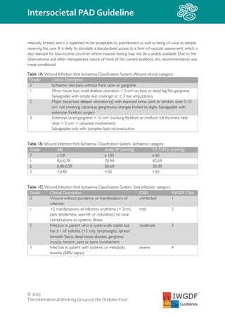
Glycaemic goals
As stated in the ADA and ESC-EASD guidelines, near-normal glycaemia with HbA1c level below 7.0%
(53 mmol/mol) will decrease microvascular complications (15, 19). Tighter glucose control initiated early
in the course of diabetes in younger individuals leads to a reduction in macrovascular complications, i.e.
cardiovascular outcomes, over a 20 year timescale. Such glucose control can have beneficial effects on
microvascular complications in a shorter period of time. However, when blood glucose lowering agents
are used that have the risk of severe hypoglycaemia, this can increase the risk of cardiovascular events
and death, as detailed in the ADA and ESC-EASD guidelines (15, 18). As many people with a DFU and
PAD also have atherosclerotic disease in other vascular beds, tight glucose control can be harmful. The
risk of hypoglycaemia is markedLy lower when people are only treated with metformin, a sodium–
glucose cotransporter 2 inhibitor or a glucagon-like peptide 1 receptor agonist. Tight glucose control is
often not indicated in persons with PAD and a DFU due to the risk of hypoglycaemia outweighing the
potential benefit. The ADA recommends in the 2022 Standards of Care to aim for an Hba1c < 8% (<](https://image.slidesharecdn.com/iwgdf-2023-05-pad-guideline-230718145745-71f141fc/85/IWGDF-2023-05-PAD-Guideline-pdf-38-320.jpg)
![© 2023
The InternationalWorking Group on the Diabetic Foot
Intersocietal PAD Guideline
IWGDF
Guidelines
REFERENCES
(1) Bus SA, Monteiro-Soares M, Game F, van Netten JJ, Apelqvist J, Fitridge R, Senneville E, Schaper NC; IWGDF
Editorial Board. Standards for the development and methodology of the 2023 International Working Group on the
Diabetic Foot guidelines. Diabetes Metab Res Rev. 2023;e3656.
(2) Guyatt GH, Oxman AD, Schünemann HJ, Tugwell P, Knottnerus A. GRADE guidelines: a new series of articles in
the Journal of Clinical Epidemiology. J Clin Epidemiol. 2011;64(4):380-2.
(3) [Current care guidelines: peripheral arterial disease]. Duodecim. 2010;126(12):1433-4.
(4) Moher D, Liberati A, Tetzlaff J, Altman DG. Preferred reporting items for systematic reviews and meta-analyses: the
PRISMA statement. Bmj. 2009;339:b2535.
(5) Lucas NP, Macaskill P, Irwig L, Bogduk N. The development of a quality appraisal tool for studies of diagnostic
reliability (QAREL). Journal of Clinical Epidemiology. 2010;63(8):854-61.
(6) Sterne JA, Hernán MA, Reeves BC, Savović J, Berkman ND, Viswanathan M, et al. ROBINS-I: a tool for assessing risk
of bias in non-randomised studies of interventions. bmj. 2016;355.
(7) Sterne JA, Savović J, Page MJ, Elbers RG, Blencowe NS, Boutron I, et al. RoB 2: a revised tool for assessing risk of
bias in randomised trials. bmj. 2019;366.
(8) Wells GA, Shea B, O’Connell D, Peterson J, Welch V, Losos M, et al. The Newcastle-Ottawa Scale (NOS) for
assessing the quality of nonrandomised studies in meta-analyses. Oxford; 2000.
(9) Whiting PF, Rutjes AW, Westwood ME, Mallett S, Deeks JJ, Reitsma JB, et al. QUADAS-2: a revised tool for the
quality assessment of diagnostic accuracy studies. Annals of Internal Medicine. 2011;155(8):529-36.
(10) Hayden JA, van der Windt DA, Cartwright JL, Côté P, Bombardier C. Assessing bias in studies of prognostic factors.
Annals of internal medicine. 2013;158(4):280-6.
(11) Schünemann H, Brożek J, Guyatt G, Oxman A. The GRADE handbook. Cochrane Collaboration London, UK; 2013.
(12) Dewidar O, Lotfi T, Langendam MW, Parmelli E, Saz Parkinson Z, Solo K, et al. Good or best practice statements:
proposal for the operationalisation and implementation of GRADE guidance. BMJ Evid Based Med. 2022.
(13) Frank U, Nikol S, Belch J, Boc V, Brodmann M, Carpentier PH, et al. ESVM Guideline on peripheral arterial disease.
Vasa. 2019;48(Suppl 102):1-79.
(14) Aboyans V, Ricco JB, Bartelink ML, Björck M, Brodmann M, Cohner T, et al. [2017 ESC Guidelines on the Diagnosis
and Treatment of Peripheral Arterial Diseases, in collaboration with the European Society for Vascular Surgery
(ESVS)]. Kardiol Pol. 2017;75(11):1065-160.
(15) Cosentino F, Grant PJ, Aboyans V, Bailey CJ, Ceriello A, Delgado V, et al. 2019 ESC Guidelines on diabetes, pre-
diabetes, and cardiovascular diseases developed in collaboration with the EASD. Eur Heart J. 2020;41(2):255-323.
(16) Mach F, Baigent C, Catapano AL, Koskinas KC, Casula M, Badimon L, et al. 2019 ESC/EAS Guidelines for the
management of dyslipidaemias: lipid modification to reduce cardiovascular risk. Eur Heart J. 2020;41(1):111-88.
(17) Conte MS, Bradbury AW, Kolh P, White JV, Dick F, Fitridge R, et al. Global Vascular Guidelines on the Management
of Chronic Limb-Threatening Ischemia. Eur J Vasc Endovasc Surg. 2019;58(1s):S1-S109.e33.
(18) 10. Cardiovascular Disease and Risk Management: Standards of Medical Care in Diabetes-2022. Diabetes Care.
2022;45(Suppl 1):S144-s74.
(19) 6. Glycemic Targets: Standards of Medical Care in Diabetes-2022. Diabetes Care. 2022;45(Suppl 1):S83-s96.
(20) Aboyans V, Ricco JB, Bartelink MEL, Bjorck M, Brodmann M, Cohnert T, et al. 2017 ESC Guidelines on the
Diagnosis and Treatment of Peripheral Arterial Diseases, in collaboration with the European Society for Vascular
Surgery (ESVS). Rev Esp Cardiol (Engl Ed). 2018;71(2):111.
(21) Van Netten JJ, Bus SA, Apelqvist J, et al. Definitions and criteria for diabetic foot disease. Diabetes Metab Res Rev.
2023;e3654.
(22) Sun H, Saeedi P, Karuranga S, Pinkepank M, Ogurtsova K, Duncan BB, et al. IDF Diabetes Atlas: Global, regional and
country-level diabetes prevalence estimates for 2021 and projections for 2045. Diabetes research and clinical
practice. 2022;183:109119.](https://image.slidesharecdn.com/iwgdf-2023-05-pad-guideline-230718145745-71f141fc/85/IWGDF-2023-05-PAD-Guideline-pdf-45-320.jpg)