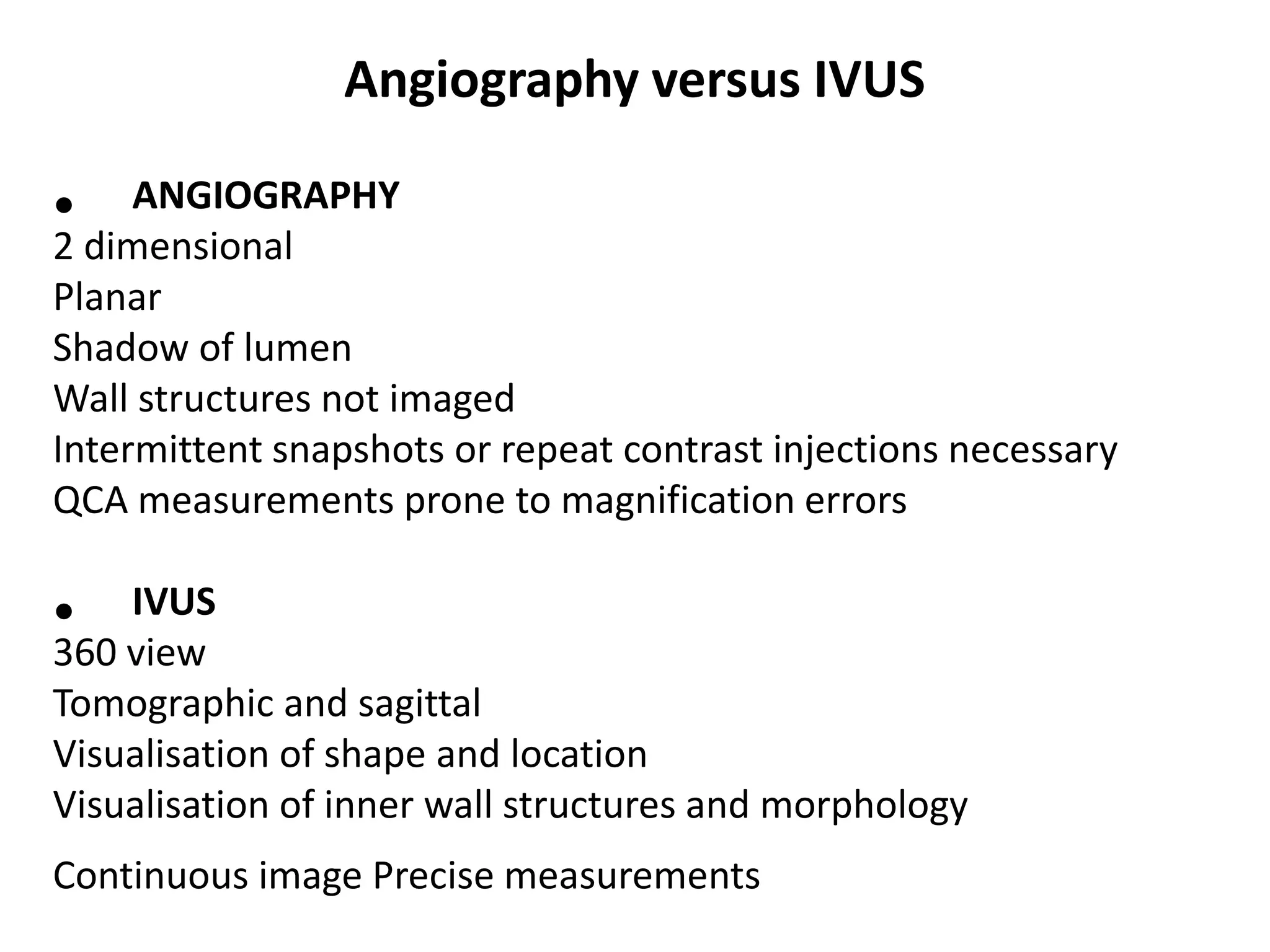
IVUS vs OCT : Relative Merits and
Drawbacks
Ankit Gupta
2nd Year DNB Resident
Introduction
• CAG has been the gold standard technique for evaluating
CAD for the past 50 years. Increasingly, however, realisation
of the limitations of CAG, mainly the inability to supply
information regarding the coronary wall, has prompted the
design and development of adjunctive technologies to better
evaluate not just luminal disease but also the burden and
character of atherosclerotic plaque within the vessel.
• The development of intracoronary imaging modalities, namely
IVUS and OCT now have established roles in the diagnosis
and treatment of CAD.
• Intracoronary devices that can assess the coronary
endothelium use either acoustic or optical signals that are
received by a coronary catheter (IVUS uses ultrasound, OCT
uses near-infrared light).
Intravascular Ultrasound
• An IVUS system consists of a flexible monorail
catheter with an ultrasound transducer at its
tip that emits ultrasound waves in the 10–40
MHz range and an electronics console to
reconstruct the image. After reflection from
tissue, part of the ultrasound energy returns
to the transducer and is converted into the
image.
• IVUS provides 3 dimensional detail of what
lies ahead
• It provides additional information not
visualized on the angio map
Diagnostic Applications of IVUS
• Identify specific disease - left main stem, ostial
lesions
• Detect angiographically silent disease
vasculopathy
• Identify plaque morphology
• Examine vessel when angiography is
inconclusive hazy lesions, presence or
absence of thrombus or dissection
• Measure plaque load
• Measure true vessel size
Angiography versus IVUS
• ANGIOGRAPHY
2 dimensional
Planar
Shadow of lumen
Wall structures not imaged
Intermittent snapshots or repeat contrast injections necessary
QCA measurements prone to magnification errors
• IVUS
360 view
Tomographic and sagittal
Visualisation of shape and location
Visualisation of inner wall structures and morphology
Continuous image Precise measurements
IVUS: Pre PCI indication
• When IVUS?
Decide strategy & sizing
• Why IVUS?
Vessel reference & % stenosis
Length of lesion
Plaque composition
IVUS: Post PCI Indication
• When IVUS?
Evaluate stent result
• Why IVUS?
Final lumen
Expansion
Apposition
Dissection or plaque shift
IVUS Transducers
Mechanical Transducer – 40 MHz Atlantis Pro (BosSci)
Solid-State Transducer – 25 MHz Eagle Eye (Volcano)
Vulnerable Plaque ?
Functional Definition
A plaque, often non-stenotic, that has a high likelihood of
becoming disrupted and forming a thrombogenic factor after
exposure to an acute risk factor
Histological Definition
Plaque containing a thin fibrous cap, lipid pools and
Macrophages
Prospective Definition
A signature that has been proven in a prospective study to
identify a plaque that is prone to disrupt and can be
recognised and measured in the cath lab
Plaque Characterization
Lesion preparation: need of rotablation
Fibrous plaque Plaque rupture Thrombus Calcification
• Optical Coherence Tomography (OCT) is a light-based imaging
modality that can provide in vivo high-resolution images of the
coronary artery with a level of resolution (axial 10-20 μm) ten
times higher than IVUS but with a penetration depth limited to
1.5-2 mm.
• The technique uses low-coherent near infrared light to create
high-resolution cross sectional images of the vessel. OCT,
originally described in the early 1990s, was first applied in the
field of ophthalmology.
Optical Coherence Tomography (OCT)
IVUS
IVUS
IVUS
IVUS
IVUS
IVUS
IVUS
IVUS
IVUS
IVUS
IVUS
IVUS
IVUS
IVUS
IVUS
IVUS

IVUS

  • 1.
    IVUS vs OCT: Relative Merits and Drawbacks Ankit Gupta 2nd Year DNB Resident
  • 2.
    Introduction • CAG hasbeen the gold standard technique for evaluating CAD for the past 50 years. Increasingly, however, realisation of the limitations of CAG, mainly the inability to supply information regarding the coronary wall, has prompted the design and development of adjunctive technologies to better evaluate not just luminal disease but also the burden and character of atherosclerotic plaque within the vessel. • The development of intracoronary imaging modalities, namely IVUS and OCT now have established roles in the diagnosis and treatment of CAD. • Intracoronary devices that can assess the coronary endothelium use either acoustic or optical signals that are received by a coronary catheter (IVUS uses ultrasound, OCT uses near-infrared light).
  • 3.
    Intravascular Ultrasound • AnIVUS system consists of a flexible monorail catheter with an ultrasound transducer at its tip that emits ultrasound waves in the 10–40 MHz range and an electronics console to reconstruct the image. After reflection from tissue, part of the ultrasound energy returns to the transducer and is converted into the image.
  • 4.
    • IVUS provides3 dimensional detail of what lies ahead • It provides additional information not visualized on the angio map
  • 5.
    Diagnostic Applications ofIVUS • Identify specific disease - left main stem, ostial lesions • Detect angiographically silent disease vasculopathy • Identify plaque morphology • Examine vessel when angiography is inconclusive hazy lesions, presence or absence of thrombus or dissection • Measure plaque load • Measure true vessel size
  • 6.
    Angiography versus IVUS •ANGIOGRAPHY 2 dimensional Planar Shadow of lumen Wall structures not imaged Intermittent snapshots or repeat contrast injections necessary QCA measurements prone to magnification errors • IVUS 360 view Tomographic and sagittal Visualisation of shape and location Visualisation of inner wall structures and morphology Continuous image Precise measurements
  • 7.
    IVUS: Pre PCIindication • When IVUS? Decide strategy & sizing • Why IVUS? Vessel reference & % stenosis Length of lesion Plaque composition
  • 8.
    IVUS: Post PCIIndication • When IVUS? Evaluate stent result • Why IVUS? Final lumen Expansion Apposition Dissection or plaque shift
  • 9.
    IVUS Transducers Mechanical Transducer– 40 MHz Atlantis Pro (BosSci) Solid-State Transducer – 25 MHz Eagle Eye (Volcano)
  • 12.
    Vulnerable Plaque ? FunctionalDefinition A plaque, often non-stenotic, that has a high likelihood of becoming disrupted and forming a thrombogenic factor after exposure to an acute risk factor Histological Definition Plaque containing a thin fibrous cap, lipid pools and Macrophages Prospective Definition A signature that has been proven in a prospective study to identify a plaque that is prone to disrupt and can be recognised and measured in the cath lab
  • 16.
    Plaque Characterization Lesion preparation:need of rotablation Fibrous plaque Plaque rupture Thrombus Calcification
  • 24.
    • Optical CoherenceTomography (OCT) is a light-based imaging modality that can provide in vivo high-resolution images of the coronary artery with a level of resolution (axial 10-20 μm) ten times higher than IVUS but with a penetration depth limited to 1.5-2 mm. • The technique uses low-coherent near infrared light to create high-resolution cross sectional images of the vessel. OCT, originally described in the early 1990s, was first applied in the field of ophthalmology. Optical Coherence Tomography (OCT)