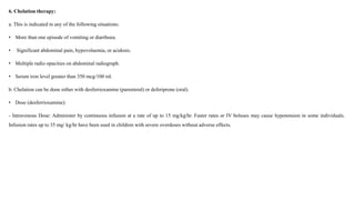
Iron is an essential element but can be toxic in high amounts. Iron poisoning most often occurs when children accidentally ingest iron supplements. Symptoms of iron toxicity can be severe and include vomiting, abdominal pain, shock, liver damage, and gastrointestinal bleeding and necrosis. Diagnosis involves measuring serum iron levels and a chelation challenge test. Treatment focuses on gastric lavage, administering charcoal or antacids, correcting dehydration and acidosis, and chelation therapy with desferrioxamine or deferiprone to remove excess iron from the body.
The kidney maintains the vital functions of clearing excess body fluid and removing metabolic and exogenous toxins from the blood
The kidney is particularly vulnerable to drugs and other agents that cause renal damage
The heart pumps approximately 25% of cardiac output into the kidneys
Any drug in the blood will eventually reach the highly vascularized kidneys
It may potentially cause drug induced renal failure
If the drug is primarily cleared by the kidney, the drug will become increasingly concentrated as it moves from the renal artery into the smaller vasculature of the kidney
The drug may be filtered or secreted into the lumen of the renal tubules
The concentrated drug exposes the kidney tissue to far greater drug concentration per surface area
Drug-induced kidney disease or nephrotoxicity (DIN) is a relatively common complication of several diagnostic and therapeutic agents.
Molecular mechanism of drug induced nephrotoxicitySuman Manandhar
This document discusses nephrotoxicity, or kidney damage, caused by drugs. It provides an introduction and overview of types of nephrotoxicity and biomarkers for detecting drug-induced renal damage. It then discusses several common pathogenic mechanisms by which drugs can cause nephrotoxicity, including altered intraglomerular hemodynamics, tubular cell toxicity, inflammation, crystal nephropathy, rhabdomyolysis, and thrombotic microangiopathy. Specific examples are given of how certain drugs like aminoglycosides, cyclosporine, and cisplatin can cause nephrotoxicity through these mechanisms.
This document discusses various medication-induced kidney injuries. It covers:
1. Risk factors for drug nephrotoxicity including patient factors like age, sex, CKD/AKI status.
2. Pathophysiology of drug nephrotoxicity including how single drugs can affect different kidney sites and how multiple drugs can affect the same site.
3. Classification of drug-induced kidney injury including prerenal AKI, acute tubular necrosis, acute/chronic interstitial nephritis, glomerular disease, and obstructive nephropathy.
4. Specific nephrotoxic drugs that can cause the different types of injury through various mechanisms are discussed along with
This document summarizes several hematological disorders and their drug-induced causes. It begins with an introduction to anemia, describing it as a deficiency of red blood cells or hemoglobin. Deep vein thrombosis is defined as a blood clot forming in a deep vein, most commonly in the legs. Various drug-induced hematological disorders are then outlined affecting red blood cells, white blood cells, and platelets. Causes, symptoms, diagnoses, and treatments are discussed for disorders like anemia, deep vein thrombosis, aplastic anemia, hemolytic anemia, neutropenia, and thrombocytopenia.
Rhabdomyolysis is a syndrome characterized by muscle necrosis and the release of intracellular muscle constituents into the circulation. It can range from asymptomatic to life-threatening acute renal failure. It was first documented in victims of bombings in London during World War II who developed crush injuries, swollen extremities, and dark urine before dying of renal failure. Early and aggressive intravenous fluid therapy is the primary treatment to prevent acute kidney injury by maintaining adequate renal perfusion and dilution of myoglobin in the kidneys.
Using Fibrinogen has been a question for many residents and physicians. This presentation has been made to solve this issue. It will make decision making easy for many of us.
Tumor lysis syndrome describes the clinical and laboratory abnormalities that result from the rapid release of intracellular contents from dying tumor cells. It is a common oncologic emergency seen by nephrologists. The rapid release of ions and metabolites causes hyperuricemia, hyperkalemia, hyperphosphatemia, and hypocalcemia. Prevention focuses on identifying at-risk patients and aggressive hydration and urate-lowering agents. Treatment involves fluid management, management of electrolyte abnormalities with agents like rasburicase, and potentially renal replacement therapy for severe cases.
Relative polycythemia, also called hemoconcentration, is caused by a decrease in plasma volume rather than an increase in red blood cells. It can result from losses of body fluids due to diarrhea, vomiting, diuretic use, or adrenal insufficiency. Absolute polycythemia is caused by an increased production of red blood cells and can be primary, such as polycythemia vera, or secondary to tissue hypoxia due to conditions like heart or lung disease. Polycythemia vera is a chronic myeloproliferative disorder characterized by increased red blood cell volume and hyperviscosity syndrome. Treatment focuses on phlebotomy, medications to suppress bone marrow, and managing symptoms like
Global Medical Cures™ | Blood disorders
DISCLAIMER-
Global Medical Cures™ does not offer any medical advice, diagnosis, treatment or recommendations. Only your healthcare provider/physician can offer you information and recommendations for you to decide about your healthcare choices.
This document discusses approaches to diagnosing and classifying anemia. It defines anemia as a reduction in red blood cell measurements like hemoglobin, hematocrit, or red blood cell count. There are two main approaches to evaluating anemia - the biologic/kinetic approach which examines red blood cell production and survival, and the morphology approach which examines red blood cell size. Key tests in the kinetic approach include the reticulocyte count to assess bone marrow response. The morphology approach uses mean corpuscular volume to classify anemias as microcytic, normocytic or macrocytic based on red blood cell size. Causes of anemia include blood loss, decreased production, and increased destruction.
This document provides an overview of anemia, including its classification, causes, signs and symptoms, diagnostic approach, and management principles. It presents a clinical case of an 18-year-old female with weakness, lethargy and heavy menstrual bleeding, then reviews epidemiology of anemia, the red blood cell lifecycle, classification of anemia by severity, etiology and morphology, diagnostic workup, and general management strategies focusing on identifying and treating underlying causes. The take-home message emphasizes that anemia requires investigation to determine its cause, and prevention through supplementation is important for reducing prevalence.
Anemia in ckd patients dr. hamed ezzatFarragBahbah
Anemia is common in patients with chronic kidney disease (CKD) due to the kidney's reduced ability to produce erythropoietin. Anemia of CKD is usually normocytic and normochromic. Evaluation of a CKD patient with anemia includes testing for iron deficiency, blood loss, inflammation, and reticulated count. Treatment involves adequate dialysis, iron supplementation, erythropoietin, nutrition, and inflammation prevention to reach a target hemoglobin level of 10 to 11.5 g/dL, rising no more than 1-2 g/dL per month. Blood transfusions should be avoided when possible.
Polycythemia vera is a chronic myeloproliferative disorder characterized by an absolute increase in red blood cells, total blood volume, leukocytosis, thrombocytosis, and splenomegaly. It is caused by a clonal proliferation of a hematopoietic stem cell. Symptoms include headaches, visual disturbances, thrombosis, pruritus, and splenomegaly. Diagnosis involves meeting certain criteria including elevated hematocrit. Treatment involves phlebotomy to reduce red blood cell mass and hydroxyurea or interferon-alpha to reduce platelet and white blood cell counts and reduce risk of thrombosis.
Polycythemia is a condition characterized by an abnormal increase in red blood cells. It can be primary, caused by bone marrow abnormalities, or secondary, caused by factors like smoking or lung diseases that result in hypoxia. Symptoms include headache, dizziness, and skin redness or itching. Diagnosis involves blood tests showing elevated red blood cell counts. Treatment may include phlebotomy to reduce blood volume, medications to suppress blood cell production, and lifestyle changes like exercise and avoiding tobacco. Nursing care focuses on monitoring for blood clots, managing pain and nutrition, and providing education.
Role of erythropoitin in chronic kidney diseaseAftab Siddiqui
1) Erythropoietin (EPO) and novel EPO stimulating proteins (NESPs) play an important role in managing anemia in chronic kidney disease (CKD). EPO therapy aims to increase hemoglobin levels by 1-2 g/dl per month and reduce need for blood transfusions.
2) Intravenous or subcutaneous iron supplementation is also important to manage iron deficiency and allow effective response to EPO. The target is to achieve transferrin saturation between 20-50% and serum ferritin above 100 ng/ml.
3) Causes of resistance to EPO action include iron, folate or carnitine deficiencies, chronic infections, inflammation, and secondary hyperpar
Polycythemia Vera is a chronic myeloproliferative disorder characterized by an overproduction of red blood cells from bone marrow due to a mutation in the JAK2 gene. This leads to thickening of the blood and complications like blood clots and spleen enlargement. It is diagnosed through blood tests showing increased red blood cells. While there is no cure, treatment focuses on reducing blood cell counts through regular blood removal and medications to suppress bone marrow production and stimulate the immune system.
The document provides an overview of anemia and presents several clinical cases involving anemia. It discusses the initial laboratory evaluation of anemia including complete blood count, peripheral smear, and reticulocyte count. Additional tests discussed include iron, B12, folate levels as well as LDH, bilirubin, and Coombs testing. Five clinical cases are then presented involving autoimmune hemolytic anemia, iron deficiency anemia, acute myeloid leukemia, thrombotic thrombocytopenic purpura, and anemia related to chronic kidney disease.
Hemochromatosis is a disease caused by excessive iron accumulation in the body's tissues. There are two main types: primary (hereditary) hemochromatosis caused by mutations in genes like HFE that result in impaired regulation of iron absorption; and secondary hemochromatosis caused by other conditions that lead to high iron levels like blood transfusions or liver disease. Symptoms include fatigue, joint pain, diabetes, heart problems and liver damage. Diagnosis involves blood tests of iron and liver enzymes and genetic testing. Treatment is by regular phlebotomy to reduce iron levels and sometimes iron chelation drugs or dietary changes.
Genetic Hemochromatosis is autosomal recessive genetic disorder that results from abnormal accumulation of iron in parenchymal organs such as the liver, pancreas, and heart leading to organ toxicity.
Rbcs & its clinical implications by Dr. Amit T. Suryawanshi, Oral Surgeon, P...All Good Things
Hi. This is Dr. Amit T. Suryawanshi. Oral & Maxillofacial surgeon from Pune, India. I am here on slideshare.com to share some of my own presentations presented at various levels in the field of OMFS. Hope this would somehow be helpful to you making your presentations. All the best.
This document discusses anemia of chronic kidney disease (CKD), including its causes, prevalence, benefits of treatment, evaluation, and treatment with iron and erythropoietic stimulating agents (ESAs). It notes that anemia is common in CKD due to reduced kidney function and erythropoietin production. While untreated anemia can cause many symptoms, managing it with iron and ESAs can improve quality of life and reduce risks. The document provides guidelines on testing, supplementing iron, dosing ESAs, monitoring treatment response, and managing side effects in patients with CKD.
Drugs affecting haematopoiesis and recent advances by swaroopaSwaroopaNallabariki
This document provides an overview of drugs affecting haematopoiesis and recent advances in the field. It begins with an introduction to haematopoiesis and haematopoietic growth factors. It then discusses erythropoiesis-stimulating agents such as erythropoietin and darbepoietin in detail, including their mechanisms of action, preparations, therapeutic uses, monitoring, and side effects. The document also summarizes myeloid growth factors and thrombopoietic growth factors. It concludes with references for further reading.
This document discusses nephrotoxic drugs and their impact on kidney function. It begins by explaining how renal damage from drugs can cause significant health issues like acute kidney injury and chronic kidney disease, as well as increasing medical costs. It then identifies several common classes of drugs that can cause nephrotoxicity, such as antibiotics, chemotherapy agents, antihypertensives, and NSAIDs. The document discusses mechanisms of nephrotoxicity for different drug classes and regions of the kidney. It also examines renal biomarkers that can help identify kidney injury earlier than serum creatinine. Finally, it provides examples of renal protective strategies like dose adjustments, monitoring, and hydration that can reduce the nephrotoxic risks of certain
Drugs acting on blood and blood forming organsUrmila Aswar
This document discusses drugs that act on blood and blood forming organs. It covers topics like hemostasis, coagulation factors, coagulation pathways, anticoagulants like heparin and warfarin, fibrinolytics, and antiplatelet drugs. Key points include that hemostasis is the process by which bleeding stops, coagulation involves intrinsic and extrinsic pathways, and anticoagulants prevent clotting through various mechanisms like inhibiting thrombin formation. Common anticoagulants discussed are heparin, low molecular weight heparins, warfarin, and fibrinolytics like streptokinase that lyse clots. Antiplatelet drugs like aspirin are also covered.
This document discusses hyperkalemia, including its definition, causes, clinical manifestations, and treatment. It provides details on potassium regulation and homeostasis in the body. The main causes of hyperkalemia are a shift of potassium from intracellular to extracellular space, excessive potassium intake, and decreased renal potassium excretion. Symptoms range from weakness to cardiac arrhythmias. Treatment involves calcium gluconate for cardiac issues, insulin with glucose to shift potassium intracellularly, sodium bicarbonate for acidosis, and diuretics or dialysis to increase renal excretion.
Iron poisoning (physical appearance, sources- dietary and environmental, uses- industrial and biological, usual fatal dose, toxicokinetics, mode of action, clinical features, diagnosis, treatment, autopsy features
Iron poisoning occurs when too much iron is ingested, usually from iron supplements. It progresses through several stages, initially causing gastrointestinal symptoms. A latent phase then occurs where symptoms improve, but toxic levels of iron are being absorbed. Multi-organ failure can then develop from shock and acidosis. Chelation therapy with desferrioxamine may be given if vomiting, abdominal pain, or high serum iron levels are present to bind the iron. Adverse effects of desferrioxamine include sepsis, visual toxicity, ototoxicity, and pulmonary toxicity.
The document discusses iron toxicity, including its chemical properties, epidemiology, sources, absorption in the body, toxicity, pathophysiology of iron poisoning, diagnosis, management, and a case study of iron toxicity in a child. It provides details on the stages of acute iron toxicity, signs and symptoms, laboratory tests, criteria for chelation therapy, and treatment approaches.
Cadmium is a heavy metal that is toxic even in low concentrations and accumulates in organisms, causing toxicity in multiple organ systems like the lungs, kidneys, bones and increasing cancer risk. It is found naturally and as a byproduct of industrial processes like mining and smelting, and also enters the body through cigarette smoke, food and water. Once absorbed, cadmium binds to proteins in the blood and is transported mainly to the liver and kidneys where it can cause damage and be stored long-term if exposure is continuous.
Iron poisoning can occur from ingesting ferrous or ferric compounds. It causes oxidative damage through reactive oxygen species, damaging cells and tissues. Clinical features are divided into 5 stages, beginning with vomiting and abdominal pain within hours. Later stages include shock, liver damage, seizures and coma over days. Management includes gastric lavage with saline, activated charcoal, magnesium hydroxide to reduce absorption, and chelation therapy with desferrioxamine or deferiprone to remove excess iron from the body. Iron poisoning can cause long term gastrointestinal scarring and obstruction.
Iron toxicity occurs when excess iron is ingested and circulating levels exceed the body's iron-binding capacity. This can lead to direct injury of the gastrointestinal tract and multiple organ failure. Symptoms progress through four stages, initially causing vomiting and diarrhea and potentially leading to coma and death from hepatic failure. A diagnosis is suggested by elevated serum iron levels above 150 mcg/100 mL or a positive chelating challenge test showing iron excretion in urine. Treatment focuses on gastric decontamination and chelation therapy with deferoxamine to remove unbound iron from circulation.
Iron deficiency anemia (IDA) is the most common form of anemia worldwide. It can be caused by blood loss or impaired iron absorption. The body needs iron to produce hemoglobin for red blood cell production. Treatment involves treating the underlying cause and replenishing iron stores, usually with oral iron supplements. Parenteral iron may be used for patients unable to tolerate or absorb oral iron. Complications of iron overload include organ damage, so excess iron intake must be avoided.
Iron deficiency anemia and acute iron poisoning are discussed. Iron deficiency can result from inadequate dietary intake or absorption and causes microcytic hypochromic anemia. It is diagnosed based on blood tests and treated with oral or parental iron supplements. Acute iron poisoning mostly affects children and can cause serious toxicity if a large amount of iron is ingested, resulting in gastrointestinal, shock, organ dysfunction, and even death. Supportive care focuses on stabilizing vital functions and gastrointestinal decontamination if needed.
This document discusses iron deficiency anemia, including its causes, signs and symptoms, diagnostic tests, and treatment options. Key points include:
- Iron deficiency anemia results from low iron levels, reducing oxygen-carrying capacity in the blood and causing tissue hypoxia.
- It has various causes including low dietary iron intake, reduced absorption, blood loss, and increased requirements during growth and pregnancy.
- Signs include fatigue, paleness, and nutritional deficiencies affecting nails, tongue, and lips. Diagnostic tests include low iron saturation and ferritin levels on blood tests.
- Treatment involves oral or intravenous iron supplementation, with adjuvants like vitamin C to aid absorption. Refractory cases may require alternative oral
This document discusses iron deficiency anemia. It covers:
- Iron's important roles in the body and common iron-containing proteins
- Factors that affect absorption of heme vs non-heme iron
- Phytates role in causing anemia in developing countries
- Iron absorption is low from cereal-based diets commonly found in developing nations
- Hepcidin regulates iron absorption and storage by inhibiting ferroportin
- Clinical manifestations of iron deficiency anemia include pallor, fatigue, and koilonychia
- Iron deficiency can affect multiple body systems beyond just hematologic effects
Approach to microcytic hypochromic anemiaShinjan Patra
This document discusses the approach to evaluating and diagnosing microcytic hypochromic anemia. It begins by covering the basics of hemoglobin synthesis and iron metabolism. It then describes the morphological classification of anemias and discusses the main causes of microcytic anemia including iron deficiency anemia, anemia of chronic disease, thalassemia, sideroblastic anemia, and lead poisoning. For each condition, it outlines the pathogenesis, clinical features, diagnostic evaluation, and treatment approach. Throughout it emphasizes the importance of obtaining a thorough history and using iron studies, blood counts, and other tests to differentiate between the various microcytic anemia etiologies.
1. The document discusses various blood diseases including different types of anemia (microcytic, macrocytic, normocytic), their causes, signs and symptoms, and treatment approaches.
2. Microcytic anemias like iron deficiency anemia result in small red blood cells, while macrocytic anemias from folate or B12 deficiency produce large cells. Normocytic anemias maintain normal cell size.
3. Diagnostic tests include complete blood counts and smears to identify cell types and sizes. Management involves treating the underlying cause, blood transfusions, and supplements.
Anemia is a condition characterized by low red blood cell count or hemoglobin levels. It can be caused by inadequate red blood cell production, increased destruction, or blood loss. Common symptoms include fatigue, pallor, and shortness of breath. Iron deficiency anemia is the most common type and results from inadequate iron intake or absorption. It can cause microcytic hypochromic anemia. Vitamin B12 deficiency can cause megaloblastic anemia due to its role in DNA synthesis. It is caused by low intake, malabsorption, or poor utilization and signs include macrocytosis, neurological symptoms, and smooth muscle problems. Diagnosis involves blood tests and treatment depends on the underlying cause.
Iron chelators in treatment of iron overload syndromesDR RML DELHI
This document reviews different iron chelators used to treat iron overload syndromes. It discusses the main iron chelators - deferoxamine, deferiprone, and deferasirox. Each chelator has advantages and disadvantages in terms of target diseases, levels of iron deposition, and patient symptoms, making the best choice complex. Proper evaluation of iron overload is important for monitoring chelation therapy effectiveness through measures like serum ferritin, liver biopsy, MRI. Chelation aims to prevent excess iron accumulation and related organ dysfunction through safely removing iron from the body.
Iron deficiency anaemia (for v year mbbs)mona aziz
Iron Deficiency Anaemia is a widespread problem globally. It affects toddlers, women of childbearing age, and school-aged children. Iron is essential for oxygen transport, cell metabolism, and immune function. Causes of iron deficiency include low dietary iron intake, blood loss, pregnancy/lactation, and malabsorption. Symptoms include pallor, fatigue, and behavioral changes. Laboratory findings show low iron stores, serum iron and transferrin saturation. Treatment involves iron supplementation orally or parenterally, and treating the underlying cause. Uncorrected iron deficiency can lead to developmental delays in children.
This document discusses various types of anemia and treatments. It covers iron deficiency anemia in detail, including causes and signs. It describes how iron is absorbed and transported in the body. Oral and intravenous iron preparations are listed to treat iron deficiency. The document also discusses vitamin B12, folic acid, ascorbic acid and erythropoietin, and their roles in anemia treatment.
This document discusses various types of anemia and treatments. It covers iron deficiency anemia in detail, including causes and signs. It describes how iron is absorbed and transported in the body. Common oral iron supplements are listed. The document also discusses vitamin B12 and folic acid deficiency anemia, their roles, and supplement options. Erythropoietin and other hematopoietic growth factors used to treat anemia are explained.
This document discusses various types of anemia and treatments. It covers iron deficiency anemia in detail, including causes and signs. It describes how iron is absorbed and transported in the body. Common oral and intravenous iron preparations are listed to treat iron deficiency. The document also discusses vitamin B12 and folic acid deficiency anemia and treatments. Erythropoietin and other hematopoietic growth factors used to treat anemia are covered.
Anemia - Types, Pathophysiology, Clinical Manifestations, Etiology, TreatmentMd Altamash Ahmad
Anaemia can be defined as a reduction from normal of the quantity of haemoglobin in the blood.
It is not one disease, but a condition that results from a number of different pathologies.
The World Health Organisation defines anaemia in adults as haemoglobin levels less than 13g/dL for males and less than 12g/dL for females.
The low haemoglobin level results in a corresponding decrease in the oxygen-carrying capacity of the blood.
Anaemia is possibly one of the most common conditions in the world and results in significant morbidity and mortality, particularly in the developing world.
Integrating Ayurveda into Parkinson’s Management: A Holistic ApproachAyurveda ForAll
Explore the benefits of combining Ayurveda with conventional Parkinson's treatments. Learn how a holistic approach can manage symptoms, enhance well-being, and balance body energies. Discover the steps to safely integrate Ayurvedic practices into your Parkinson’s care plan, including expert guidance on diet, herbal remedies, and lifestyle modifications.
Does Over-Masturbation Contribute to Chronic Prostatitis.pptxwalterHu5
In some case, your chronic prostatitis may be related to over-masturbation. Generally, natural medicine Diuretic and Anti-inflammatory Pill can help mee get a cure.
Here is the updated list of Top Best Ayurvedic medicine for Gas and Indigestion and those are Gas-O-Go Syp for Dyspepsia | Lavizyme Syrup for Acidity | Yumzyme Hepatoprotective Capsules etc
TEST BANK For An Introduction to Brain and Behavior, 7th Edition by Bryan Kol...rightmanforbloodline
TEST BANK For An Introduction to Brain and Behavior, 7th Edition by Bryan Kolb, Ian Q. Whishaw, Verified Chapters 1 - 16, Complete Newest Versio
TEST BANK For An Introduction to Brain and Behavior, 7th Edition by Bryan Kolb, Ian Q. Whishaw, Verified Chapters 1 - 16, Complete Newest Version
8 Surprising Reasons To Meditate 40 Minutes A Day That Can Change Your Life.pptxHolistified Wellness
We’re talking about Vedic Meditation, a form of meditation that has been around for at least 5,000 years. Back then, the people who lived in the Indus Valley, now known as India and Pakistan, practised meditation as a fundamental part of daily life. This knowledge that has given us yoga and Ayurveda, was known as Veda, hence the name Vedic. And though there are some written records, the practice has been passed down verbally from generation to generation.
Rasamanikya is a excellent preparation in the field of Rasashastra, it is used in various Kushtha Roga, Shwasa, Vicharchika, Bhagandara, Vatarakta, and Phiranga Roga. In this article Preparation& Comparative analytical profile for both Formulationon i.e Rasamanikya prepared by Kushmanda swarasa & Churnodhaka Shodita Haratala. The study aims to provide insights into the comparative efficacy and analytical aspects of these formulations for enhanced therapeutic outcomes.
- Video recording of this lecture in English language: https://youtu.be/kqbnxVAZs-0
- Video recording of this lecture in Arabic language: https://youtu.be/SINlygW1Mpc
- Link to download the book free: https://nephrotube.blogspot.com/p/nephrotube-nephrology-books.html
- Link to NephroTube website: www.NephroTube.com
- Link to NephroTube social media accounts: https://nephrotube.blogspot.com/p/join-nephrotube-on-social-media.html
share - Lions, tigers, AI and health misinformation, oh my!.pptxTina Purnat
• Pitfalls and pivots needed to use AI effectively in public health
• Evidence-based strategies to address health misinformation effectively
• Building trust with communities online and offline
• Equipping health professionals to address questions, concerns and health misinformation
• Assessing risk and mitigating harm from adverse health narratives in communities, health workforce and health system
3. Introduction
• Iron is the fourth most abundant atomic element in the earth's crust and is found in the minerals hematite, magnetite, and siderite.
Biologically, iron is an essential element for most living organisms because it is a component or cofactor of many critical proteins such as
hemoglobin, myoglobin and enzymes such as catalase, xanthine oxidase, aconitase, reduced nicotinamide adenine dinucleotide,
ribonucleotide reductase, peroxidases, cytochrome oxidase etc.
4. Epidemiology of Iron Poisoning
•The American Association of Poison Control Centers (AAPCC) is a consortium of 66 regional poison control and information centers located throughout the
United States
• The AAPCC reported 1.5 million poisoning exposures for children and adolescents younger than 20 years.
•Children younger than 6 years accounted for 77% of the pediatric exposures and 50% of all reported exposures.
•Several reviews of poisoning hospitalizations suggest that iron poisoning is an infrequent cause of hospitalization;
the hospitalization rate for iron poisoning among children aged 0 to 4 years has been estimated at 8.7 per 100,000 children.
5. Physical Appearance of iron:
•Metallic iron is silvery white in colour, occurring naturally as haematite, magnetite, etc. and usually causes no problems.
• It is an essential element and deficiency results in anaemia.
• It is more than the required intake daily, the excess is excreted. But in some individuals with inborn errors, even normal dietary iron can cause toxic effects
due to accumulation, e.g. haemochromatosis (bronze diabetes).
• Iron salts are administered therapeutically in individuals with iron deficiency anaemia.
•Iron poisoning is related in most instances to overdose of iron salts.
•For instance ferrous sulfate (green vitriol) which occurs as bluish green crystals whereas ferric oxide, i.e. rust does not cause iron poisoning.
6. Uses/Sources:
Dietary Sources: The required daily amount of iron of 10–20 mg for adults is supplied through average diet.
• The required intake increases to 25–30 mg in pregnancy.
•The average daily intake for adults is 15 mg.
Environmental Sources: Iron is found in 5.1% of the earth’s crust.
• It is the second most abundant heavy metal, and the fourth most abundant element.
Industrial uses: Iron is used mainly in powder metallurgy and serves as a catalyst in chemical reactions.
• Iron is a component of carbon steels, cast iron, high-speed steels, high-strength low-alloy steels, manganese alloy steels, and stainless steels.
• Steel is the most important alloy of iron. It contains 0.25–2% of carbon. Alloyed with carbon (C), manganese (Mn), chromium (Cr), nickel (Ni) and other elements, iron is
used to form steel.
• Wrought iron is almost pure iron.
Other uses:
Iron uses include magnets, dyes, pigments, and abrasives.
7. Fatal dose:
The usual fatal dose corresponds to about 200 to 250 mg of elemental iron per kg of body weight. This can be calculated from the percentage of
elemental iron in a particular preparation, e.g. a single 150 mg tablet of anhydrous ferrous sulfate which contains 37% of elemental iron will
contain a total of 55 mg of elemental iron. But such calculations can be misleading since serious hepatotoxicity can result at much lower
concentrations of iron in the body which can lead to death
8. Clinical Features :
Most cases occur in children. There are 5 stages:
■ Stage I (0.5 to 2 hours) includes vomiting, haematemesis, abdominal pain, diarrhoea, haematochezia, lethargy, shock, acidosis, and coagulopathy. Necrosis
to the GI tract occurs from the direct effect of iron on GI mucosa. Severe gastrointestinal haemorrhagic necrosis with large losses of fluid and blood contribute
to shock. Free iron and ferritin produce vasodilatation that may also contribute to shock.
■ Stage II (after Stage I) includes apparent recovery and may contribute to a false sense of security. Observe closely.
■ Stage III (2 to 12 hours after Stage I) includes profound shock, severe acidosis, cyanosis and fever. Increased total peripheral resistance, decreased plasma
volume, haemoconcentration, decrease in total blood volume, hypotension, CNS depression, and metabolic acidosis have been reported.
9. ■ Stage IV (2 to 4 days) includes possible hepatotoxicity, convulsions, and coma. Thought to be a direct action of iron on mitochondria. Monitor
liver function tests and bilirubin. Acute lung injury may also occur. The primary site of hepatic injury is the periportal areas of the hepatic lobule
(the principal site for hepatic regeneration), which may explain the increase in mortality and poorer prognosis. Iron induced hepatotoxicity is a
presumed result of free radical generation and lipid peroxidation. Iron catalyses hydroxyl radical formation (the most potent-free radical), which
initiates lipid peroxidation. Based on limited data, antioxidants may have a hepatoprotective role in iron poisoning.
■ Stage V (days to weeks) includes GI scarring and strictures. GI obstruction secondary to gastric or pyloric scarring may occur due to
corrosive effects of iron. Evaluate with barium contrast studies. Sustained-release preparations have resulted in small intestinal necrosis with
resultant scarring and obstruction.
10. Causes:
• There are several causes of iron poisoning, including overdose, iron overload, and genetic predisposition.
• Overdose
• Acute iron toxicity is usually the result of an accidental overdose.
• Most cases occur in children younger than 5 years old who accidentally eat iron supplements or adult multivitamins.
• Iron overload
• Iron overload is also known as chronic iron toxicity. Causes include:
• repeated blood transfusions to treat anemia.
• excessive iron therapy, either intravenously for anemia, or with supplements
• liver diseases, such as chronic hepatitis C or alcoholism
• Genetic causes
• Iron overload can occur naturally due to certain diseases. One example is hereditary hemochromatosis, which is a genetic condition that leads to
abnormally increased absorption of iron in the body from food.
11. Diagnosis:
1. x-ray: Like all other heavy metals, iron and its compounds are radiopaque. However, chewable iron tablets and liquid iron formulations
are usually not visualised on x-ray. Completely dissolved iron tablets/capsules may also not be radiopaque.
2. Serum iron level: Poisoning is indicated if this exceeds 150 mcg/100 ml, and serious toxicity is usually associated with levels beyond 500
to 600 mcg/100 ml. Peak levels are seen around 4 hours after ingestion. Measuring the total iron binding capacity and relating it to the
serum iron level is often misleading and unreliable.
3. Total leucocyte count (TLC), electrolytes, glucose, blood gas, clotting studies, liver function and renal function tests are useful estimates.
12. 4. Chelation challenge test: Desferrioxamine in a dose of 25 mg/kg (maximum 1 gm) is given IM. If the serum iron has exceeded iron binding
capacity, the excess iron is chelated to desferrioxamine and the complex is excreted as a pinkish (vin rosé) colour in the urine (Fig 9.20). But a
negative result does not rule out iron poisoning.
5. Qualitative desferrioxamine colour test (QDCT): 2 ml of gastric fluid and 2 drops of 30% hydrogen peroxide are placed in 2 plastic tubes. 0.5
ml of solution of desferrioxamine (500 mg in 4 ml distilled water) is added into one tube and the resulting colour change is compared with the
other tube (control). If the test is positive, an orange to red colour will develop in the tube in which desferrioxamine was added. The test must be
done within 2 hours of ingestion of iron.
24. Toxicokinetics:
Iron poisoning occurs when serum iron level exceeds the total iron-binding capacity (TIBC), resulting in free circulating iron in the bloodstream.
Mode of Action Free iron causes:
a. Massive postarteriolar dilatation which results in venous pooling.
b. b. Increased capillary permeability resulting in decreased plasma volume.
c. c. Oxidation of ferrous to ferric iron releasing hydrogen ions. Subsequent hydration of ferric iron results in metabolic acidosis.
d. d. Inhibits mitochondrial function leading to hepatic damage, hypoglycaemia, and hypoprothrombinaemia.
e. e. Inhibits thrombin-induced conversion of fibrinogen into fibrin.
f. f. Has a direct corrosive action on the GI mucosa.
25. Treatment:
1. Stomach wash with normal saline performed gently may be of benefit in massive ingestions. Desferrioxamine must not be used for lavage.
2. Activated charcoal is ineffective.
3. Magnesium hydroxide solution (1%) administered orally may help reduce absorption of iron by precipitating theformation of ferrous
hydroxide. Magnesium hydroxide and calcium carbonate containing antacids may safely be used in therapeutic doses to help reduce iron
absorption.
4. Obtain serum iron levels, creatinine, electrolytes, blood haemoglobin concentration, blood prothrombin time, baseline liver function tests, and
arterial blood gases in seriously poisoned patients.
5. Correction of hypovolaemia, and metabolic acidosis.
26. 6. Chelation therapy:
a. This is indicated in any of the following situations:
• More than one episode of vomiting or diarrhoea.
• Significant abdominal pain, hypovolaemia, or acidosis.
• Multiple radio opacities on abdominal radiograph.
• Serum iron level greater than 350 mcg/100 ml.
b. Chelation can be done either with desferrioxamine (parenteral) or deferiprone (oral).
• Dose (desferrioxamine):
- Intravenous Dose: Administer by continuous infusion at a rate of up to 15 mg/kg/hr. Faster rates or IV boluses may cause hypotension in some individuals.
Infusion rates up to 35 mg/ kg/hr have been used in children with severe overdoses without adverse effects.
27. - Intramuscular Dose: Administer 90 mg/kg, up to a maximum of 1 gm/dose, every 8 hours as needed. Pain and induration at the injection site
are often experienced.
- Total Daily Dose: The recommended total intravenous or intramuscular daily dose should not generally exceed 6 grams.
- Duration of Infusion: Duration of infusion is guided by the patient’s clinical condition. Patients with moderate toxicity are generally treated for
8 to 12 hours, those with severe toxicity may require desferrioxamine for 24 hours or longer. Patients should be re-evaluated for evidence of
recurrent toxicity (hypotension, metabolic acidosis) several hours after the infusion is discontinued. Infusion duration of greater than 24 hours
has been associated with the development of ARDS.
28. Adverse Effects:
Sepsis: desferrioxamine induces Yersinia enterocolitica septicaemia and mucormycosis in children.
Visual Toxicity: Continuous intravenous administration of desferrioxamine produce visual toxicity (decreased visual acuity, night blindness,
colour blindness, retinal pigmentary abnormalities).
Sometimes Visual toxicity can be associated in patients with rheumatoid arthritis and chronic renal failure.
Ototoxicity:is risk factor of desferrioxamine (dose, duration of therapy and the presence of a low serum ferritin).