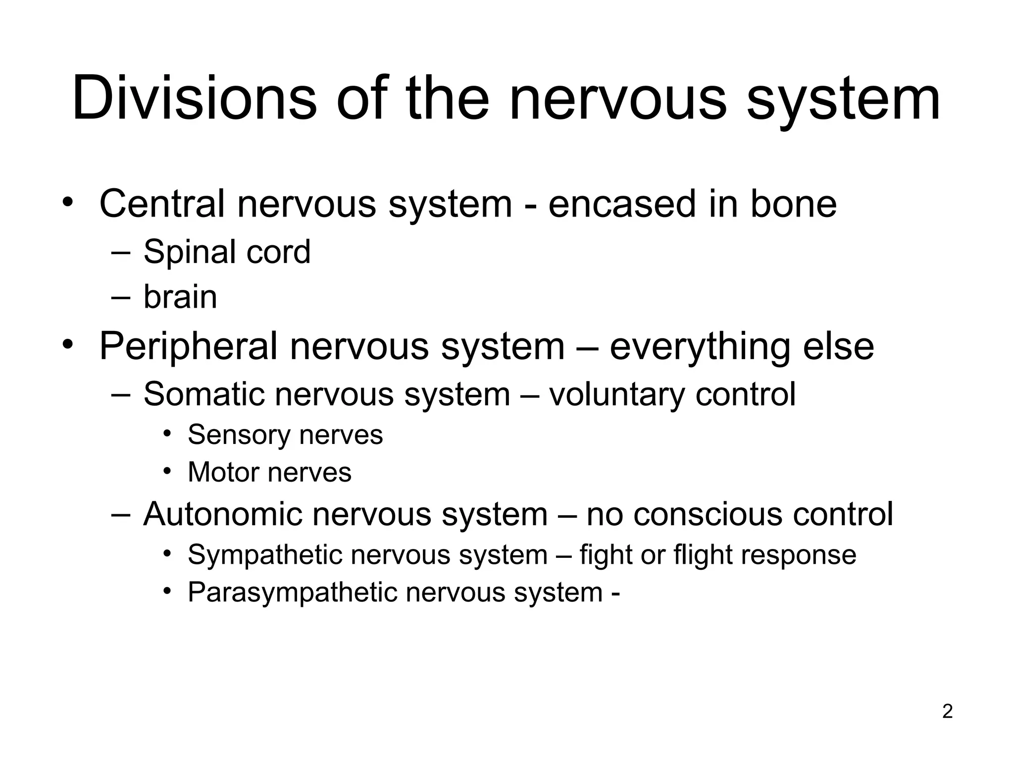
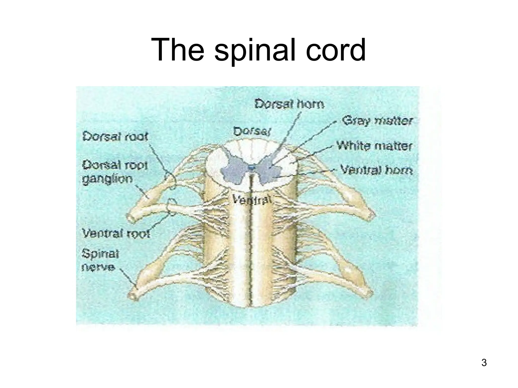
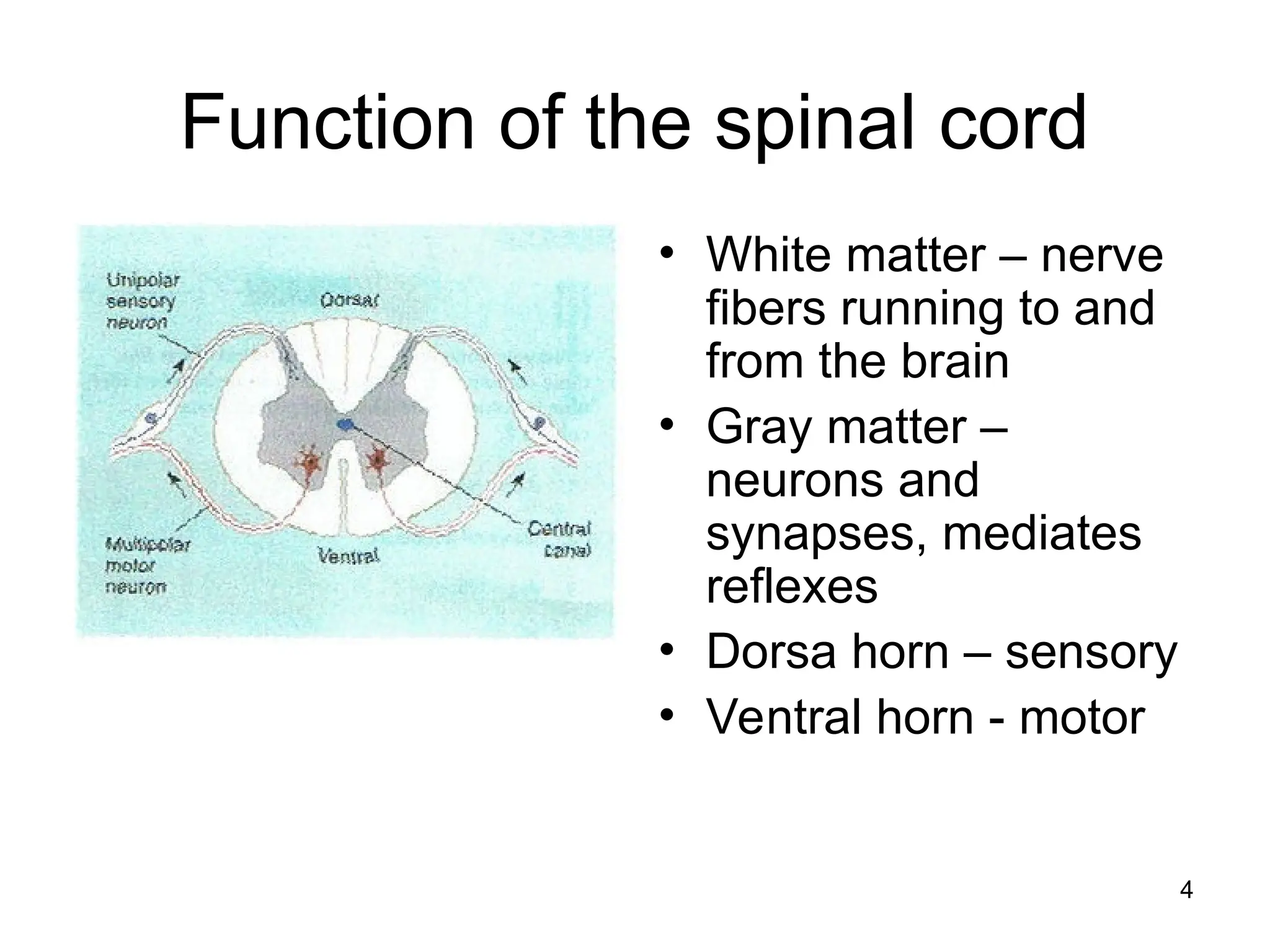
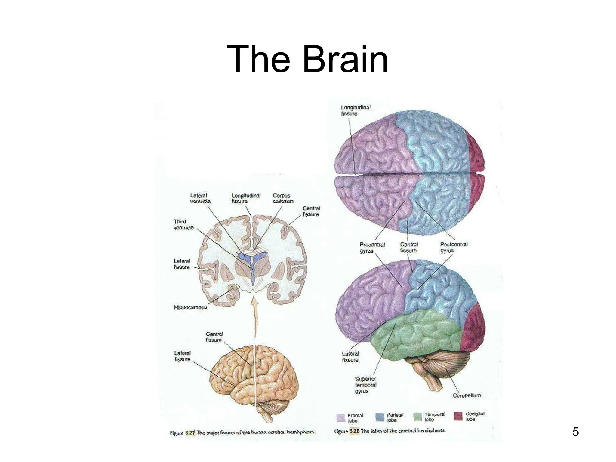
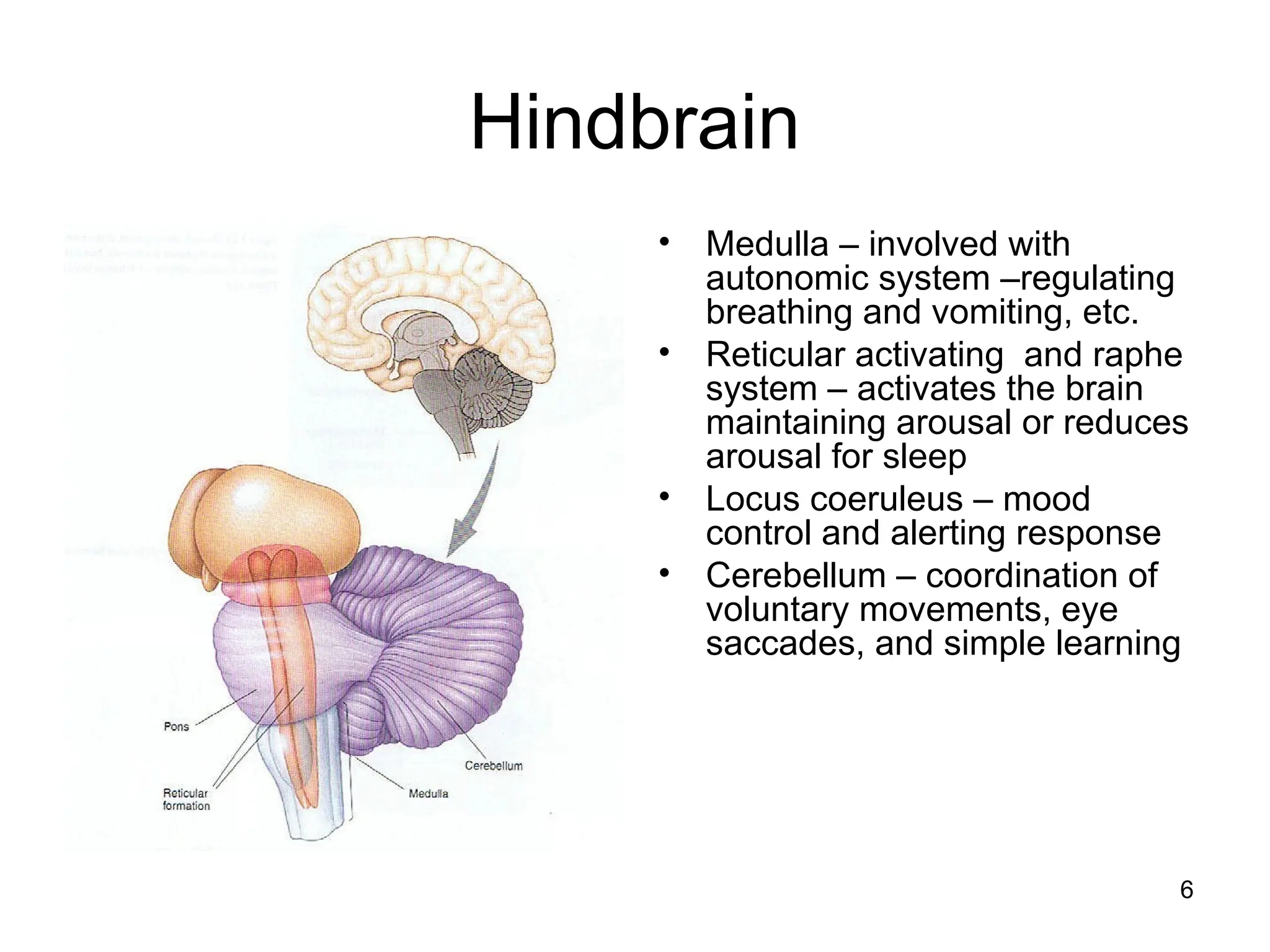
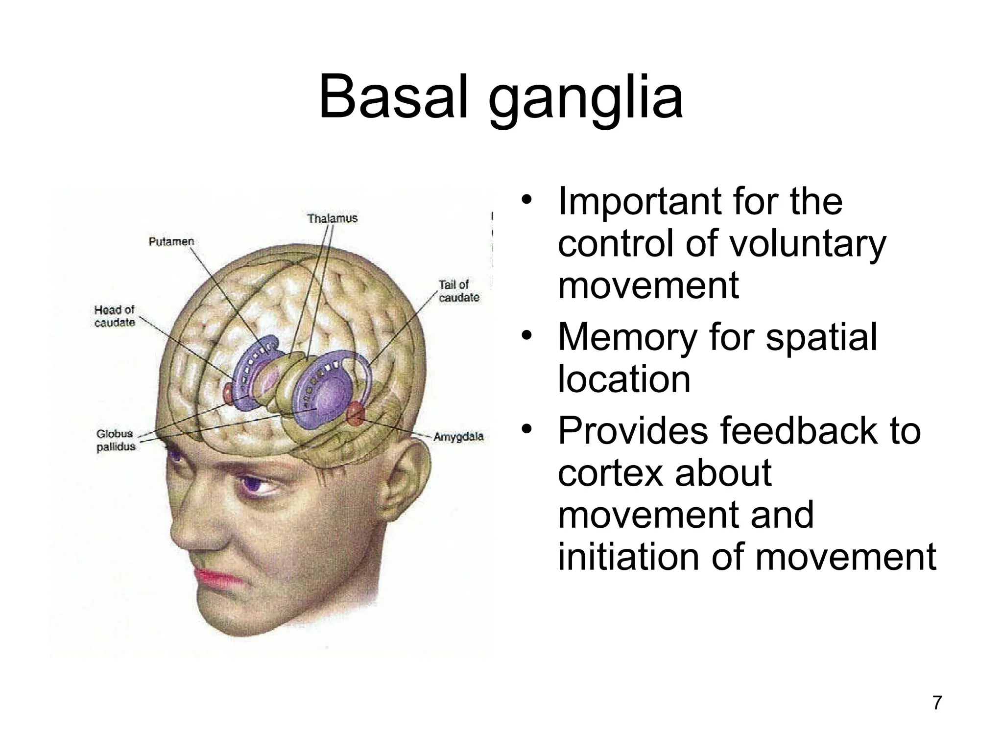
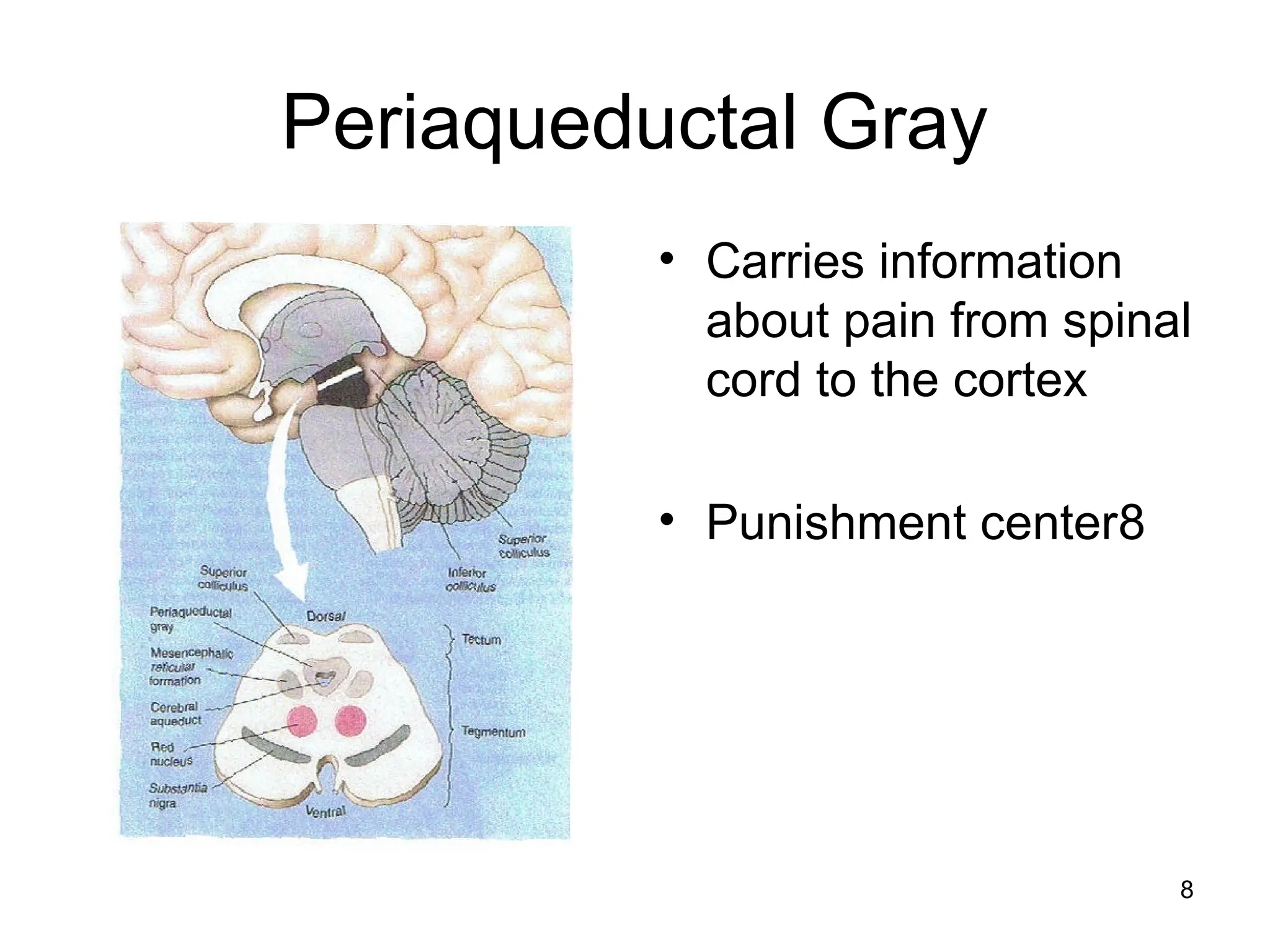
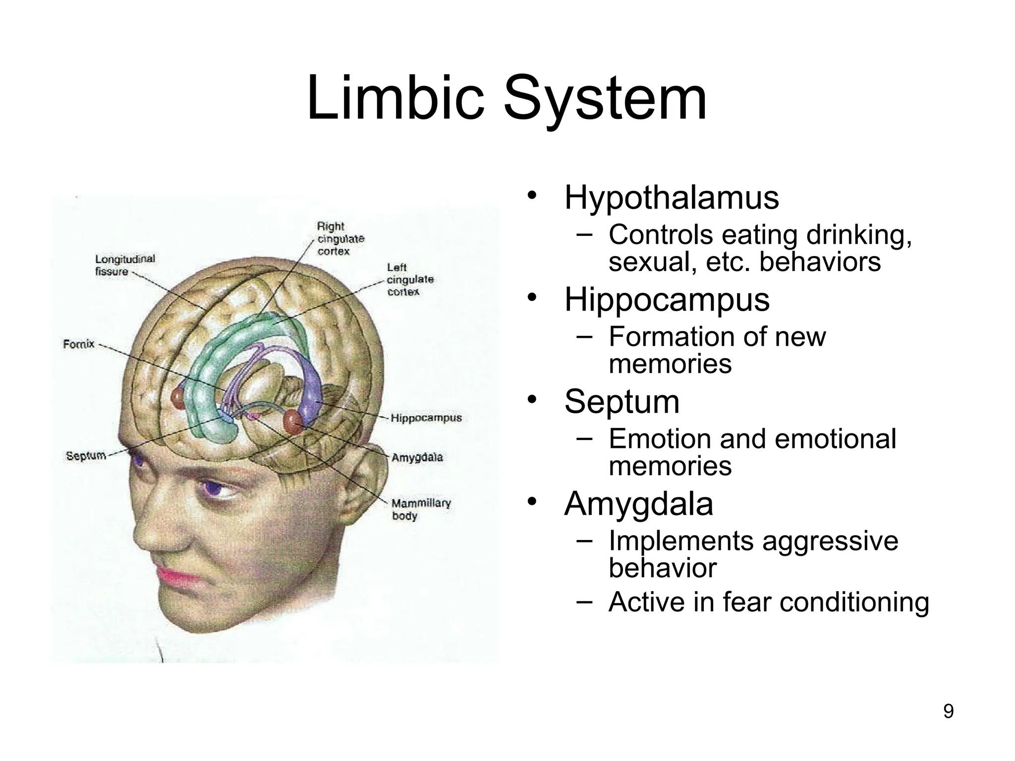
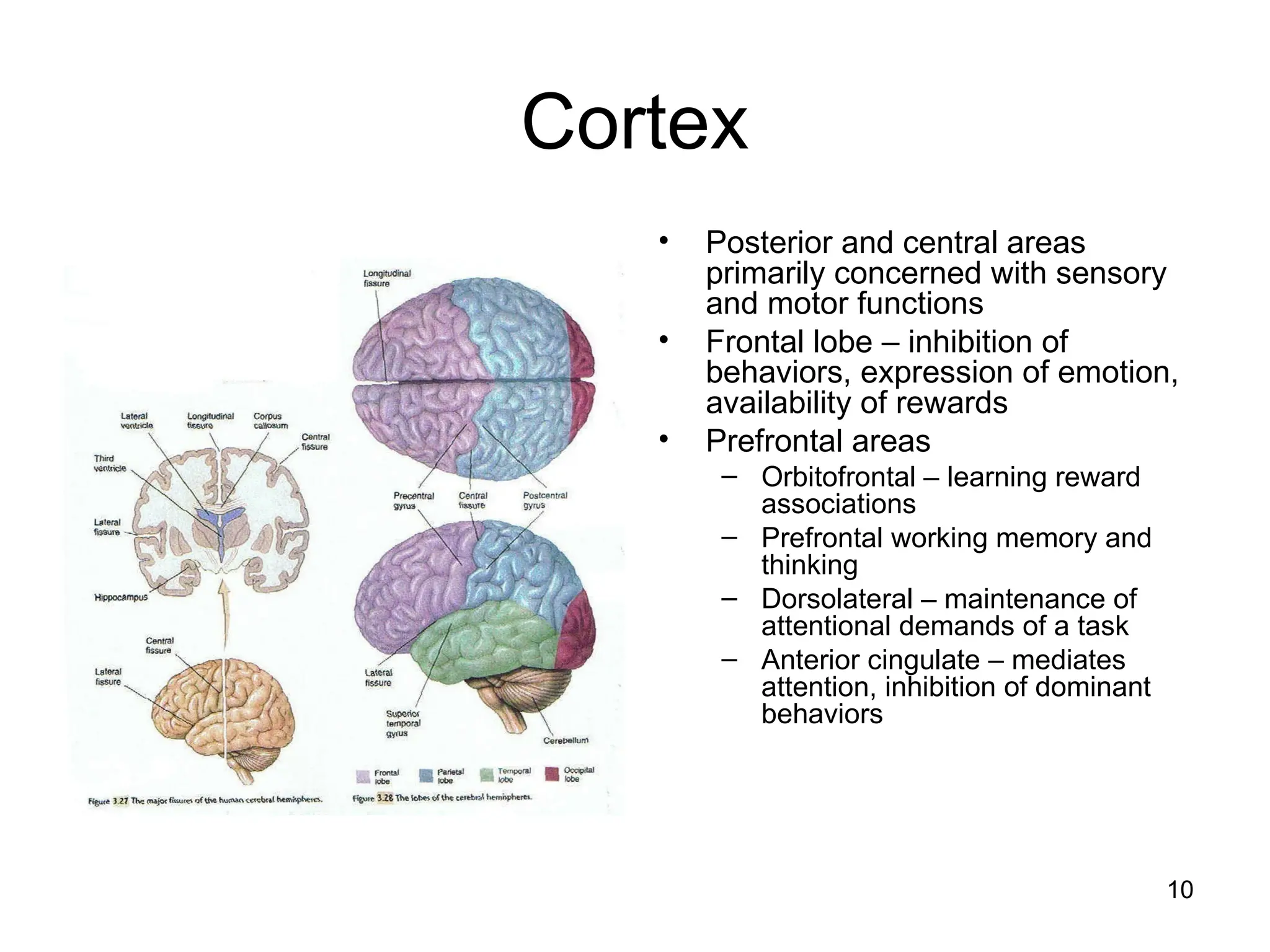
1
Introduction to the nervous
System
2
Divisions of the nervous system
• Central nervous system - encased in bone
– Spinal cord
– brain
• Peripheral nervous system – everything else
– Somatic nervous system – voluntary control
• Sensory nerves
• Motor nerves
– Autonomic nervous system – no conscious control
• Sympathetic nervous system – fight or flight response
• Parasympathetic nervous system -
3
The spinal cord
4
Function of the spinal cord
• White matter – nerve
fibers running to and
from the brain
• Gray matter –
neurons and
synapses, mediates
reflexes
• Dorsa horn – sensory
• Ventral horn - motor
5
The Brain
6
Hindbrain
• Medulla – involved with
autonomic system –regulating
breathing and vomiting, etc.
• Reticular activating and raphe
system – activates the brain
maintaining arousal or reduces
arousal for sleep
• Locus coeruleus – mood
control and alerting response
• Cerebellum – coordination of
voluntary movements, eye
saccades, and simple learning
7
Basal ganglia
• Important for the
control of voluntary
movement
• Memory for spatial
location
• Provides feedback to
cortex about
movement and
initiation of movement
8
Periaqueductal Gray
• Carries information
about pain from spinal
cord to the cortex
• Punishment center8
9
Limbic System
• Hypothalamus
– Controls eating drinking,
sexual, etc. behaviors
• Hippocampus
– Formation of new
memories
• Septum
– Emotion and emotional
memories
• Amygdala
– Implements aggressive
behavior
– Active in fear conditioning
10
Cortex
• Posterior and central areas
primarily concerned with sensory
and motor functions
• Frontal lobe – inhibition of
behaviors, expression of emotion,
availability of rewards
• Prefrontal areas
– Orbitofrontal – learning reward
associations
– Prefrontal working memory and
thinking
– Dorsolateral – maintenance of
attentional demands of a task
– Anterior cingulate – mediates
attention, inhibition of dominant
behaviors
11
Development of the Nervous
System
• Formation of neurons – neurogenesis- largely prenatal
– Majority of cells develop in first 3 months gestation - up to
150,000 cells per minute easily disrupted by chemicals and
radiation
• Migration pre and post natal
– Cells move from where they were formed to their target area
– They move either toward or away from chemicals released by
areas of the brain
• Interconnections develop – largely postnatal
• Programmed cell death
12
Methods of investigating drug
effects on the nervous system
• Electroencephalograms (EEG):
Electrodes are attached to the subject’s
scalp, and the device records the patterns
of brain waves.
• Event-Related Potentials: The
momentary changes in electrical activity of
the brain when a particular stimulus is
presented to the subject.
13
Methods of investigating drug
effects on the nervous system
(cont.)
• Imaging Technology
• Magnetic Resonance Imaging (MRI): Gives clear
pictures of the structure of the brain.
• Functional MRI: Image shows regions of the brain
with heightened neural activity, with different colors
reflecting high or low levels of blood flow, oxygen
uptake, and the like.
• PET scans- use radioactive labels attached to
glucose

Introduction to the nervo+.pptgukhfdgjkf

  • 1.
    1 Introduction to thenervous System
  • 2.
    2 Divisions of thenervous system • Central nervous system - encased in bone – Spinal cord – brain • Peripheral nervous system – everything else – Somatic nervous system – voluntary control • Sensory nerves • Motor nerves – Autonomic nervous system – no conscious control • Sympathetic nervous system – fight or flight response • Parasympathetic nervous system -
  • 3.
  • 4.
    4 Function of thespinal cord • White matter – nerve fibers running to and from the brain • Gray matter – neurons and synapses, mediates reflexes • Dorsa horn – sensory • Ventral horn - motor
  • 5.
  • 6.
    6 Hindbrain • Medulla –involved with autonomic system –regulating breathing and vomiting, etc. • Reticular activating and raphe system – activates the brain maintaining arousal or reduces arousal for sleep • Locus coeruleus – mood control and alerting response • Cerebellum – coordination of voluntary movements, eye saccades, and simple learning
  • 7.
    7 Basal ganglia • Importantfor the control of voluntary movement • Memory for spatial location • Provides feedback to cortex about movement and initiation of movement
  • 8.
    8 Periaqueductal Gray • Carriesinformation about pain from spinal cord to the cortex • Punishment center8
  • 9.
    9 Limbic System • Hypothalamus –Controls eating drinking, sexual, etc. behaviors • Hippocampus – Formation of new memories • Septum – Emotion and emotional memories • Amygdala – Implements aggressive behavior – Active in fear conditioning
  • 10.
    10 Cortex • Posterior andcentral areas primarily concerned with sensory and motor functions • Frontal lobe – inhibition of behaviors, expression of emotion, availability of rewards • Prefrontal areas – Orbitofrontal – learning reward associations – Prefrontal working memory and thinking – Dorsolateral – maintenance of attentional demands of a task – Anterior cingulate – mediates attention, inhibition of dominant behaviors
  • 11.
    11 Development of theNervous System • Formation of neurons – neurogenesis- largely prenatal – Majority of cells develop in first 3 months gestation - up to 150,000 cells per minute easily disrupted by chemicals and radiation • Migration pre and post natal – Cells move from where they were formed to their target area – They move either toward or away from chemicals released by areas of the brain • Interconnections develop – largely postnatal • Programmed cell death
  • 12.
    12 Methods of investigatingdrug effects on the nervous system • Electroencephalograms (EEG): Electrodes are attached to the subject’s scalp, and the device records the patterns of brain waves. • Event-Related Potentials: The momentary changes in electrical activity of the brain when a particular stimulus is presented to the subject.
  • 13.
    13 Methods of investigatingdrug effects on the nervous system (cont.) • Imaging Technology • Magnetic Resonance Imaging (MRI): Gives clear pictures of the structure of the brain. • Functional MRI: Image shows regions of the brain with heightened neural activity, with different colors reflecting high or low levels of blood flow, oxygen uptake, and the like. • PET scans- use radioactive labels attached to glucose