ANATOMICAL TERMS
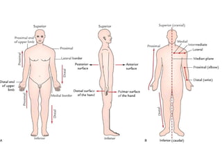
1. Anterior:towards the front aspect of the body.
2. Posterior: towards the back aspect of the body.
3. Superior: towards the head.
4. Inferior: towards the feet.
5. Central: towards the center of the mass of the body.
6. Peripheral: away from the center of the mass of the body.
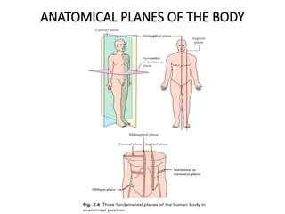
7. Median: along the midsagittal (median) plane.
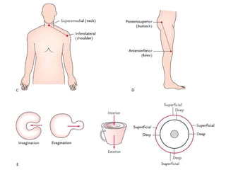
8. Medial: towards the median plane.
9. Lateral: away from the median plane.
15.
10. Intermediate: betweenmedial and lateral.
11. External: close to the surface of the body.
12. Internal: close to the center of the body.
13. Superficial and deep: these terms are used to describe
the relative positions of the two structures with respect to
the surface of the body.
14. Ventral: towards the belly.
15. Dorsal: towards the back.
16. Cranial/rostral: towards the head.
17. Caudal: towards the tail.
18.
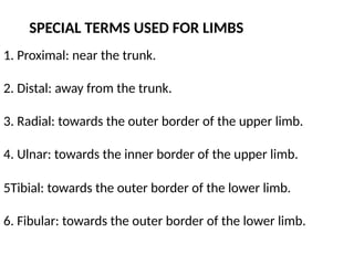
1. Proximal: nearthe trunk.
2. Distal: away from the trunk.
3. Radial: towards the outer border of the upper limb.
4. Ulnar: towards the inner border of the upper limb.
SPECIAL TERMS USED FOR LIMBS
5Tibial: towards the outer border of the lower limb.
6. Fibular: towards the outer border of the lower limb.
19.
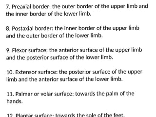
7. Preaxial border:the outer border of the upper limb and
the inner border of the lower limb.
8. Postaxial border: the inner border of the upper limb
and the outer border of the lower limb.
9. Flexor surface: the anterior surface of the upper limb
and the posterior surface of the lower limb.
10. Extensor surface: the posterior surface of the upper
limb and the anterior surface of the lower limb.
11. Palmar or volar surface: towards the palm of the
hands.
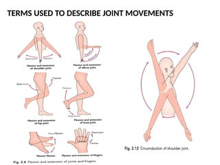
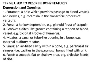
TERMS USED TODESCRIBE BONY FEATURES
Depression and Openings
1. Foramen: a hole which provides passage to blood vessels
and nerves, e.g. foramina in the transverse process of
vertebra.
2. Fossa: a hollow depression, e.g. glenoid fossa of scapula.
3. Groove: a ditch-like groove containing a tendon or blood
vessel, e.g. bicipital groove of humerus.
4. Meatus: a canal or tube-like opening in a bone, e.g.
external auditory meatus.
5. Sinus: an air-filled cavity within a bone, e.g. paranasal air
sinuses (i.e. cavities in the paranasal bones filled with air).
6. Facet: a smooth, flat or shallow area, e.g. articular facets
of ribs.
24.
Projections or Processes
1.Condyle:rounded knuckle-like projection, e.g. condyles of
femur.
2. Eminence: projecting prominent part of bone, e.g. inter-
condylar eminence of tibia.
3. Head: rounded articular projection beyond a narrow neck
like portion of bone, e.g. head of femur, head of humerus.
4. Crest: sharp ridge or border.
5. Epicondyle: prominence above or on a condyle, e.g.
medial and lateral epicondyles of humerus.
25.
5. Epicondyle: prominenceabove or on a condyle, e.g.
medial and lateral epicondyles of humerus.
6. Line: less prominent ridge, e.g. linea aspera of femur.
7. Spine: long thin projection, e.g. spines of vertebrae.
8. Trochanter: very large prominence for muscle attachment,
e.g. trochanters of femur.
9. Tubercle: small rounded projection, e.g. greater and lesser
tubercles of humerus.
10. Tuberosity: large rounded projection, e.g. ischial
tuberosity.
26.
TERMS USED TODESCRIBE NERVES AND ASSOCIATED
STRUCTURES
1. Nerves: whitish cords consisting of large number of
ex-ceedingly fine filaments called nerve fibers, bound
together in bundles by fibrous tissue.
2. Plexus: braided structure resulting from network of
nerve fibers.
3. Ganglion: group/collection of nerve cells outside the
central nervous system.
4. Nucleus: group/collection of nerve cells within the
central nervous system.
27.
Suffixes:
(a)itis inflammation, e.g.tonsillitis (inflammation of
tonsil), appendicitis (inflammation of appendix).
(b)ectomy removal from the body, e.g. tonsillec-tomy
(removal of tonsil), appendicectomy (removal of
appendix).
(c) -otomy to open and then close a hollow viscus or re-
gion, e.g. laparotomy (opening and then closing
abdominal cavity), hysterotomy (opening and then closing
uterus).
(d)tomy act of cutting, in a surgical operation.
28.
(e) -ostomy toopen a hollow organ and then leave it
open for a desired period, e.g. tracheostomy (opening
of trachea and then leaving it open), colostomy
(opening of the colon and then leaving it open).
(f) oma a tumor, e.g. lipoma (tumor of fat cells),
osteoma (bone tumor), hemangioma (tumor of blood
vessels).
29.
TERMS USED TODESCRIBE FASCIAE
1. Superficial fascia: a mixture of loose areolar and
adipose tissue lying deep to the dermis of the skin.
2. Deep fascia: dense inelastic membrane investing struc-
tures deep to the superficial fascia. It sends septa
between the muscle groups.
3. Fibrous sheath: derived from deep fascia to form
sheath around the tendons.
4. Retinaculum: thickening of deep fascia that retains the
underlying tendons in position.
30.
TERMS USED TODESCRIBE BLOOD VESSELS
1. Arteries: vessels that carry oxygenated blood away from
the heart.
2. Veins: vessels that carry deoxygenated blood towards the
heart.
3. Anastomosis: communication between the branches of
arteries and tributaries of veins.
4. End artery: an artery whose terminal branches do not
anastomose with the branches of the arteries supplying the
adjacent areas.
31.
5. Venae comitantes:two veins that accompany an artery.
6. Sinusoid: small thin-walled blood vessels (capillaries)
with irregular lumen lined by endothelial cells.
7. Collateral circulation: anastomotic alternate pathways
(collateral channels) through which the blood can reach
the field of distribution of the main artery if it is blocked.
8. Capillaries: microscopic vessels forming a network
through which the arterioles (smallest arteries) dis-charge
blood into smallest tributaries of veins.
32.
TERMS USED TODESCRIBE MUSCLES
1.Belly: fleshy contractile part of the muscle.
2. Tendon: fibrous noncontractile, cord-like part of the
muscle.
3. Aponeurosis: flattened tendon/flat sheet of collagen fibers
extending from muscle to its attachment on bone.
4. Raphe: stretchable fibrous band made up of interdigitating
fibers of the tendons or aponeurosis.
5. Origin: end of muscle which remains fixed during its
contraction.
33.
6. Insertion: endof muscle which moves during its
contraction.
7. Bursa: sacs of connective tissue filled with synovial fluid,
found where tendon slides over the bone.
8. Synovial sheath of tendons: similar to bursae in structure,
consisting of double-layered synovial membrane tubes
surrounding the tendons.
9. Prime movers: group of muscles initiating and main-
taining a particular movement.
34.
10. Antagonists: groupof muscles which oppose the move-
ment initiated by prime movers.
11. Synergists:
muscles which assist prime movers.
12. Fixators: muscles which contract isometrically to stabilize
the origin of prime movers so that it acts efficiently.
35.
ANATOMICAL NOMENCLATURE
Claudius Galen(AD 130-201) and Andreas Vesalius (1514-
1564), two great anatomists, wrote books on anatomy in
Greek and Latin respectively. Therefore, most of the ana-
tomical terms have Greek or Latin origin. Initially,
anatomi-cal terms used in textbooks and journals were
about 30,000 in number. In 1895, the German Anatomical
Society held its meeting at Basale and approved a list of
about 5000 terms, called Basle Nomina Anatomica (BNA).
It also laid down six strict rules, which are as follows:
36.
1. Each partshall have only one name.
2. Each term shall be in Latin.
3. Each term shall be as short and simple as possible.
4. The terms shall be merely memory signs.
5. The related terms shall be similar, viz. femoral artery,
femoral vein, and femoral nerve.