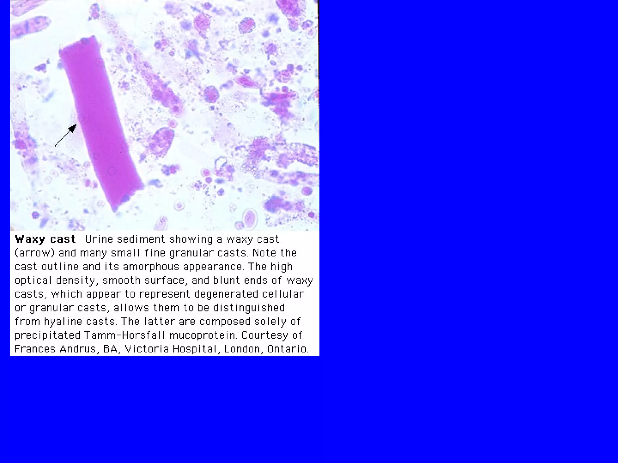
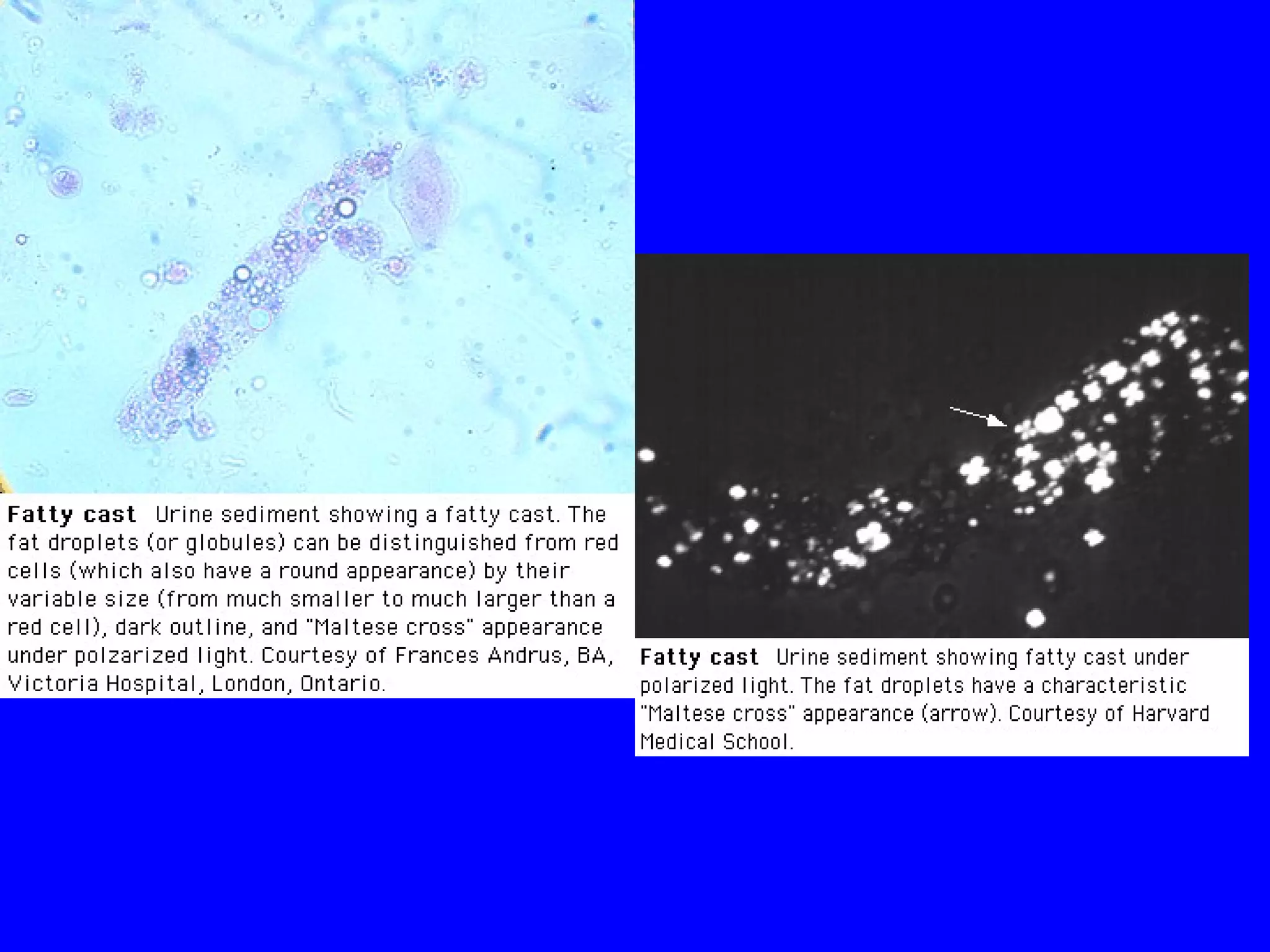
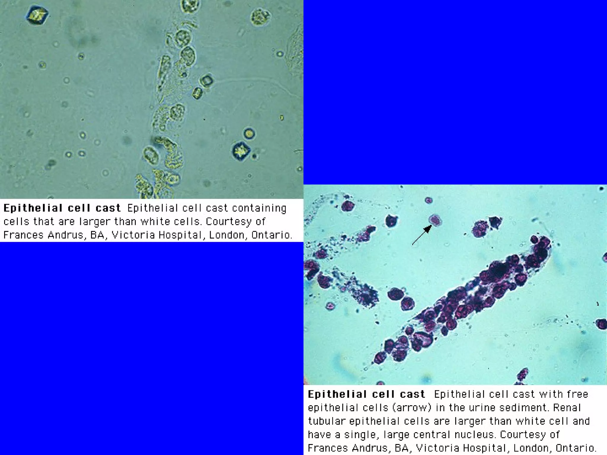
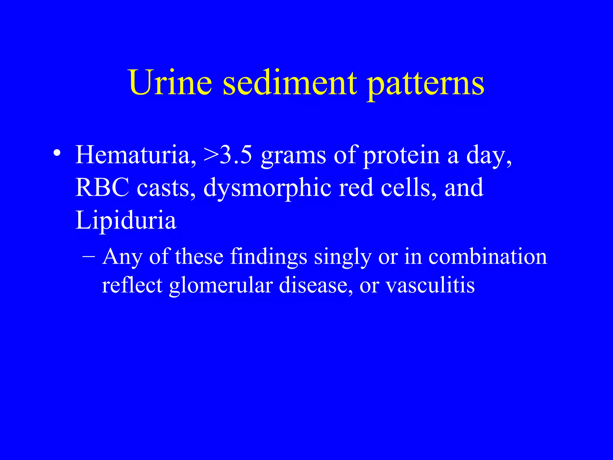
Tests of Renal Function
Cyrus Cryst MD FASN
VMMC
Estimates of Glomerular function
• Serum Creatinine
• BUN
• eGFR
• Cockroft-Gault Formula or MDRD
• 24 hour Creatinine Clearance
• Iothalamate Clearance
Plasma Creatinine
• Creatinine is derived from metabolism of creatine
in skeletal muscle and in dietary meat protein
• It is released constantly into the circulation
• It is freely filtered at the glomerulus and is neither
absorbed nor metabolized by the kidney
• 15% of urinary creatinine is derived from tubular
secretion
Plasma Creatinine
• GFR x PCr = Ucr x Volume
• GFR = Ucr x Volume / PCr
• This formula overestimates the true GFR by
10-15% (due to tubular secretion of
creatinine) and that is balanced by the error
in creatinine measurement.
Calculation of CCR
• If : PCr = 1.2 GFR = U V/P
• Ucr = 100 mg/dl
• V = 1.2 Liters per day
• GFR = 100 mg/dl x 1.2mg/dl /1.2L/day= 100 L/day
• This must be multiplied by 1000 ml/liter and again by
1/1440 (minutes per day) To yield conventional units:
70 ml/min
Normal is 130 ml/min in men and 120 ml/min
in women
Limitations of eGFR
• Only applies in “steady state”
• Only for those with impaired GFR < 60
• Only for those under 65
• Depends on accurate measurement of creat
• CKD-EPI formula for “normal population”
Plasma Creatinine
• Is the most useful
blood test to monitor
change in renal
function.
• Change in plasma
creatinine means a
change in rate of
production OR a
change in GFR
• Don’t neglect
conditions that cause
increase in plasma
creatinine production:
– Rhabdomyalysis
– Protein ingestion
• Drugs affect creatinine
secretion can raise
creat 10% (trimethaprim)
eGFR to Creat
eGFR and Creatinine
• Nomogram – takes creat and ‘assigns’ an
estimate of GFR
• No account of age, muscle mass, prior
condition – two values for race.
• Very concerning for patients to be told “stage 3
kidney failure”
• No context can go up and down
• At extremes of age, weight, muscle mass may
be inaccurate
Heat map: stages of renal
impairment
When Creatinine Elevation is
Misleading
• Pts who have lost or donated a kidney
• People taking sulfa-TRIMETHOPRIM
• Volume depletion – ‘fasting’
• ACEI and ARB – lead to ~30% decline in
eGFR that is reversible with discontinuation
of the drug
Clinical tips
• A plasma creatinine rising 1.0 mg/dl/24
hours reflects GFR = 0
• RAAS inhibitors can raise serum creat by
0.2-0.6 depending on volume depletion
• Getting a complete 24 hour urine for Creat
Clearance in the hospital is very difficult.
Urinalysis in renal disease
• Color
• Specific Gravity
• Leucocyte Esterase
• Protein
• pH
• Osmolality
• Glucose
• Part 2
– Sediment
– Patterns of injury
• Part 3
– Urine electrolytes
– Settings where useful
Urine Color
• Usually clear, light yellow
• Lighter when dilute, darker when concentrated
• White due to phosphate crystals or pyuria
• Green due to methylene blue dye, propofol
• Black due to malignancy
• Red or brown due to Heme or pigments
• Turbidity may indicate infection, crystals or
phosphates
Red urine
• Hematuria - the sediment is red, supernatent
is clear
• If supernatent is RED, test for heme.
– Heme Positive - RHABDOMYALYSIS
– Heme Negative? - RARE- Porphyuria, Beets,
phenazopyridine.
Urine Specific Gravity
• Surrogate for urine osmolality
• Indicates dilute or concentrated urine
• SG of 1.010 is about isosomolar
• Highest in extreme vol depletion 1.030
• May be falsely high in cases of contrast
excretion or glucose
• Cannot be falsely low
Leucocyte Esterase
• Enzyme released by neutrophils – marker for
leucocytes in urine.
• False positive if urine is VERY dilute or
concentrated
• False Negative if urine is very concentrated
• Nitrates- test for presence of nitrate reductase
– Also has false pos, negs and not elaborated by all bugs
Leucocyte Esterase
• Leukocyte estrase and nitrite - surrogate
markers for pyuria and enterobacteriaceae
• Postive results may be an important clue:
– May detect WBC or bacteria not associated with UTI!
– “sterile pyuria?”
• TB,
• Interstitial nephritis,
• Stones
Urine Protein
• Patients usually only excrete 150 mg
protein /day or less
• Dipstick usually only picks up ALBUMIN
• Dipstick usually positive only for >300
mg/day
• Scores 1-2-3+
• Sulfosalicylic acid test - for light chains
Microalbuminuria
• Screening test for diabetic nephropathy
• Risk factor for cardiac disease
• Also used with microalbumin/creat ratio
Quantitative protein excretion
• 24 hour collection determines if patient has
“nephrotic range” proteinuria - evidence of
glomerular disease - >3.5 gm/1.73 M2
• Alternative method is to use a spot Protein-
to-creatinine ratio in mg/mg which
correlates with g/1.73 M2
protein excretion.
Urine pH
• Range is 4.5-8.0
• Best use is in Metabolic acidosis - normally
patients should excrete an acid urine pH <5
– If urine pH is >5 In a patient with ACIDOSIS
suspect a renal tubular acidosis
– If urine pH is > 7.0 - 9.0 - suspect a urease
producing organism such as P. miriabilis
(associated with staghorn calculi)
Urine Osmolality
• Plasma osmolality is maintained in a narrow range
normally by the kidney’s ability to elaborate urine
that varies widely in osmolality.
• Urine osmolality measurements are key to
unraveling the DDX of
• hypernatremia,
• hyponatremia,
• polyuria
Urine Glucose
• Indicates High plasma glucose levels (but is
insensitive)
• Can indicate a proximal tubular
reabsorptive problem - Fanconi’s syndrome
• Urinary glucose measurements reflect mean
glucose levels, not peak concentrations.
PART II
• The Urine Sediment Exam
Sediment
• Crystals
• Cells
– Heme
– Wbc
– epithelial
• Bacteria
• Casts
• Patterns
Crystals
• Uric Acid
• Calcium
• Cystine
• Struvite - magnesium/ammonium/phosphate
Crystals
Sediment - Cells
• Pyuria
– May reflect infection
– Usually accompanies any renal disease
– May be stained for eosinophils
• Tubular epithelial cells
– In casts reflect ATN
– Individually are seen in many situations
Sediment - Cells
• Hematuria -
– Always abnormal in older men
– Can be transient in younger patients
– RBC morphology can reflect upper or lower
tract bleeding
– Persistent heme needs to be evaluated
– Always present if foley in place
Urine sediment - Casts
• Hyaline
• Red Cell Casts
• WBC casts
• Epithelial cell casts
• Fatty casts
• Granular cell casts
– Muddy brown casts
• Waxy casts
• Broad casts
Urine sediment patterns
• Hematuria, >3.5 grams of protein a day,
RBC casts, dysmorphic red cells, and
Lipiduria
– Any of these findings singly or in combination
reflect glomerular disease, or vasculitis
Urine sediment patterns
• Multiple granular and epithelial cell casts
with free epithelial cells.
– Coarse granular casts in “dirty” background
– Signifies ATN - acute toxic or ischemic injury
– Beware of hyperbilirubinemia
Urine sediment patterns
• Pyuria with WBC and Granular or waxy
casts and no proteinuria
– Suggests interstitial disease or Obstruction
– Can be seen with other signs of glomerular
disease
Urine sediment patterns
• Hematuria alone
– Variable significance - depends on the setting
• Stone? Flank pain
• Tumor? Age, imaging studies
• Mild glomerular disease - IgA, thin basement
membrane disease, Alport’s,
• PCKD
Urine sediment patterns
• Hematuria and Pyuria with no or variable
casts-can be seen with any of the following:
– Interstitial nephritis
– Glomerular disease
– Vasculitis
– Obstruction
– Renal infarction
Urine sediment patterns
• Pyuria alone?
• Usually a UTI
• Sterile pyuria suggests TB, analgesic nephropathy
Tests of renal function
Part 3 - Urine Electrolytes
• Sodium excretion
• FENa
• Chloride excretion
• Potassium excretion
• Urine osmolality and pH

Tests of renal function 3

  • 1.
    Tests of RenalFunction Cyrus Cryst MD FASN VMMC
  • 2.
    Estimates of Glomerularfunction • Serum Creatinine • BUN • eGFR • Cockroft-Gault Formula or MDRD • 24 hour Creatinine Clearance • Iothalamate Clearance
  • 3.
    Plasma Creatinine • Creatinineis derived from metabolism of creatine in skeletal muscle and in dietary meat protein • It is released constantly into the circulation • It is freely filtered at the glomerulus and is neither absorbed nor metabolized by the kidney • 15% of urinary creatinine is derived from tubular secretion
  • 4.
    Plasma Creatinine • GFRx PCr = Ucr x Volume • GFR = Ucr x Volume / PCr • This formula overestimates the true GFR by 10-15% (due to tubular secretion of creatinine) and that is balanced by the error in creatinine measurement.
  • 5.
    Calculation of CCR •If : PCr = 1.2 GFR = U V/P • Ucr = 100 mg/dl • V = 1.2 Liters per day • GFR = 100 mg/dl x 1.2mg/dl /1.2L/day= 100 L/day • This must be multiplied by 1000 ml/liter and again by 1/1440 (minutes per day) To yield conventional units: 70 ml/min Normal is 130 ml/min in men and 120 ml/min in women
  • 6.
    Limitations of eGFR •Only applies in “steady state” • Only for those with impaired GFR < 60 • Only for those under 65 • Depends on accurate measurement of creat • CKD-EPI formula for “normal population”
  • 7.
    Plasma Creatinine • Isthe most useful blood test to monitor change in renal function. • Change in plasma creatinine means a change in rate of production OR a change in GFR • Don’t neglect conditions that cause increase in plasma creatinine production: – Rhabdomyalysis – Protein ingestion • Drugs affect creatinine secretion can raise creat 10% (trimethaprim)
  • 8.
  • 9.
    eGFR and Creatinine •Nomogram – takes creat and ‘assigns’ an estimate of GFR • No account of age, muscle mass, prior condition – two values for race. • Very concerning for patients to be told “stage 3 kidney failure” • No context can go up and down • At extremes of age, weight, muscle mass may be inaccurate
  • 10.
    Heat map: stagesof renal impairment
  • 11.
    When Creatinine Elevationis Misleading • Pts who have lost or donated a kidney • People taking sulfa-TRIMETHOPRIM • Volume depletion – ‘fasting’ • ACEI and ARB – lead to ~30% decline in eGFR that is reversible with discontinuation of the drug
  • 12.
    Clinical tips • Aplasma creatinine rising 1.0 mg/dl/24 hours reflects GFR = 0 • RAAS inhibitors can raise serum creat by 0.2-0.6 depending on volume depletion • Getting a complete 24 hour urine for Creat Clearance in the hospital is very difficult.
  • 13.
    Urinalysis in renaldisease • Color • Specific Gravity • Leucocyte Esterase • Protein • pH • Osmolality • Glucose • Part 2 – Sediment – Patterns of injury • Part 3 – Urine electrolytes – Settings where useful
  • 14.
    Urine Color • Usuallyclear, light yellow • Lighter when dilute, darker when concentrated • White due to phosphate crystals or pyuria • Green due to methylene blue dye, propofol • Black due to malignancy • Red or brown due to Heme or pigments • Turbidity may indicate infection, crystals or phosphates
  • 15.
    Red urine • Hematuria- the sediment is red, supernatent is clear • If supernatent is RED, test for heme. – Heme Positive - RHABDOMYALYSIS – Heme Negative? - RARE- Porphyuria, Beets, phenazopyridine.
  • 17.
    Urine Specific Gravity •Surrogate for urine osmolality • Indicates dilute or concentrated urine • SG of 1.010 is about isosomolar • Highest in extreme vol depletion 1.030 • May be falsely high in cases of contrast excretion or glucose • Cannot be falsely low
  • 18.
    Leucocyte Esterase • Enzymereleased by neutrophils – marker for leucocytes in urine. • False positive if urine is VERY dilute or concentrated • False Negative if urine is very concentrated • Nitrates- test for presence of nitrate reductase – Also has false pos, negs and not elaborated by all bugs
  • 19.
    Leucocyte Esterase • Leukocyteestrase and nitrite - surrogate markers for pyuria and enterobacteriaceae • Postive results may be an important clue: – May detect WBC or bacteria not associated with UTI! – “sterile pyuria?” • TB, • Interstitial nephritis, • Stones
  • 20.
    Urine Protein • Patientsusually only excrete 150 mg protein /day or less • Dipstick usually only picks up ALBUMIN • Dipstick usually positive only for >300 mg/day • Scores 1-2-3+ • Sulfosalicylic acid test - for light chains
  • 21.
    Microalbuminuria • Screening testfor diabetic nephropathy • Risk factor for cardiac disease • Also used with microalbumin/creat ratio
  • 22.
    Quantitative protein excretion •24 hour collection determines if patient has “nephrotic range” proteinuria - evidence of glomerular disease - >3.5 gm/1.73 M2 • Alternative method is to use a spot Protein- to-creatinine ratio in mg/mg which correlates with g/1.73 M2 protein excretion.
  • 23.
    Urine pH • Rangeis 4.5-8.0 • Best use is in Metabolic acidosis - normally patients should excrete an acid urine pH <5 – If urine pH is >5 In a patient with ACIDOSIS suspect a renal tubular acidosis – If urine pH is > 7.0 - 9.0 - suspect a urease producing organism such as P. miriabilis (associated with staghorn calculi)
  • 24.
    Urine Osmolality • Plasmaosmolality is maintained in a narrow range normally by the kidney’s ability to elaborate urine that varies widely in osmolality. • Urine osmolality measurements are key to unraveling the DDX of • hypernatremia, • hyponatremia, • polyuria
  • 25.
    Urine Glucose • IndicatesHigh plasma glucose levels (but is insensitive) • Can indicate a proximal tubular reabsorptive problem - Fanconi’s syndrome • Urinary glucose measurements reflect mean glucose levels, not peak concentrations.
  • 26.
    PART II • TheUrine Sediment Exam
  • 28.
    Sediment • Crystals • Cells –Heme – Wbc – epithelial • Bacteria • Casts • Patterns
  • 29.
    Crystals • Uric Acid •Calcium • Cystine • Struvite - magnesium/ammonium/phosphate
  • 31.
  • 33.
    Sediment - Cells •Pyuria – May reflect infection – Usually accompanies any renal disease – May be stained for eosinophils • Tubular epithelial cells – In casts reflect ATN – Individually are seen in many situations
  • 35.
    Sediment - Cells •Hematuria - – Always abnormal in older men – Can be transient in younger patients – RBC morphology can reflect upper or lower tract bleeding – Persistent heme needs to be evaluated – Always present if foley in place
  • 38.
    Urine sediment -Casts • Hyaline • Red Cell Casts • WBC casts • Epithelial cell casts • Fatty casts • Granular cell casts – Muddy brown casts • Waxy casts • Broad casts
  • 43.
    Urine sediment patterns •Hematuria, >3.5 grams of protein a day, RBC casts, dysmorphic red cells, and Lipiduria – Any of these findings singly or in combination reflect glomerular disease, or vasculitis
  • 44.
    Urine sediment patterns •Multiple granular and epithelial cell casts with free epithelial cells. – Coarse granular casts in “dirty” background – Signifies ATN - acute toxic or ischemic injury – Beware of hyperbilirubinemia
  • 46.
    Urine sediment patterns •Pyuria with WBC and Granular or waxy casts and no proteinuria – Suggests interstitial disease or Obstruction – Can be seen with other signs of glomerular disease
  • 47.
    Urine sediment patterns •Hematuria alone – Variable significance - depends on the setting • Stone? Flank pain • Tumor? Age, imaging studies • Mild glomerular disease - IgA, thin basement membrane disease, Alport’s, • PCKD
  • 48.
    Urine sediment patterns •Hematuria and Pyuria with no or variable casts-can be seen with any of the following: – Interstitial nephritis – Glomerular disease – Vasculitis – Obstruction – Renal infarction
  • 49.
    Urine sediment patterns •Pyuria alone? • Usually a UTI • Sterile pyuria suggests TB, analgesic nephropathy
  • 51.
    Tests of renalfunction Part 3 - Urine Electrolytes • Sodium excretion • FENa • Chloride excretion • Potassium excretion • Urine osmolality and pH