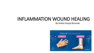
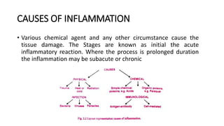
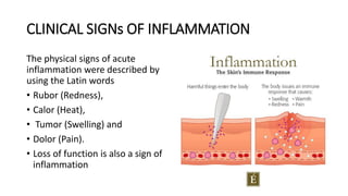
This document discusses inflammation and wound healing. It begins by defining inflammation as the body's protective response to tissue injury. The causes of inflammation include microbial infections, hypersensitivity reactions, physical trauma, chemicals, and tissue necrosis. The signs of acute inflammation are redness, heat, swelling, pain, and loss of function. Mediators of inflammation such as histamine, prostaglandins, and complement proteins are released in response to injury and stimulate the inflammatory response. This includes increased blood flow, vascular permeability, and recruitment of immune cells to the injured site to clear infection and initiate repair. Both acute and chronic inflammation aim to remove harmful stimuli and initiate healing, though chronic inflammation can lead to fibrosis and tissue damage if the stimulus
inflammation is the body's immune system's response to an irritant. The irritant might be a germ, but it could also be a foreign object, such as a splinter in your finger.
Inflammation- General Pathology seminar PG 1st yearDr. Ritu Gupta
this seminar includes general inflammation, its etiology, acute inflammation, features, events, fate, chronic inflammation, causes, features, types, granulomatous inflammation, acute v/s chronic inflammation, inflammatory disorders of pulp and periradicular tissues
Inflammation is the body's mechanism for coping with agents that could damage it.
In other words, inflammation is a protective response to rid the body of the cause of cell injury and the resultant necrotic cells that cell injury produces.
Although the processes of acute and chronic inflammation are an important protective mechanism used by the body to deal with potentially damaging agents, they are potentially damaging to the body and must be closely regulated.
Title: Sense of Taste
Presenter: Dr. Faiza, Assistant Professor of Physiology
Qualifications:
MBBS (Best Graduate, AIMC Lahore)
FCPS Physiology
ICMT, CHPE, DHPE (STMU)
MPH (GC University, Faisalabad)
MBA (Virtual University of Pakistan)
Learning Objectives:
Describe the structure and function of taste buds.
Describe the relationship between the taste threshold and taste index of common substances.
Explain the chemical basis and signal transduction of taste perception for each type of primary taste sensation.
Recognize different abnormalities of taste perception and their causes.
Key Topics:
Significance of Taste Sensation:
Differentiation between pleasant and harmful food
Influence on behavior
Selection of food based on metabolic needs
Receptors of Taste:
Taste buds on the tongue
Influence of sense of smell, texture of food, and pain stimulation (e.g., by pepper)
Primary and Secondary Taste Sensations:
Primary taste sensations: Sweet, Sour, Salty, Bitter, Umami
Chemical basis and signal transduction mechanisms for each taste
Taste Threshold and Index:
Taste threshold values for Sweet (sucrose), Salty (NaCl), Sour (HCl), and Bitter (Quinine)
Taste index relationship: Inversely proportional to taste threshold
Taste Blindness:
Inability to taste certain substances, particularly thiourea compounds
Example: Phenylthiocarbamide
Structure and Function of Taste Buds:
Composition: Epithelial cells, Sustentacular/Supporting cells, Taste cells, Basal cells
Features: Taste pores, Taste hairs/microvilli, and Taste nerve fibers
Location of Taste Buds:
Found in papillae of the tongue (Fungiform, Circumvallate, Foliate)
Also present on the palate, tonsillar pillars, epiglottis, and proximal esophagus
Mechanism of Taste Stimulation:
Interaction of taste substances with receptors on microvilli
Signal transduction pathways for Umami, Sweet, Bitter, Sour, and Salty tastes
Taste Sensitivity and Adaptation:
Decrease in sensitivity with age
Rapid adaptation of taste sensation
Role of Saliva in Taste:
Dissolution of tastants to reach receptors
Washing away the stimulus
Taste Preferences and Aversions:
Mechanisms behind taste preference and aversion
Influence of receptors and neural pathways
Impact of Sensory Nerve Damage:
Degeneration of taste buds if the sensory nerve fiber is cut
Abnormalities of Taste Detection:
Conditions: Ageusia, Hypogeusia, Dysgeusia (parageusia)
Causes: Nerve damage, neurological disorders, infections, poor oral hygiene, adverse drug effects, deficiencies, aging, tobacco use, altered neurotransmitter levels
Neurotransmitters and Taste Threshold:
Effects of serotonin (5-HT) and norepinephrine (NE) on taste sensitivity
Supertasters:
25% of the population with heightened sensitivity to taste, especially bitterness
Increased number of fungiform papillae
New Directions in Targeted Therapeutic Approaches for Older Adults With Mantl...i3 Health
i3 Health is pleased to make the speaker slides from this activity available for use as a non-accredited self-study or teaching resource.
This slide deck presented by Dr. Kami Maddocks, Professor-Clinical in the Division of Hematology and
Associate Division Director for Ambulatory Operations
The Ohio State University Comprehensive Cancer Center, will provide insight into new directions in targeted therapeutic approaches for older adults with mantle cell lymphoma.
STATEMENT OF NEED
Mantle cell lymphoma (MCL) is a rare, aggressive B-cell non-Hodgkin lymphoma (NHL) accounting for 5% to 7% of all lymphomas. Its prognosis ranges from indolent disease that does not require treatment for years to very aggressive disease, which is associated with poor survival (Silkenstedt et al, 2021). Typically, MCL is diagnosed at advanced stage and in older patients who cannot tolerate intensive therapy (NCCN, 2022). Although recent advances have slightly increased remission rates, recurrence and relapse remain very common, leading to a median overall survival between 3 and 6 years (LLS, 2021). Though there are several effective options, progress is still needed towards establishing an accepted frontline approach for MCL (Castellino et al, 2022). Treatment selection and management of MCL are complicated by the heterogeneity of prognosis, advanced age and comorbidities of patients, and lack of an established standard approach for treatment, making it vital that clinicians be familiar with the latest research and advances in this area. In this activity chaired by Michael Wang, MD, Professor in the Department of Lymphoma & Myeloma at MD Anderson Cancer Center, expert faculty will discuss prognostic factors informing treatment, the promising results of recent trials in new therapeutic approaches, and the implications of treatment resistance in therapeutic selection for MCL.
Target Audience
Hematology/oncology fellows, attending faculty, and other health care professionals involved in the treatment of patients with mantle cell lymphoma (MCL).
Learning Objectives
1.) Identify clinical and biological prognostic factors that can guide treatment decision making for older adults with MCL
2.) Evaluate emerging data on targeted therapeutic approaches for treatment-naive and relapsed/refractory MCL and their applicability to older adults
3.) Assess mechanisms of resistance to targeted therapies for MCL and their implications for treatment selection
TEST BANK for Operations Management, 14th Edition by William J. Stevenson, Ve...kevinkariuki227
TEST BANK for Operations Management, 14th Edition by William J. Stevenson, Verified Chapters 1 - 19, Complete Newest Version.pdf
Pulmonary Thromboembolism - etilogy, types, medical- Surgical and nursing man...VarunMahajani
Disruption of blood supply to lung alveoli due to blockage of one or more pulmonary blood vessels is called as Pulmonary thromboembolism. In this presentation we will discuss its causes, types and its management in depth.
These lecture slides, by Dr Sidra Arshad, offer a quick overview of physiological basis of a normal electrocardiogram.
Learning objectives:
1. Define an electrocardiogram (ECG) and electrocardiography
2. Describe how dipoles generated by the heart produce the waveforms of the ECG
3. Describe the components of a normal electrocardiogram of a typical bipolar leads (limb II)
4. Differentiate between intervals and segments
5. Enlist some common indications for obtaining an ECG
Study Resources:
1. Chapter 11, Guyton and Hall Textbook of Medical Physiology, 14th edition
2. Chapter 9, Human Physiology - From Cells to Systems, Lauralee Sherwood, 9th edition
3. Chapter 29, Ganong’s Review of Medical Physiology, 26th edition
4. Electrocardiogram, StatPearls - https://www.ncbi.nlm.nih.gov/books/NBK549803/
5. ECG in Medical Practice by ABM Abdullah, 4th edition
6. ECG Basics, http://www.nataliescasebook.com/tag/e-c-g-basics
The prostate is an exocrine gland of the male mammalian reproductive system
It is a walnut-sized gland that forms part of the male reproductive system and is located in front of the rectum and just below the urinary bladder
Function is to store and secrete a clear, slightly alkaline fluid that constitutes 10-30% of the volume of the seminal fluid that along with the spermatozoa, constitutes semen
A healthy human prostate measures (4cm-vertical, by 3cm-horizontal, 2cm ant-post ).
It surrounds the urethra just below the urinary bladder. It has anterior, median, posterior and two lateral lobes
It’s work is regulated by androgens which are responsible for male sex characteristics
Generalised disease of the prostate due to hormonal derangement which leads to non malignant enlargement of the gland (increase in the number of epithelial cells and stromal tissue)to cause compression of the urethra leading to symptoms (LUTS
Ethanol (CH3CH2OH), or beverage alcohol, is a two-carbon alcohol
that is rapidly distributed in the body and brain. Ethanol alters many
neurochemical systems and has rewarding and addictive properties. It
is the oldest recreational drug and likely contributes to more morbidity,
mortality, and public health costs than all illicit drugs combined. The
5th edition of the Diagnostic and Statistical Manual of Mental Disorders
(DSM-5) integrates alcohol abuse and alcohol dependence into a single
disorder called alcohol use disorder (AUD), with mild, moderate,
and severe subclassifications (American Psychiatric Association, 2013).
In the DSM-5, all types of substance abuse and dependence have been
combined into a single substance use disorder (SUD) on a continuum
from mild to severe. A diagnosis of AUD requires that at least two of
the 11 DSM-5 behaviors be present within a 12-month period (mild
AUD: 2–3 criteria; moderate AUD: 4–5 criteria; severe AUD: 6–11 criteria).
The four main behavioral effects of AUD are impaired control over
drinking, negative social consequences, risky use, and altered physiological
effects (tolerance, withdrawal). This chapter presents an overview
of the prevalence and harmful consequences of AUD in the U.S.,
the systemic nature of the disease, neurocircuitry and stages of AUD,
comorbidities, fetal alcohol spectrum disorders, genetic risk factors, and
pharmacotherapies for AUD.
Tom Selleck Health: A Comprehensive Look at the Iconic Actor’s Wellness Journeygreendigital
Tom Selleck, an enduring figure in Hollywood. has captivated audiences for decades with his rugged charm, iconic moustache. and memorable roles in television and film. From his breakout role as Thomas Magnum in Magnum P.I. to his current portrayal of Frank Reagan in Blue Bloods. Selleck's career has spanned over 50 years. But beyond his professional achievements. fans have often been curious about Tom Selleck Health. especially as he has aged in the public eye.
Follow us on: Pinterest
Introduction
Many have been interested in Tom Selleck health. not only because of his enduring presence on screen but also because of the challenges. and lifestyle choices he has faced and made over the years. This article delves into the various aspects of Tom Selleck health. exploring his fitness regimen, diet, mental health. and the challenges he has encountered as he ages. We'll look at how he maintains his well-being. the health issues he has faced, and his approach to ageing .
Early Life and Career
Childhood and Athletic Beginnings
Tom Selleck was born on January 29, 1945, in Detroit, Michigan, and grew up in Sherman Oaks, California. From an early age, he was involved in sports, particularly basketball. which played a significant role in his physical development. His athletic pursuits continued into college. where he attended the University of Southern California (USC) on a basketball scholarship. This early involvement in sports laid a strong foundation for his physical health and disciplined lifestyle.
Transition to Acting
Selleck's transition from an athlete to an actor came with its physical demands. His first significant role in "Magnum P.I." required him to perform various stunts and maintain a fit appearance. This role, which he played from 1980 to 1988. necessitated a rigorous fitness routine to meet the show's demands. setting the stage for his long-term commitment to health and wellness.
Fitness Regimen
Workout Routine
Tom Selleck health and fitness regimen has evolved. adapting to his changing roles and age. During his "Magnum, P.I." days. Selleck's workouts were intense and focused on building and maintaining muscle mass. His routine included weightlifting, cardiovascular exercises. and specific training for the stunts he performed on the show.
Selleck adjusted his fitness routine as he aged to suit his body's needs. Today, his workouts focus on maintaining flexibility, strength, and cardiovascular health. He incorporates low-impact exercises such as swimming, walking, and light weightlifting. This balanced approach helps him stay fit without putting undue strain on his joints and muscles.
Importance of Flexibility and Mobility
In recent years, Selleck has emphasized the importance of flexibility and mobility in his fitness regimen. Understanding the natural decline in muscle mass and joint flexibility with age. he includes stretching and yoga in his routine. These practices help prevent injuries, improve posture, and maintain mobilit
Explore natural remedies for syphilis treatment in Singapore. Discover alternative therapies, herbal remedies, and lifestyle changes that may complement conventional treatments. Learn about holistic approaches to managing syphilis symptoms and supporting overall health.
These simplified slides by Dr. Sidra Arshad present an overview of the non-respiratory functions of the respiratory tract.
Learning objectives:
1. Enlist the non-respiratory functions of the respiratory tract
2. Briefly explain how these functions are carried out
3. Discuss the significance of dead space
4. Differentiate between minute ventilation and alveolar ventilation
5. Describe the cough and sneeze reflexes
Study Resources:
1. Chapter 39, Guyton and Hall Textbook of Medical Physiology, 14th edition
2. Chapter 34, Ganong’s Review of Medical Physiology, 26th edition
3. Chapter 17, Human Physiology by Lauralee Sherwood, 9th edition
4. Non-respiratory functions of the lungs https://academic.oup.com/bjaed/article/13/3/98/278874
HOT NEW PRODUCT! BIG SALES FAST SHIPPING NOW FROM CHINA!! EU KU DB BK substit...GL Anaacs
Contact us if you are interested:
Email / Skype : kefaya1771@gmail.com
Threema: PXHY5PDH
New BATCH Ku !!! MUCH IN DEMAND FAST SALE EVERY BATCH HAPPY GOOD EFFECT BIG BATCH !
Contact me on Threema or skype to start big business!!
Hot-sale products:
NEW HOT EUTYLONE WHITE CRYSTAL!!
5cl-adba precursor (semi finished )
5cl-adba raw materials
ADBB precursor (semi finished )
ADBB raw materials
APVP powder
5fadb/4f-adb
Jwh018 / Jwh210
Eutylone crystal
Protonitazene (hydrochloride) CAS: 119276-01-6
Flubrotizolam CAS: 57801-95-3
Metonitazene CAS: 14680-51-4
Payment terms: Western Union,MoneyGram,Bitcoin or USDT.
Deliver Time: Usually 7-15days
Shipping method: FedEx, TNT, DHL,UPS etc.Our deliveries are 100% safe, fast, reliable and discreet.
Samples will be sent for your evaluation!If you are interested in, please contact me, let's talk details.
We specializes in exporting high quality Research chemical, medical intermediate, Pharmaceutical chemicals and so on. Products are exported to USA, Canada, France, Korea, Japan,Russia, Southeast Asia and other countries.
Lung Cancer: Artificial Intelligence, Synergetics, Complex System Analysis, S...Oleg Kshivets
RESULTS: Overall life span (LS) was 2252.1±1742.5 days and cumulative 5-year survival (5YS) reached 73.2%, 10 years – 64.8%, 20 years – 42.5%. 513 LCP lived more than 5 years (LS=3124.6±1525.6 days), 148 LCP – more than 10 years (LS=5054.4±1504.1 days).199 LCP died because of LC (LS=562.7±374.5 days). 5YS of LCP after bi/lobectomies was significantly superior in comparison with LCP after pneumonectomies (78.1% vs.63.7%, P=0.00001 by log-rank test). AT significantly improved 5YS (66.3% vs. 34.8%) (P=0.00000 by log-rank test) only for LCP with N1-2. Cox modeling displayed that 5YS of LCP significantly depended on: phase transition (PT) early-invasive LC in terms of synergetics, PT N0—N12, cell ratio factors (ratio between cancer cells- CC and blood cells subpopulations), G1-3, histology, glucose, AT, blood cell circuit, prothrombin index, heparin tolerance, recalcification time (P=0.000-0.038). Neural networks, genetic algorithm selection and bootstrap simulation revealed relationships between 5YS and PT early-invasive LC (rank=1), PT N0—N12 (rank=2), thrombocytes/CC (3), erythrocytes/CC (4), eosinophils/CC (5), healthy cells/CC (6), lymphocytes/CC (7), segmented neutrophils/CC (8), stick neutrophils/CC (9), monocytes/CC (10); leucocytes/CC (11). Correct prediction of 5YS was 100% by neural networks computing (area under ROC curve=1.0; error=0.0).
CONCLUSIONS: 5YS of LCP after radical procedures significantly depended on: 1) PT early-invasive cancer; 2) PT N0--N12; 3) cell ratio factors; 4) blood cell circuit; 5) biochemical factors; 6) hemostasis system; 7) AT; 8) LC characteristics; 9) LC cell dynamics; 10) surgery type: lobectomy/pneumonectomy; 11) anthropometric data. Optimal diagnosis and treatment strategies for LC are: 1) screening and early detection of LC; 2) availability of experienced thoracic surgeons because of complexity of radical procedures; 3) aggressive en block surgery and adequate lymph node dissection for completeness; 4) precise prediction; 5) adjuvant chemoimmunoradiotherapy for LCP with unfavorable prognosis.
2. Introduction
• Is a local physiological response to tissue injury. It is not, in itself, a disease, but is usually a
manifestation of disease.
• The exogenous and endogenous stimulus is lead to a cell injury, cause complex reactions ín
vascularized connective tissue leading to inflammation.
• The inflammation is a protective response to eliminate the initial cause of cell injury as well as
necrotic cells and tissue, resulting from triggered stimuli.
• Inflammation may have beneficial effects, it works by diluting, destroying or neutralizing harmful
agents i.e. microbes and toxins.
• It set in motion that events that eventually heal and reconstitute the site of injury,
• Hence the inflammation help in the repair processes whereby the damaged tissue is replaced by
regeneration of parenchymal cells or by repair of the defects with fibrous scar tissue.
• Inflammation thus helps in clear infection and repairs and also does the healing of the wound.
• The inflammation responses are sometimes life threatening like anaphylactic reactions, due to
the insect bite or drugs and also due to certain chronic diseases such as rheumatoid arthritis and
atherosclerosis.
• On the other hand it may produce disease; for example, an abscess in the brain would act as a
space-occupying lesion compressing vital surrounding structures, or fibrosis resulting from
chronic inflammation may distort the tissues and permanently alter their function.
3. CAUSES OF INFLAMMATION
• Various chemical agent and any other circumstance cause the
tissue damage. The Stages are known as initial the acute
inflammatory reaction. Where the process is prolonged duration
the inflammation may be subacute or chronic
4. CAUSES OF INFLAMMATION
1. Microbial infections: One of the commonest causes of inflammation is microbial
infection.
• Viruses lead to the death of individual cells by intracellular multiplication.
• Bacteria release specific exotoxins which are chemicals synthesised by them,
which specifically initiate inflammation associated with their cell walls.
• Additionally, some organisms´ cause immunologically-mediated inflammation
through hypersensitivity reactions.
2. Hypersensitivity reactions: hypersensitivity reaction occurs when an altered state of
immunological responsiveness causes an inappropriate or excessive immune reaction
which damages the tissues. The types of reaction are due to chemical mediators similar to
those involved in inflammation.
3. Physical agents: Tissue damage leading to inflammation may occur through physical
trauma. ultraviolet or other ionising radiation, burns or excessive cooling (frostbite).
4. Irritant and corrosive chemicals: Corrosive chemicals (acids, alkalis, oxidising agents)
provoke inflammation through tissue damage.
5. Tissue necrosis: Death of tissues from lack of Oxygen or nutrients resulting from
inadequate blood flow is potent inflammatory stimulus.
5. CLINICAL SIGNs OF INFLAMMATION
The physical signs of acute
inflammation were described by
using the Latin words
• Rubor (Redness),
• Calor (Heat),
• Tumor (Swelling) and
• Dolor (Pain).
• Loss of function is also a sign of
inflammation
6. CLINICAL SIGNs OF INFLAMMATION
• Redness: An acutely inflamed tissue appears red, for example, skin affected by sunburn,
cellulitis due to bacterial infection or acute conjunctivitis. This is due to dilatation of small
blood vessels within the damaged area.
• Heat: Increase in temperature is seen only in peripheral parts of the body, such as the skin. It
is due to increased blood flow through the region, resulting in vascular dilatation and the
delivery of warm blood to the area. Systemic fever, which results from some of the chemical
mediators of inflammation, also contributes to the local temperature.
• Swelling: Swelling results from oedema - the accumulation of fluid in the extravascular space
as part of the fluid exudate - and, to a much lesser extent, from the physical mass of the
inflammatory cells migrating into the area
• Pain: Pain is one of the known features of acute inflammation, It results partly from the
stretching and distortion of tissues due to inflammatory oedema and, in particular, from pus
under pressure in an abscess cavity.
• Loss of function
7. MEDIATORS OF INFLAMMATION
• A chemical mediator is any messenger that acts on blood vessels, inflammatory
cells or other cells to contribute to an inflammatory response.
• Mediator of inflammation originates from plasma or cells.
• When mediators in plasma, are in an inactive state and must be activated and
when in cells, they are often within granules and need to be secreted or they are
synthesized in response to a stimulus.
• The production of active mediators is triggered by microbial products or host
proteins (eg cytokines).
• However, some inflammatory mediators have direct enzymatic activity; most
require binding to specific receptors on target cells for biologic activity.
• Some mediator can stimulate the release of other mediators by target cells (ie
provide amplification).
10. • Few inflammation mediators details have been given:
• Histamine: it causes vascular dilation and the immediate transient phase of increased vascular
permeability. It stored in mast cells, basophil and eosinophil, leukocytes, and platelets.
• Lysosomal compounds: These are released from neutrophils and include cationcc proteins, which
may increase vascular permeability, and neutral proteases, which may activate complement.
• Prostaglandins: These are a group of long-chain fatty acids derived from arachidonic acid and
synthesised by many cell types. Some prostaglandins potentiate the increase in vascular
permeability cause by other compound.
• Leukotrienes: These are also synthesized from arachidonic acid, especially in neutrophils, and
appear to have vasoactive properties. SRS-A (slow reacting substance of anaphylaxis), involved in
type I hypersensitivity is a mixture of leukotrienes.
• 5-hydroxytryptamine (serotonin): This is present in high concentration in mast cells and platelets.
It is a potent vasoconstrictor.
• Plasma factors: The plasma contains four enzymatic cascade systems - complement, kinins,
coagulation factors and the fibrinolytic system - which are inter-related and produce Various
inflammatory mediators.
• Complement System: it is cascade system of enzymatic proteins. It can be activated during acute
inflammatory reaction in various ways
• Tissue necrosis, enzymes capable of activating complement are released from dying 2. During
cells; infection, the formation of antigen-antibody complexes can activate complement via the
classical pathway, while the endotoxins of Gram-negative bacteria activate via the alternative
pathway.
11. TYPE OF INFLAMMATION
• I. Acute inflammation: it is a short duration lasting from a few
minutes to few days which is characterized by fluid and plasma
proteins exudation and accumulation. predominantly neutrophilic
leukocyte
• 2. Chronic inflammation: It is of long duration, lasting from the days
to years leads to an influx of lymphocytes and macrophages
associated with vascular proliferation and scarring.
12. MECHANISM OF INFLAMMATION
• When the tissue injury releases the inflammation mediators cause inflammation.
• These chemicals release from the site of injury such as histamine, kinin, prostaglandins,
leukotriene's and white blood cells are released to protect or repair the body from
foreign substances.
• The inflammatory responses are due to circulating cells and plasma proteins, vascular
cells and extracellular matrix of the surrounding connective tissue.
• The circulating cell include neutrophils, eosinophil, basophils, lymphocytes, monocytes
and platelets.
• The circulating proteins includes : clotting factor, kininogen and complement
components largely synthesized by the liver.
• The vascular cells wall include: endothelial cells, which come in direct contact wit blood
vessels.
• The connective tissue cells includes :mast cell, macrophages, and Iymphocytes and
fibroblasts that synthesized the extracellular matrix, which can proliferate to fill in the
wound.
• The extracellular matrix (ECM) consists of fibrous structural proteins i.e. collagen and
elastin. gel forming proteoglycans and adhesive glycoproteins i.e. fibronectin.
• All these parameters interact to or resolve a tissue function. Local injury and restore
normal tissue functions.
14. Pathogenesis of Acute Inflammation
• Acute inflammatory response by host to any etiologic event is a
continuous process. It can be divided into two component
1. Vascular Event: Includes alteration in vascular permeability and
blood flow
2. Cellular Event: Cellular phase includes
• A. recruitments of leucocytes or migration of WBC at site of infection,
• B. their activation to recognise the microbes
• C. Phagocytosis & clearing off the offending agent.
15. Alteration in vascular Permeability
• In the early stages, edema fluid, fibrin and neutrophil polymorphs
accumulate in the extracellular spaces of the damaged tissue.
• The presence of the cellular component, neuophil polymorph is
essential for a histological diagnosis of acute inflammation.
• The acute inflammatory response involves three processes:
• 1. Changes in vessel caliber and consequently flow
• 2. Increased vascular permeability and formation of the fluid
exudates
• 3. Formation of the cellular exudate - emigration of the neutrophil
polymophs into the extravascular space.
16. Alteration in vascular Permeability: Changes in
vessel caliber:
• The microcirculation consists of the network of small capillaries lying between arterioles,
which have a thick muscular wall, and thin-walled venules.
• Capillaries have no smooth muscle in their walls to control their caliber and are so narrow
that red blood cells must pass through them in single tile.
• The smooth muscle of arteriolar walls forms pre-capillary sphincters which regulate blood
flow through the capillary bed. Flow through the capillaries is intermittent, and some form
preferential channels for flow while others are usually shut down.
• In blood vessels larger than capillaries, blood cells flow mainly in the center of the lumen
(axial low), while the area near the vessel wall carries only plasma (plasmatic zone). This
feature of normal blood flow keeps blood cells away from the vessel wall.
• Changes in micro-circulation occurs after injury, which is called triple response to injury.
• a.The Flush: dull red line due to capillary dilation
• b.The Flare: red irregular surrounding zone due to arteriol dilation
• c.The Wheal: Zone of edema due to fluid exudation into extra-cellular space.
17. Changes in vessel caliber:
• Phase of vasodilation may last for 15 mins to several hours,
depending upon severity of injury.
• The experimentally it is evident that blood flow to injured area may
increase upto ten folds.
• As the blood flow begins to slow again. Blood cells begins to flow
nearest to vessel wall, in plasma zone rather axial zone. This allow the
escape of leacocytes and emigration of WBCs to extracellular space.
• The slowing of blood flow, which follows phase of hyperemia is due to
increased vascular permeability allowing plasma to escape into
injured issue while blood cell are remain within cell and increased the
blood viscosity.
18. Increased the vascular permeability
• Small blood vessel are lined by single layer of epithelial cell.
• In some tissue these form complete layer of uniform thickness around
vessel wall while other tissue there are areas of endothelial cell lining
known as fenetrations.
• Wall of small blood vessel ac as microfilters, allowing the passage of
water and nutrients but blocs the larger molecules and cells.
• Under normal circumstances high hydrostatic pressure at arteriols,
end of capillary force the fluid out into extravascular spaces. But this
fluid return into capillary at their veinous end, where hydrostatic
pressure is low.
• In acute inflammation, not only capillary hydrostatic pressure
increased but also escape of plasma protein into extracellular space.
Consequently much more fluid leaves the vessel. The net escape of
protein rich fluid is called exudation. The fluid is called fluid exudate.
19. Formation of Cellular Fluid exudate
• Increased vascular permeability results in large molecules such as
protein, can escape from vessels. Hence exudate fluid has high conc.
Of proteins upto 50 g/L.
• This proteins includes immunoglobulines which are important for
destruction of invading micro-organism and coagulation factors
including fibrinogen. Which results in fibrin deposition on contact
with extravascular tissue.
• The inflame organ surface are covered with fibrin. The inflammatory
exudates is constantly drain by local lymphatic channel and are
replace by new exudates.
20. Cellular Event: Recruitments of leucocytes or
migration of WBC at site of infection
• The main inflammatory cells are poly-morphonulear leucocytes (Neutrophils,
eosinophil's, basophils), mast cells, mononuclear cells (macrophages and
lymphocytes) and platelets.
• The total leucocytes counts in blood and relative proportion of different white
blood cells may greatly modified the systemic response to inflammation.
• The migration of leucocytes involves the
• following steps.
1.Margination
2. Rolling & Adhesion
3. Emigration
4. Chemotaxis
5. Phagocytosis.
21. Migration of WBC at site of infection
1. Margination: in normal blood circulation, WBC flow in central stream of
blood vessel and do no flow in peripheral zone near to endothelial.
However, due to Loss intracellular fluid and increased plasma blood
viscosity. WBC fall out of central column and tumble slowly to periphery
of vascular lumen until they come in contact with endothelial cell surface
of capillaries.
2. Rolling and Adhesion: marginated leucocytes lines of endothelial.
Leucocyte starts to become adhere to surface of endothelial cell through
various adhesion molecules. The adhesion of leucocyte to endothelium is
loose first, allowing leucocytes to roll along with endothelial surface. The
adhesion become firmer, leucocytes migrate through endothelium and
into site of injury. Leucocytes randomly contacted with endothelium but
not get attached in normal condition. Four adhesion molecules play
important roll in adhesion process. Selectins(P& E Selectin), Mucin,
Integrine(CDH/CD18) and immunoglobuline molecultes.
22. 3. Emigration of leukocyte: The process by which leukocytes escape from the blood
to perivascular tissues; moving to the site of inflammation. After firm adhesion
(using integrins) the leukocytes insert large cytoplasmic extensions into endothelial
gap. The vascular gaps have been created by actions of histamine and other
chemical mediator as well as by the leukocytes themselves. The leukocyte pass
through the basement membrane of the vessel, the emigration occurs in the post-
capillary venule because it is there that adequate number of inter-endothelial gaps
and receptors are found (particularly histamine receptors).
4. Chemotoxins: The initial margination of neutrophils and mononuclears is
potentiated by slowing of blood flow and by increased 'stickiness' of the
endothelial surface. After penetration of the vessel wall, the subsequent
movement of the leucocytes is controlled by chemotaxis. The cell moves in
response to an increasing concentration gradient of the particular chemotactic
agent, usually a protein or polypeptide
23. The process of chemotaxins can be exogenous or endogenous Bacteria
a.Exogenous chemo attractants: LPS in the wall of Gram-negative bacteria; attract neutrophils
eosinophils, monocytes/macrophages. Foreign material (eg wood splinter).
b.Endogenous chemno attractants: are from plasma and/or necrotic tissues, Histamine
Complement (particularly C5a) - attracts neutrophils, eosinophils, monocytes, basophils, Fibrin-
degradation products (FDPs)- attracts neutrophils, Leukotrienes.
5. Phagocytosis: is a process of engulf, kill and degrade foreign material: most commonly bacteria.
In this process, the neutrophils and macrophages clear the injurious agent. The aimed at engulfing
an injurious agent include following steps.
• Recognition and attachment of agent (in case of bacteria): Mannose on the bacterial wall is
recognized directly by the leukocyte's mannose receptor or bacteria are opsonized by antibodies
and complement (C3b) fragments that are then recognized by specific receptors on leukocytes.
• Engulfment: wrap Small cytoplasmic extensions (pseudopods) project from the leukocyte.
Pseudopo around the attached particle until it is engulfed. Pseudopods meet and fuse, forming
phagosome
• Phagolysosome formation: The fusion of lysosomal granules with phagosome to for
phagolysosome. phagolysome killed and digested the bacteria.
24. CHRONIC INFLAMMATION
• Chronic inflammation is an inflammatory response of prolonged duration:
weeks, months or even indefinitely by a continuous causative stimulus to
inflammation in the tissue.
• The inflammatory process causes tissue damage and is accompanied by
simultaneous healing and repair.
• The exact nature and extent time course of chronic inflammation is
variable and depend on the balance between the causative agent and the
attempts of the body to remove it.
• The Characteristics of chronic Inflammation are
(1)Mononuclear inflammatory cells are the most numerous leukocytes and
plasma cells.
(2) Tissue destruction is present largely induced by the inflammatory cells.
(3) Repair is underway through the proliferation of fibroblasts and
endothelial cells (angiogenesis and fibrosis).
25. Etiology of chronic inflammation
• May progress from acute inflammation, if the acute process cannot be recovered
because of a persistence of the agent or interference with normal healing.
• However, Some specific injurious agents which typically cause chronic
inflammation, such as
(1) Persistent viral infections (eg. caprine arthritis encephalitis virus).
(2) Persistent microbial infections (eg. Mycobacterium spp. and fungi).
(3) Prolonged exposure to some toxic agents (eg. asbestos).
(4) Few autoimmune diseases (eg. lupus erythematosus).
Infectious organisms that can avoid or resist host defences and so persist in the
tissue for a prolonged period. Examples include Mycobacterium tuberculosis,
Actinomycetes, and numerous fungi, protozoa and metazoal parasites. Such
organisms are able to avoid phagocytosis or survive within phagocytic cells,
26. Inflammation caused by food
• The food can either promote or prevent inflammation. Foods containing arachidonic
acid, such as eggs, organ meats (including liver, heart and giblets), beef and dairy
products promote inflammation.
• Through a complicated process the body breaks down arachidonic acid into inflammatory
compounds, including the hormones, prostaglandins and leukotrienes that control the
mechanisms of inflammation, constrict blood vessels and promote blood clotting.
• Overcooked food incite the inflammatory response because they create Advance
glycation end products (AGES), something which the body treats as an invader.
• AGES are produced when a protein binds to a glucose molecule, resulting in damaged,
cross-linked proteins.
• As the body tries to break these AGES apart, immune cells secrete large amounts of
inflammatory cytokines.
• Depending on where the AGES occur, the result can be arthritis, heart disease, cataracts,
memory loss, wrinkled skin or diabetes complications.
27. Remedies :
• Eat at least vegetables and fruit every day.
• These are foods which have a low rating on the glycemic index meaning the body takes
longer to break them down into blood glucose and are the best choices for reducing
inflammation.
• The standard, high-carbohydrate, low protein diet disrupts our bodies' ability to regulate
blood sugar adequately. It forces our bodies to pump out too much insulin in order to
reduce abnormally high blood glucose, and our bodies cells become resistant to insulin's
action.
• In addition, those high blood-glucose levels increase inflammation. Foods low on the
glycemic index chart calm the inflammatory process.
• One should eat moderate amounts of organic chicken and plenty of fish. Do not eat
margarine, shortening or highly processed oils. Avoid all foods containing trans fats,
which promote inflammation.
• Extra virgin olive oil is the safest oil. Avoid processed foods of all types, they should be
labeled, "Warning inflammation will occur if you eat this."
28. Pathogenesis of Chronic Inflammation
• Chronic inflammation, also known as
systemic inflammation, is delayed
and ongoing inflammation that can
occur internally and externally. E.g.
chronic arthritis is an example of
chronic inflammation.
• Persistent release of chemical
mediators induces, the tissue
destruction and increase the blood
flow & increased vascular
permeability. The recruitment of
inflammatory cell are macrophages,
lymphocytes and plasma cells.
• The entire process occurs in
repetitive cycles.
29.
30.
31. Pathogenesis of Chronic Inflammation
• Macrophages are activated by microbial products, cytokines and other mediators
also activated macrophages become immobilized and long-lived at the sites of
chronic inflammation. So the Macrophages cause tissue destruction if properly
activated.
• Activation of lymphocytes and plasma cell such as T and B lymphocytes migrates
into inflammatory sites.
• The T lymphocytes have a reciprocal relationship with macrophages, which
drives chronic inflammation. The macrophages present "processed'" antigen
fragments on their surface antigen-presenting macrophages interact with
lymphocytes.
• Lymphocyte-derived mediators are produced (eg. IFN-) activation of additional
macrophages cycle of lymphocyte and macrophage stimulation persists until
triggering antigen is removed or reaction otherwise modulated.
• The B lymphocytes are also stimulated by macrophages and T helper lymphocytes
(CD4) to become plasma cells and produce antigen-specific antibodies. So the
whole entire process proceeds in repetitive cycles.
32. PRINCIPLE OF WOUND HEALING IN THE SKIN
• Wound healing is a complex cellular and biochemical process that leads to restitution of
integrity and function. Although individual tissues may have unique healing
characteristics, all tissues heal by similar mechanisms and the process undergoes phases
of inflammation.
1. Healing by First Intention (primary union): This occurs where the tissue surfaces have
been approximated (closed). This can be with stitches, or staples, or skin glue (like Derma
bond), Or even with tapes (like steri-strips).
• This kind of closure is used when there has been very little tissue loss. It is also called
"primary union or first intention healing." An example of wound healing by primary
intention is a surgical incision.
2. Healing by the Second intention: A wound is extensive and involves considerable tissue
loss and in which the edges cannot be brought together heals in this manner. Secondary
intention healing differs from primary intention healing in three ways: (i) The repair time is
longer. (ii) The scarring is greater. (iii) The chances of infection are far greater.
• The mechanism of second intention healing is by granulation, eventual re-
epithelialization, and wound contraction which occurs due to myo-fibroblasts, it is rather
than with suturing closed by first intention. The wound heals from the bottom up; it
heals spontaneously if the dermal base is preserved.
33. PRINCIPLE OF WOUND HEALING IN THE SKIN
3. Healing by the third intention: This type of wound healing is also
known as delayed" or "secondary closure" and is indicated where there
is a reason to delay suturing or closing a wound some other way, for
example when there is poor circulation to the injured area. The edges
are closed 4 to 6 days postoperatively after meticulous debridement.
34. Mechanism of wound healing
• It is a complex process of
wound healing normally
proceeds from coagulation
and inflammation through
fibroplasia, matrix deposition,
angiogenesis, epithelialization,
collagen maturation, and
finally wound contraction.
35. Mechanism of wound healing
1. Hemostasis Phase: in this phase stops the bleeding of an sub-dermal plexus
and deeper incision. The blood vessels in the in the subcutaneous planes are
cut, and bleeding occurs. The cellular and molecular elements involved in
hemostasis which is a signal of tissue repa1r. In injury release, the fibrin,
fibrino-peptides, thrombin split products for coagulation the blood, and
complement components attract inflammatory cells into the wound.
2. Inflammation: In is the second phase of healing which involves inflammation,
which starts at the moment of tissue disruption. Inflammatory cells are the
presence during the phase of healing is polymorphonuclear leukocytes (PMNs)
and macrophages. The PMNs are initial to appear and clear the devitalized
tissue, blood clot, foreign material, and bacteria from the wound. PMNs are
also a part of host defenses that destroy bacteria by phagocytosis mechanism
and secreting oxygen free radicals. large numbers of macrophages appear
within 48 hours of wounding and play a central role in the inflammatory phase
36. • 3. Migration and Proliferation: The proliferative phase begins with formation of a
provisional ECM composed of fibrin and fibronectin precipitated from blood
extravasated into the wound at the time of the initial injury.
• The provisional matrix is a protein scaffold that stabilizes the wound edges and
provides a framework for migration of PMNs, macrophages, fibroblasts, and
other cells into the wound from surrounding tissues.
• The fibroblasts replace macrophages as the most numerous cell type such as
macrophages, fibroblasts that are multifunctional.
• These are responsible for new tissue formation, collagen production,
4. Angiogenesis: In this stage, the bleeding is under control, the body then starts
the process of rebuilding tissue is called Angiogenesis. It involves the formation of
new blood vessels. This process occurs when the cells begin to replace the veins
and arteries that were damaged, either creating new sections or adding to existing
portions. In the process of angiogenesis or neovascularization is stimulated by
VEGF, basic fibroblast growth factor (bFGF),
These factors are secreted by several cell types including vascular endothelial cells,
epidermal cells, fibroblasts, and macrophages.
37. 5. Re-epithelialization: In this duration, the veins and damage skin began to
regrow. The epidermis are comprised of cells called keratinocytes and it involves
the creation of several layers, each working in tandem to offer protection and
prevent fluid loss.
6. Remodeling: In this process, involves certain proteins form blood clots, which
helps further prevent bleeding as new skin and veins are formed. The remodeling
phase is characterized by continued synthesis and degradation of the ECM
components. There are various cells are secreted the specific collagenases such as
fibroblasts, neutrophils, and macrophages each o which can cleave the collagen
molecule.
As the scar matures, late remodeling occurs (that takes up to I year); the scar
contracts and thins out