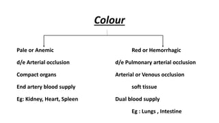
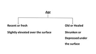
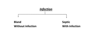
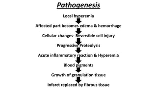
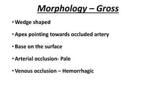
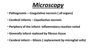
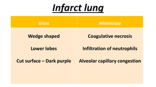
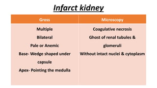
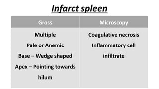
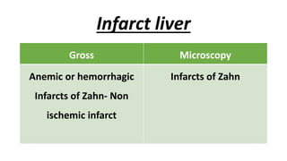
INFARCT
Dr . Sridevi
Post Graduate
Department of Pathology
Plan
1. Definition
2. Causes
3. Types
4. Pathogenesis
5. Gross
6. Microscopy
Definition
•Infarct is an area of ischemic necrosis
caused by occlusion of either the arterial
supply or the venous drainage
Causes
1. Thrombotic or Embolic arterial occlusion
2. Local vasospasm
3. Hemorrhage into the atheromatous plaque
4. Extrinsic vessel compression by tumour
5. Torsion of vessels
6. Traumatic rupture
7. Anterior compartment syndrome
Types
1. Colour
2. Age
3. Infection
Colour
Pale or Anemic Red or Hemorrhagic
d/e Arterial occlusion d/e Pulmonary arterial occlusion
Compact organs Arterial or Venous occlusion
End artery blood supply soft tissue
Eg: Kidney, Heart, Spleen Dual blood supply
Eg : Lungs , Intestine
Age
Recent or fresh Old or Healed
Slightly elevated over the surface Shrunken or
Depressedunder
the surface
Infection
Bland Septic
Without infection With infection
Pathogenesis
Local hyperemia
Affected part becomes edema & hemorrhage
Cellular changes- Reversible cell injury
Progressive Proteolysis
Acute inflammatory reaction & Hyperemia
Blood pigments
Growth of granulation tissue
Infarct replaced by fibrous tissue
Morphology – Gross
• Wedge shaped
• Apex pointing towards occluded artery
• Base on the surface
• Arterial occlusion- Pale
• Venous occlusion – Hemorrhagic
Microscopy
• Pathognomic – Coagulative necrosis ( all organs)
• Cerebral infarcts – Liquefactive necrosis
• Periphery of the infarct- Inflammatory reaction noted
• Generally infarct replaced by fibrous tissue
• Cerebral infarct – Gliosis ( replacement by microglial cells)
Infarct lung
Gross Microscopy
Wedge shaped
Lower lobes
Cut surface – Dark purple
Coagulative necrosis
Infiltration of neutrophils
Alveolar capillary congestion
Infarct kidney
Gross Microscopy
Multiple
Bilateral
Pale or Anemic
Base- Wedge shaped under
capsule
Apex- Pointing the medulla
Coagulative necrosis
Ghost of renal tubules &
glomeruli
Without intact nuclei & cytoplasm
Infarct spleen
Gross Microscopy
Multiple
Pale or Anemic
Base – Wedge shaped
Apex – Pointing towards
hilum
Coagulative necrosis
Inflammatory cell
infiltrate
Infarct liver
Gross Microscopy
Anemic or hemorrhagic
Infarcts of Zahn- Non
ischemic infarct
Infarcts of Zahn
Most commonly affected organs
Location Gross Outcome
Myocardium Pale Frequently lethal
Lungs Hemorrhagic Less fatal
Cerebrum Hemorrhagic or pale Fatal if massive
Intestine Hemorrhagic Frequently lethal
Renal Pale No lethal
Spleen Pale Not lethal
Liver Pale Not lethal
Lower extremities Pale Not lethal

infarct-190709060623.pdf

  • 1.
    INFARCT Dr . Sridevi PostGraduate Department of Pathology
  • 2.
    Plan 1. Definition 2. Causes 3.Types 4. Pathogenesis 5. Gross 6. Microscopy
  • 3.
    Definition •Infarct is anarea of ischemic necrosis caused by occlusion of either the arterial supply or the venous drainage
  • 4.
    Causes 1. Thrombotic orEmbolic arterial occlusion 2. Local vasospasm 3. Hemorrhage into the atheromatous plaque 4. Extrinsic vessel compression by tumour 5. Torsion of vessels 6. Traumatic rupture 7. Anterior compartment syndrome
  • 5.
  • 6.
    Colour Pale or AnemicRed or Hemorrhagic d/e Arterial occlusion d/e Pulmonary arterial occlusion Compact organs Arterial or Venous occlusion End artery blood supply soft tissue Eg: Kidney, Heart, Spleen Dual blood supply Eg : Lungs , Intestine
  • 7.
    Age Recent or freshOld or Healed Slightly elevated over the surface Shrunken or Depressedunder the surface
  • 8.
  • 9.
    Pathogenesis Local hyperemia Affected partbecomes edema & hemorrhage Cellular changes- Reversible cell injury Progressive Proteolysis Acute inflammatory reaction & Hyperemia Blood pigments Growth of granulation tissue Infarct replaced by fibrous tissue
  • 10.
    Morphology – Gross •Wedge shaped • Apex pointing towards occluded artery • Base on the surface • Arterial occlusion- Pale • Venous occlusion – Hemorrhagic
  • 11.
    Microscopy • Pathognomic –Coagulative necrosis ( all organs) • Cerebral infarcts – Liquefactive necrosis • Periphery of the infarct- Inflammatory reaction noted • Generally infarct replaced by fibrous tissue • Cerebral infarct – Gliosis ( replacement by microglial cells)
  • 12.
    Infarct lung Gross Microscopy Wedgeshaped Lower lobes Cut surface – Dark purple Coagulative necrosis Infiltration of neutrophils Alveolar capillary congestion
  • 13.
    Infarct kidney Gross Microscopy Multiple Bilateral Paleor Anemic Base- Wedge shaped under capsule Apex- Pointing the medulla Coagulative necrosis Ghost of renal tubules & glomeruli Without intact nuclei & cytoplasm
  • 14.
    Infarct spleen Gross Microscopy Multiple Paleor Anemic Base – Wedge shaped Apex – Pointing towards hilum Coagulative necrosis Inflammatory cell infiltrate
  • 15.
    Infarct liver Gross Microscopy Anemicor hemorrhagic Infarcts of Zahn- Non ischemic infarct Infarcts of Zahn
  • 16.
    Most commonly affectedorgans Location Gross Outcome Myocardium Pale Frequently lethal Lungs Hemorrhagic Less fatal Cerebrum Hemorrhagic or pale Fatal if massive Intestine Hemorrhagic Frequently lethal Renal Pale No lethal Spleen Pale Not lethal Liver Pale Not lethal Lower extremities Pale Not lethal