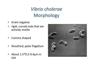
Department of pathology
MORPHOLOGY
CULTURAL CHARACTERISTICS
They are aerobics facultative
anaerobes growing well in the
temperature of 37 degree C and in the
Ph of 7.4 - 9.6
ORDINARY MEDIA
 NUTRIENTAGAR MEDIUM
 MACCONKEYAGAR MEDIUM
 BLOODAGAR MEDIUM
 GELATIN STAB CULTURE
SPECIAL MEDIA
 TRANSPORT MEDIA
 VR medium
 Bile peptone transport medium
 Cary blair medium
 ENRICHMENT MEDIA
 Alkaline peptone water
 Monsur’s medium
 PLATING MEDIA
 Alkaline bile salt agar medium
 Monsur’s gelatin taurocholate trypticase tellurite agar medium
 Thiosulphate citrate bile sucrose agar medium
ORDINARY MEDIA
 NUTRIENT AGAR MEDIUM
After overnight incubation round,
moist, translucent, bluish colonies will be
appear with 1-2mm size.
MACCONKEY AGAR MEDIUM
 Colorless colonies will be form after that it
will change to pink color.
BLOOD AGAR MEDIUM
 A zone of green discoloration appears
around the colonies at first and later it
become clear.
GELATIN STAB CULTURE
 After three days of incubation a white line of
growth appears in the medium
TRANSPORT MEDIA
 VENKATRAMAN - RAMAKRISHNAN MEDIUM
⚫This is an ideal transport medium.
⚫The organism will be viable in this
medium for several weeks without
multiplication
ENRICHMENT MEDIA
 ALKALINE PEPTONE WATER
Rapid growth occurs in about 6 hours with
formation of thick surface pellicle.
PLATING MEDIA
 ALKALINE BILE SALTAGAR MEDIUM
It is a modified nutrient agar medium and the
colonies are similar that appears in the nutrient agar
medium.
MONSUR’S GELATIN TAUROCHOLATE
TRYPTICASE TELLURITE AGAR MEDIUM
 After 24 hours of incubation small colonies will be
formed with 1-2 mm in size and grayish color with
black centers.
 The size will be increased to 3-4 mm after 48 hours
of incubation.
THIOSULPHATE CITRATE BILE SUCROSE AGAR
MEDIUM
 In this medium large colonies will be formed in
yellow color.
 Later the yellow color will be changed to green
color.
PATHOGENESIS
 Vibrio cholerae is the causative agent of the
diarrheal disease cholera.
 The disease is characterized by sudden
effortless vomiting, profuse rice water stool
followed by rapid dehydration.
 Infection occurs in humans via oral route by
ingestion of contaminated food or water.
 Incubation period is 6hours to 3 days.
 The ingested micro organism reaches to the
stomach and the multiplication occurs in the small
intestine.
 After the growth and multiplication it starts to
produce toxins namely, “cholera toxin” or “cholera
enterotoxin” or “choleragen”
 This toxins cause the hyper secretion of glands in
the intestine and resulting in large amount of
intestinal fluids in the intestinal lumens.
 The toxin also increases the capillary permeability
leading to edema.
 On the same hand it produces proteolytic enzymes,
which help the organism to penetrate the covering
of GI tract and causes the diarrheal disease among
humans.
 The stools are rice water similar which containing
mucous, epithelial cells and large number of
vibrios.
 The massive loss of electrolytes from the body
leads to metabolic acidosis, muscle cramps, anuria,
acute tubular necrosis, shock and some times
death.
 The disease may last for 4-5 days with an average
fluid loss of 15-20 liters per day.
LABORATORY DIAGNOSIS
 Hematological investigations:
no significant abnormalities
 Bacteriological examination
1, microscopic examination
2, cultural studies
3, biochemical test
4, slide agglutination test
MICROSCOPIC EXAMINATION
 Under the microscope can observe gram negative
comma shaped bacilli.
CULTURAL STUDIES
 Ordinary media, transport media and enrichment
media can be use for the culture studies.
 In macconkeys medium pale color colonies will
developed.
 In mansur medium colonies will be formed with
black centers.
BIOCHEMICAL TESTS
 Sugar fermentation test: fermentation of glucose,
sucrose and maltose will occur with the production
of gas.
 Nitrate reduction test: positive
 Iodole test: positive
 Catalase test: positive
 Oxidase test: positive
 Motility test: positive
SLIDE AGGLUTINATION TEST
 The specimen from a selective media is placed on
a microscopic slide and add a drop of normal saline
over that and then observe under a microscope
after adding one drop of antiserum over the
specimen.
 Presence of clumps indicates the positive test.
TREATMENT
 Adequate fluid and electrolyte replacement is
necessary for such patients which is known as fluid
replacement therapy.
 Oral rehydration therapy.
16-190630113629.pptx

16-190630113629.pptx

  • 2.
  • 4.
  • 5.
    CULTURAL CHARACTERISTICS They areaerobics facultative anaerobes growing well in the temperature of 37 degree C and in the Ph of 7.4 - 9.6
  • 6.
    ORDINARY MEDIA  NUTRIENTAGARMEDIUM  MACCONKEYAGAR MEDIUM  BLOODAGAR MEDIUM  GELATIN STAB CULTURE
  • 7.
    SPECIAL MEDIA  TRANSPORTMEDIA  VR medium  Bile peptone transport medium  Cary blair medium  ENRICHMENT MEDIA  Alkaline peptone water  Monsur’s medium  PLATING MEDIA  Alkaline bile salt agar medium  Monsur’s gelatin taurocholate trypticase tellurite agar medium  Thiosulphate citrate bile sucrose agar medium
  • 8.
    ORDINARY MEDIA  NUTRIENTAGAR MEDIUM After overnight incubation round, moist, translucent, bluish colonies will be appear with 1-2mm size.
  • 9.
    MACCONKEY AGAR MEDIUM Colorless colonies will be form after that it will change to pink color.
  • 10.
    BLOOD AGAR MEDIUM A zone of green discoloration appears around the colonies at first and later it become clear.
  • 11.
    GELATIN STAB CULTURE After three days of incubation a white line of growth appears in the medium
  • 12.
    TRANSPORT MEDIA  VENKATRAMAN- RAMAKRISHNAN MEDIUM ⚫This is an ideal transport medium. ⚫The organism will be viable in this medium for several weeks without multiplication
  • 13.
    ENRICHMENT MEDIA  ALKALINEPEPTONE WATER Rapid growth occurs in about 6 hours with formation of thick surface pellicle.
  • 14.
    PLATING MEDIA  ALKALINEBILE SALTAGAR MEDIUM It is a modified nutrient agar medium and the colonies are similar that appears in the nutrient agar medium.
  • 15.
    MONSUR’S GELATIN TAUROCHOLATE TRYPTICASETELLURITE AGAR MEDIUM  After 24 hours of incubation small colonies will be formed with 1-2 mm in size and grayish color with black centers.  The size will be increased to 3-4 mm after 48 hours of incubation.
  • 16.
    THIOSULPHATE CITRATE BILESUCROSE AGAR MEDIUM  In this medium large colonies will be formed in yellow color.  Later the yellow color will be changed to green color.
  • 17.
    PATHOGENESIS  Vibrio choleraeis the causative agent of the diarrheal disease cholera.  The disease is characterized by sudden effortless vomiting, profuse rice water stool followed by rapid dehydration.
  • 18.
     Infection occursin humans via oral route by ingestion of contaminated food or water.  Incubation period is 6hours to 3 days.  The ingested micro organism reaches to the stomach and the multiplication occurs in the small intestine.  After the growth and multiplication it starts to produce toxins namely, “cholera toxin” or “cholera enterotoxin” or “choleragen”
  • 19.
     This toxinscause the hyper secretion of glands in the intestine and resulting in large amount of intestinal fluids in the intestinal lumens.  The toxin also increases the capillary permeability leading to edema.  On the same hand it produces proteolytic enzymes, which help the organism to penetrate the covering of GI tract and causes the diarrheal disease among humans.
  • 20.
     The stoolsare rice water similar which containing mucous, epithelial cells and large number of vibrios.  The massive loss of electrolytes from the body leads to metabolic acidosis, muscle cramps, anuria, acute tubular necrosis, shock and some times death.  The disease may last for 4-5 days with an average fluid loss of 15-20 liters per day.
  • 21.
    LABORATORY DIAGNOSIS  Hematologicalinvestigations: no significant abnormalities  Bacteriological examination 1, microscopic examination 2, cultural studies 3, biochemical test 4, slide agglutination test
  • 22.
    MICROSCOPIC EXAMINATION  Underthe microscope can observe gram negative comma shaped bacilli.
  • 23.
    CULTURAL STUDIES  Ordinarymedia, transport media and enrichment media can be use for the culture studies.  In macconkeys medium pale color colonies will developed.  In mansur medium colonies will be formed with black centers.
  • 24.
    BIOCHEMICAL TESTS  Sugarfermentation test: fermentation of glucose, sucrose and maltose will occur with the production of gas.  Nitrate reduction test: positive  Iodole test: positive  Catalase test: positive  Oxidase test: positive  Motility test: positive
  • 25.
    SLIDE AGGLUTINATION TEST The specimen from a selective media is placed on a microscopic slide and add a drop of normal saline over that and then observe under a microscope after adding one drop of antiserum over the specimen.  Presence of clumps indicates the positive test.
  • 27.
    TREATMENT  Adequate fluidand electrolyte replacement is necessary for such patients which is known as fluid replacement therapy.  Oral rehydration therapy.