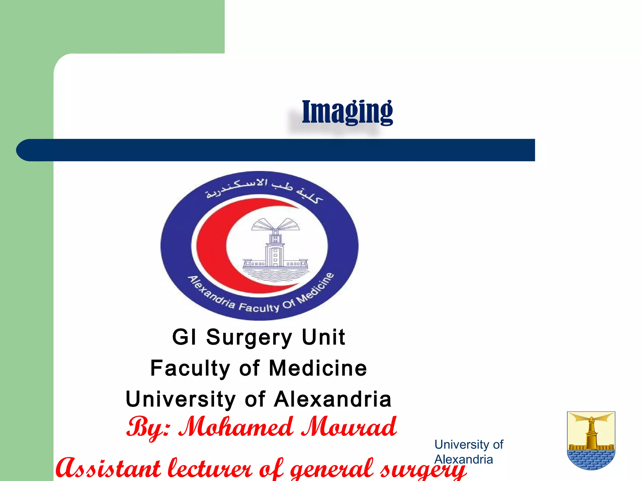
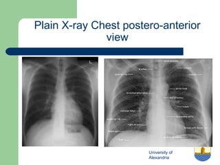
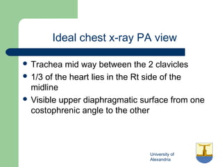
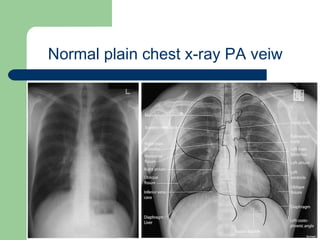
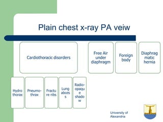
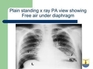
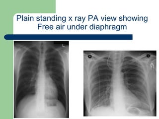
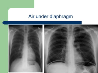
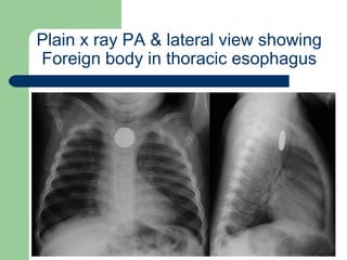
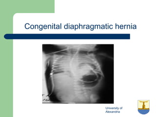
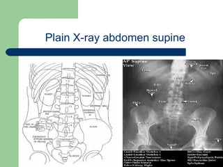
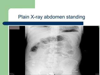
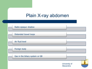
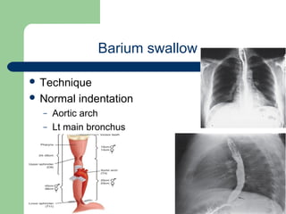
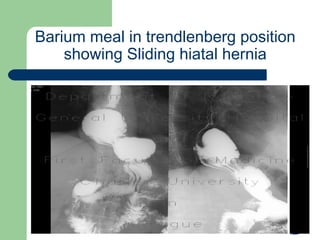
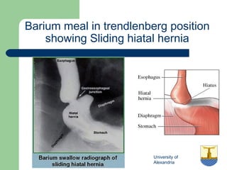
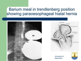
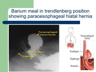
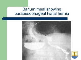
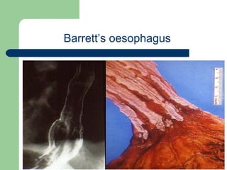
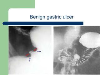
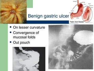
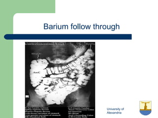
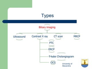
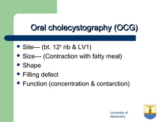
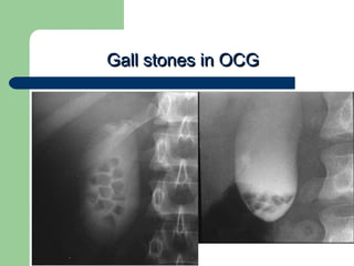
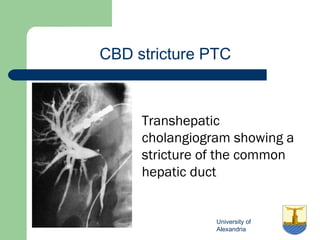
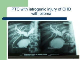
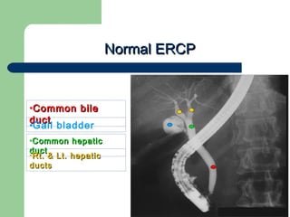
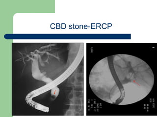
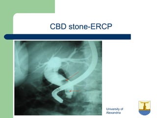
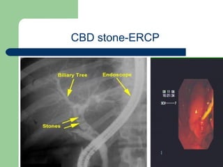
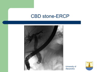
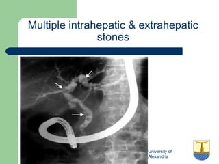
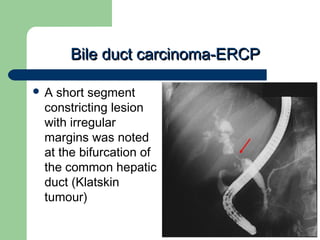
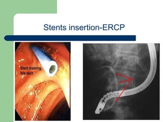
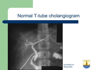
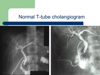
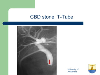
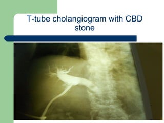
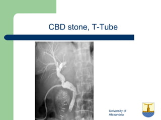
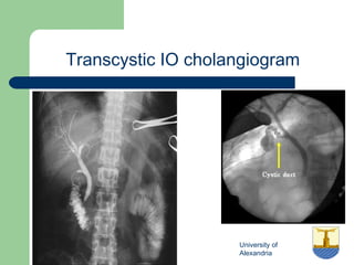
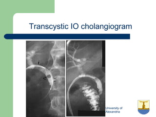
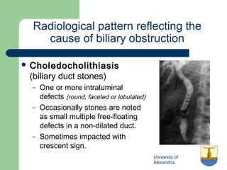
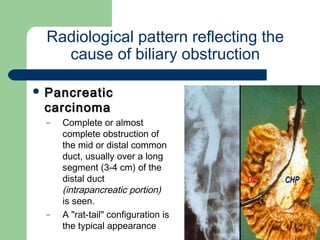
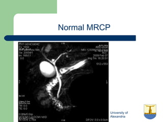
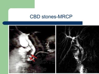
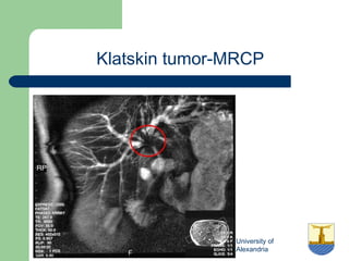
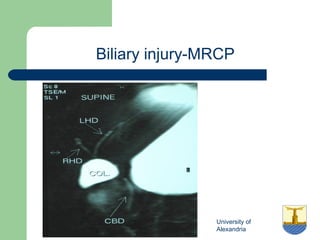
University of
Alexandria
Imaging
GI Surgery Unit
Faculty of Medicine
University of Alexandria
By: Mohamed Mourad
Assistant lecturer of general surgery
University of
Alexandria
Different imaging modalities
University of Alexandria
Plain chest x-ray
University of
Alexandria
Plain X-ray Chest postero-anterior
view
University of
Alexandria
Ideal chest x-ray PA view
 Trachea mid way between the 2 clavicles
 1/3 of the heart lies in the Rt side of the
midline
 Visible upper diaphragmatic surface from one
costophrenic angle to the other
University of
Alexandria
Normal plain chest x-ray PA veiw
University of
Alexandria
Plain chest x-ray PA veiw
University of
Alexandria
Plain standing x ray PA view showing
Free air under diaphragm
University of
Alexandria
Plain standing x ray PA view showing
Free air under diaphragm
University of
Alexandria
Air under diaphragm
University of
Alexandria
Plain x ray PA & lateral view showing
Foreign body in thoracic esophagus
University of
Alexandria
Congenital diaphragmatic hernia
University of Alexandria
Plain abdomen x-ray
University of
Alexandria
Plain X-ray abdomen supine
University of
Alexandria
Plain X-ray abdomen standing
University of
Alexandria
Plain X-ray abdomen
University of
Alexandria
Plain x ray abdomen and pelvis PA
view showing Radio-opaque shadow
University of
Alexandria
DD radio-opaque shadow of
Rt. hypochondrium
 Gall bladder stones
 Renal stones (Dx Lat X-ray &
US)
 Porcelain gall bladder
 Calcified LN
 Fracture tr. Process of
vertebra
 FB
 Fecolith
 Calcified hydatid cyst
University of
Alexandria
DD: renal & gall bladder stones
University of
Alexandria
Radio-opaque shadow of the
abdomen x ray
University of
Alexandria
Chronic panreatitis
University of
Alexandria
Erect Plain x-ray abdomen &pelvis
PA view showing Distended bowel
loops and Multiple air fluid levels
 Distended
Jejunal loops
– Central
– Valvae
conniventes
University of
Alexandria
Plain x-ray abdomen &pelvis PA view
showing Distended bowel loops
 Distended Ileal
loops
– Central
– featureless
University of
Alexandria
Plain x-ray abdomen &pelvis PA view
showing Distended bowel loops
 Distended
Colon
– peripheral
– Colonic
hausteration
University of
Alexandria
Plain x-ray abdomen & pelvis
showing distended colonic loops
University of
Alexandria
Ileo-ceacal valve
University of
Alexandria
Post operative adynamic ileus
•SupineSupine •ErectErect
University of
Alexandria
Sigmoid Volvulus
University of
Alexandria
Plain x-ray abdomen and pelvis
showing pneumatic tyre appearance
University of
Alexandria
Plain x-ray abdomen and pelvis
showing pneumatic tyre appearance
University of
Alexandria
Plain x-ray abdomen and pelvis
showing coffee bean appearance
 Sigmoid volvulus--
bowel loop points
to RUQ
 Caecal volvulus--
bowel loop points
to LUQ 
University of
Alexandria
Caecal volvulus
University of
Alexandria
Plain x ray abdomen & pelvis
showing double bubble sign
University of
Alexandria
Plain x-ray abdomen and pelvis
Foreign body
University of
Alexandria
Plain x ray abdomen showing
aeroblia
University of
Alexandria
Invertogram
University of Alexandria
Barium swallow
University of
Alexandria
Barium swallow
 Technique
 Normal indentation
– Aortic arch
– Lt main bronchus
University of
Alexandria
Pathologies by barium swallow
University of
Alexandria
Achalasia of the cardia
University of
Alexandria
Barium swallow showing smooth
narrowing of distal end of the
oesophagus with proximal dilatation
University of
Alexandria
Barium swallow showing smooth
long narrowing of middle 1/3
oesophagus
 Corrosive stricture
– Single or multiple
– Long segment
– Smooth tapering
– Usually in children
– Starts at aortic arch
level
University of
Alexandria
Corrosive stricture
University of
Alexandria
Barium swallow showing Corkscrow
oesophagus
University of
Alexandria
Irregular filling defect mostly Cancer
oesophagus
University of
Alexandria
Oesophageal malignancy
University of
Alexandria
Oesophageal malignancy
University of
Alexandria
DD between achalasia and
malignancy
•The cardia is normally
below the diaphragm
•In X-ray 11, the
oesophagus is
interrupted above
the diaphragm
•In X-ray 22, the
cardia below the
diaphragm is
closed with “bird
beak-like” shape
University of
Alexandria
serpiginous filling defects in the
oesophagus Oesophageal
varices
University of
Alexandria
Barium swallow with outpouch of the
barium--Oesophageal diverticula
University of
Alexandria
Epiphrenic and Zenker’s diverticulum
University of
Alexandria
Oesophageal web
University of Alexandria
Barium meal
University of
Alexandria
Barium meal
University of
Alexandria
Barium meal
 Large volume low
density
 Distension of
stomach by
volume
 Small volume high
density+ effervescent
 Distension of stomach
by buscopan
University of
Alexandria
Barium meal (Single & Double)
University of
Alexandria
Barium meal
 1, Gastric antrum
 2, pyloric canal
 3, duodenal cap
 4, duodenal loop
University of
Alexandria
Pathological findings in barium meal
University of
Alexandria
Hiatal hernia
University of
Alexandria
Barium meal in trendlenberg position
showing Sliding hiatal hernia
University of
Alexandria
Barium meal in trendlenberg position
showing Sliding hiatal hernia
University of
Alexandria
Barium meal in trendlenberg position
showing Sliding hiatal hernia
University of
Alexandria
Barium meal in trendlenberg position
showing paraoesophageal hiatal hernia
University of
Alexandria
Barium meal in trendlenberg position
showing paraoesophageal hiatal hernia
University of
Alexandria
Barium meal showing
paraoesophageal hiatal hernia
University of
Alexandria
Barrett’s oesophagus
University of
Alexandria
Benign gastric ulcer
University of
Alexandria
Benign gastric ulcer
 On lesser curvature
 Convergence of
mucosal folds
 Out pouch
University of
Alexandria
Barium meal showing ulcer nich at the middle of
lesser curvature with mucosal convergence
Benign gastric ulcer
University of
Alexandria
Hour glass deformity
University of
Alexandria
Normal duodenal cap and DU
University of
Alexandria
2 kissing duodenal ulcers
University of
Alexandria
Trifoliate deformity of DU
University of
Alexandria
Gastric tumors
University of
Alexandria
Malignant gastric ulcer
University of
Alexandria
Malignant gastric ulcer
 Any site of stomach mainly antrum and
greater curvature
 Ulcer dose not protrude to the outside
 Mucosal folds obliterate before the edges of
the ulcer
University of
Alexandria
Lintis plastica
University of
Alexandria
Pyloric filling defect of malignant
origin
University of
Alexandria
Pre-pyloric tumor
University of
Alexandria
Duodenal diverticulum
University of
Alexandria
Barium meal showing dilated stomach reaching
pelvis with mottled dye without passage of dye
to dudenum (Gastric outlet obstruction)
University of
Alexandria
Barium meal showing Soap dish
appearance
University of
Alexandria
Congenital hypertrophic pyloric
stenosis
University of
Alexandria
GOO by compression from outside
University of
Alexandria
GOO by pseudopancreatic cyst
University of
Alexandria
Volvulus of the stomach
•Organoaxial volvulusOrganoaxial volvulus •Mesenteroaxial volvulus
University of
Alexandria
Organo-axial volvulus
University of
Alexandria
Duodenal atresia
University of Alexandria
Barium follow through
University of
Alexandria
Barium follow through
University of
Alexandria
Stricture of small intestine
•Chron’s
•Lymphoma
•TB
•Radiation
University of
Alexandria
Chron’s stricture
 Commonly at terminal
ileum
 Multiple, rigid, irregular
stricture (string sign of
Kantor)
 Skip lesions
 Cobble stones
 Polyps
 Fistula
University of Alexandria
Barium enema
University of
Alexandria
Barium enema
University of
Alexandria
Normal barium enema filling phase
University of
Alexandria
Pathological findings in barium enema
University of
Alexandria
Transverse colon filling defect
University of
Alexandria
Transverse colon filling defect
(colonic malignancy)
University of
Alexandria
Cancer ascending colon
University of
Alexandria
Barium enema showing arrest of the
dye at the level of rectosegmoid
junction due obstruction of the lumen
University of
Alexandria
Diverticulosis
University of
Alexandria
Chron’s disease
 Commonly at terminal ileum
 Multiple, rigid, irregular
 Skip lesions
 Cobble stones
 Polyps
 Fistula
 Loss of hausterations
University of
Alexandria
Ulcerative colitis
 Rectosegmoid
 Loss of hausteration
 Polyps
 No skip lesion
 Narrow colon (lead pipe)
University of
Alexandria
Ischemic colitis
 segmental narrowing
of the entire
transverse colon.
 Within the narrowed
segment, there are
multiple nodular
indentations, many of
which have the
appearance of
thumbprinting
University of
Alexandria
DD of colonic stricture
 Chron’s caecum
 TB iliocecal
 Cancer segmoid
 Diverticulitis segmoid
 Ischemic colitis splenic flexure
University of
Alexandria
Hirschsprung disease
 Stenotic segment
 Dilated rectum
 Transitional zone
(funnel)
 Delayed
evacuation
University of
Alexandria
Rounded filling defect (polyps)
University of
Alexandria
Rectosegmoid polyps
University of
Alexandria
Barium enema showing hundreds of
filling defects of the entire colon (FAP)
University of
Alexandria
Intussusception
University of
Alexandria
Intussusception
University of
Alexandria
Barium reduction of intussusception
•Head of intussusception
is at hepatic flexure
•Free flow of contrast into
distal small bowel indicates
complete reduction
•Partial reduction
University of Alexandria
Biliary imaging
University of
Alexandria
Types
University of Alexandria
Oral Cholecystography (OCG)Oral Cholecystography (OCG)
University of
Alexandria
Steps of oral cholecystographySteps of oral cholecystography
(OCG)(OCG)
 Control X-ray
 Ingestion of telepaque
 Another X-ray 12hrs latter
 Intake of fatty meal
 Another X-ray 2hrs latter
Remember that 85% of gallstones are
radiolucent
Remember that 85% of gallstones are
radiolucent
University of
Alexandria
Oral cholecystography (OCG)Oral cholecystography (OCG)
University of
Alexandria
Oral cholecystography (OCG)Oral cholecystography (OCG)
 Site--- (bt. 12th
rib & LV1)
 Size--- (Contraction with fatty meal)
 Shape
 Filling defect
 Function (concentration & contarction)
University of
Alexandria
Gall stones in OCGGall stones in OCG
University of
Alexandria
Non visualized gall bladder in OCG
 Absent gall bladder
 Stone in cystic duct
 Non functioning gall
bladder
University of Alexandria
Cholangiogram
University of Alexandria
PTC
University of
Alexandria
Normal PTCNormal PTC
University of
Alexandria
CBD stone-PTCCBD stone-PTC
University of
Alexandria
CBD stone-PTCCBD stone-PTC
 One or more intraluminal
defects (round, faceted or
lobulated) which produce
varying degrees of biliary
obstruction are seen.
 Occasionally stones are
noted as small multiple
free-floating defects in a
non-dilated duct.
University of
Alexandria
CBD stricture PTC
Transhepatic
cholangiogram showing a
stricture of the common
hepatic duct
University of
Alexandria
PTC with iatrogenic injury of CHD
with biloma
University of Alexandria
ERCP
University of
Alexandria
Normal ERCPNormal ERCP
•Common bileCommon bile
ductduct
•Gall bladder
•Common hepaticCommon hepatic
ductduct
•Rt. & Lt. hepaticRt. & Lt. hepatic
ductsducts
University of
Alexandria
CBD stone-ERCP
University of
Alexandria
CBD stone-ERCP
University of
Alexandria
CBD stone-ERCP
University of
Alexandria
CBD stone-ERCP
University of
Alexandria
Multiple intrahepatic & extrahepatic
stones
University of
Alexandria
Bile duct carcinoma-ERCPBile duct carcinoma-ERCP
 A short segment
constricting lesion
with irregular
margins was noted
at the bifurcation of
the common hepatic
duct (Klatskin
tumour)
University of
Alexandria
Stents insertion-ERCP
University of Alexandria
T-tube cholangiogram
University of
Alexandria
Normal T-tube cholangiogram
University of
Alexandria
Normal T-tube cholangiogram
University of
Alexandria
CBD stone, T-Tube
University of
Alexandria
T-tube cholangiogram with CBD
stone
University of
Alexandria
CBD stone, T-Tube
University of
Alexandria
Transcystic IO cholangiogram
University of
Alexandria
Transcystic IO cholangiogram
University of
Alexandria
Radiological pattern reflecting the
cause of biliary obstruction
 CholedocholithiasisCholedocholithiasis
(biliary duct stones)
– One or more intraluminal
defects (round, faceted or lobulated)
– Occasionally stones are noted
as small multiple free-floating
defects in a non-dilated duct.
– Sometimes impacted with
crescent sign.
University of
Alexandria
Radiological pattern reflecting the
cause of biliary obstruction
 PancreaticPancreatic
carcinomacarcinoma
– Complete or almost
complete obstruction of
the mid or distal common
duct, usually over a long
segment (3-4 cm) of the
distal duct
(intrapancreatic portion)
is seen.
– A "rat-tail" configuration is
the typical appearance
University of
Alexandria
Radiological pattern reflecting the
cause of biliary obstruction
 CholangiocarcinomCholangiocarcinom
aa
– The narrowing may
occur at any level and
typically presents as a
segmental stenosis.
– When the lesion is
located at the
bifurcation, it is referred
to as a Klatskin tumor
University of
Alexandria
Radiological pattern reflecting the
cause of biliary obstruction
 Benign strictureBenign stricture
– There is a short segmental
circumferential stricture in
the mid-common duct in a
patient with a previous
cholecystectomy.
– Most benign strictures are
secondary to ductal injury
during cholecystectomy.
(iatrogenic)
University of
Alexandria
Radiological pattern reflecting the
cause of biliary obstruction
 AmpullaryAmpullary
carcinomacarcinoma
– Focal obstruction of very
distal CBD is noted.
– A smooth constriction or
an irregular polypoid mass
growing into distal CBD
may be seen.
University of
Alexandria
Radiological pattern reflecting the
cause of biliary obstruction
 Ascariasis
– Worms are seen
extending through the
common bile duct and
major hepatic ducts
University of Alexandria
MRCP
University of
Alexandria
Normal MRCP
University of
Alexandria
CBD stones-MRCP
University of
Alexandria
Klatskin tumor-MRCP
University of
Alexandria
Biliary injury-MRCP
University of Alexandria
Liver imaging
University of
Alexandria
Hydatid liver cyst
 Fluid attenuation
 No peripheral
enhancement
 Daughter cysts
 Well defined
 Rounded or oval
University of
Alexandria
Hydatid cyst
University of Alexandria
Imaging
Thank You

Imaging