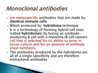
Definitions, polyclonal and monoclonal Ab,
types of samples
 IHC is a method for localizing specific antigens
in tissues or cells based on Ag-Ab recognition
 Immunohistochemistry (IHC) Combines
histological, immunological and biochemical
techniques for the identification of specific
tissue components by means of a specific
antigen/antibody reaction tagged with a
visible label
 1945 – Albert Coons 1st used an Ab labeled
with a fluorescent dye to visualize tissues
 1966 – 1 st developed enzyme labeling
instead of fluorescent label
 1974 – IHC was performed for the 1st time on
routine formalin fixed paraffin embedded
sections
 1981 – developed avidin-biotin labeling
 1991 – Heat induced antigen retrieval
technique in IHC was done
 1995 – Polymer technology introduced
 is a molecule that stimulate the formation of
an antibody and it has one or more antibody
binding sites.
 antigen (Ag), abbreviation of
antibody generator
 These sites are highly specific region being
known as antigenic determinant groups or
epitopes.
 Antigens have two main properties.
 • immunogenicity, which is the ability to
induce antibody formation.
 • specific reactivity, which means that the
antigen can react “with the antibody it caused
to be produced.
 proteins of high molecular weight known as
immunoglobulins and are produced after
stimulation by an antigen.
 act specifically against the antigen in an
immune response
 Antibodies are divided into five major classes,
IgM, IgG, Iga, IgD, and IgE, based on their
constant region structure and immune
function.
 The frequently used Ab is IgG molecule
 it is composed of two pairs of light and heavy
polypeptide chains form a Y-shaped
structure.
 Abs are also proteins - thus any part of the
Ab may itself serve as epitope to induce Ab
formation (to which secondary Ab binds)
 IHC technique prove that Ig molecules can
serve both as Ab and Ag
 The amino acid side-chains of the variable
domain of an antibody form a cavity which is
geometrically and chemically complementary
to a single type of antigen epitope.
 lock (antibody) and key (antigen)
 The associated antibody and antigen are held
together by a combination of hydrogen
bonds, electrostatic interactions, and van der
Waals’ forces.
 Antibody specificity
This refers to the characteristics of an
antibody to bind selectively to a single
epitope on an antigen.
 Sensitivity
This refers to the relative amount of antigen
that an immunohistochemical technique is
able to detect.
A technique with high sensitivity is able to
detect smaller amounts of antigen than a
technique with low sensitivity.
 Polyclonal antibodies
 Polyclonal antibodies are produced by immunizing an
animal with a purified specific molecule (immunogen)
bearing the antigen of interest.
 B cells will produce immunoglobulin specific for the
antigen
 The IgG is produced from different clones of plasma
cells (polyclonal).
 Each clone will produce an antibody with a slightly
different specificity to the variety of epitopes present
on the immunogen.
 A polyclonal antiserum is therefore a mixture of
antibodies to different epitopes on the immunogen.
 are monospecific antibodies that are made by
identical immune cells
 Which produced by hybridoma technique
 It is a technology of forming hybrid cell lines
(called hybridomas) by fusing an antibody-
producing B cell with a myeloma (B cell cancer)
cell that is selected for its ability to grow in
tissue culture and for an absence of antibody
chain synthesis.
 The antibodies produced by the hybridoma are
all of a single specificity and are therefore
monoclonal antibodies
 Polyclonal antibodies
 –Mixtures of different antibodies to a single
antigen are called polyclonal antibodies.
 (Large complex antigens may have multiple
epitopes and elicit several antibody types)
 Monoclonalantibodies – Antibodies specific
for a single epitope and produced by a single
clone are called monoclonal antibodies
 Polyclonal antibody
 greater potential for false positive staining
due to antibodies cross-reacting to undesired
targets.(background staining)
 Monoclonal antibody
 highly specific
 less background
steps
-Preparation of sample
-Immunohistochemical reaction
and Visualizing the reaction
-Counter stain.
 Is the type of sample and the fixation
affecting the IHC reactions ?
 Adequate and appropriate fixation is the
cornerstone of all histological and
immunohistochemical preparations.
 The demonstration of many antigens depends
heavily on the fixative employed.
 There is no one fixative that is ideal for all
antigens.
 Most laboratories use fixation based on
formalin, such as unbuffered 10% formal
saline or 10% neutral buffered formalin.
 Some groups prefer picric acid fixation
(Bouin’s) or mercuric fixation.

 Good fixation is the delicate balance between
under-fixation and over-fixation
 Ideal fixation is the balance between good
morphology and good antigenicity.
 Poor fixation or delay in fixation causes loss
of antigenicity or diffusion of antigens into
the surrounding tissue.
 Although the use of frozen sections for
diagnostic purposes is decreasing,
 immunohistochemistry on frozen sections
remains an important histological tool
Although frozen sections have certain
disadvantages compared to paraffin sections,
including poor morphology, limited
retrospective studies, and storage of material,
the technique should be considered the gold
standard when evaluating and assessing new
antibodies.
 Acetone-fixed smears are often preferred by
the immunohistochemist, since acetone
allows a wide range of primary antibodies to
be employed without destroying the target
epitopes.

IHC 2

  • 1.
    Definitions, polyclonal andmonoclonal Ab, types of samples
  • 2.
     IHC isa method for localizing specific antigens in tissues or cells based on Ag-Ab recognition  Immunohistochemistry (IHC) Combines histological, immunological and biochemical techniques for the identification of specific tissue components by means of a specific antigen/antibody reaction tagged with a visible label
  • 3.
     1945 –Albert Coons 1st used an Ab labeled with a fluorescent dye to visualize tissues  1966 – 1 st developed enzyme labeling instead of fluorescent label  1974 – IHC was performed for the 1st time on routine formalin fixed paraffin embedded sections  1981 – developed avidin-biotin labeling  1991 – Heat induced antigen retrieval technique in IHC was done  1995 – Polymer technology introduced
  • 4.
     is amolecule that stimulate the formation of an antibody and it has one or more antibody binding sites.  antigen (Ag), abbreviation of antibody generator  These sites are highly specific region being known as antigenic determinant groups or epitopes.
  • 5.
     Antigens havetwo main properties.  • immunogenicity, which is the ability to induce antibody formation.  • specific reactivity, which means that the antigen can react “with the antibody it caused to be produced.
  • 6.
     proteins ofhigh molecular weight known as immunoglobulins and are produced after stimulation by an antigen.  act specifically against the antigen in an immune response
  • 7.
     Antibodies aredivided into five major classes, IgM, IgG, Iga, IgD, and IgE, based on their constant region structure and immune function.  The frequently used Ab is IgG molecule  it is composed of two pairs of light and heavy polypeptide chains form a Y-shaped structure.
  • 8.
     Abs arealso proteins - thus any part of the Ab may itself serve as epitope to induce Ab formation (to which secondary Ab binds)  IHC technique prove that Ig molecules can serve both as Ab and Ag
  • 9.
     The aminoacid side-chains of the variable domain of an antibody form a cavity which is geometrically and chemically complementary to a single type of antigen epitope.  lock (antibody) and key (antigen)  The associated antibody and antigen are held together by a combination of hydrogen bonds, electrostatic interactions, and van der Waals’ forces.
  • 10.
     Antibody specificity Thisrefers to the characteristics of an antibody to bind selectively to a single epitope on an antigen.  Sensitivity This refers to the relative amount of antigen that an immunohistochemical technique is able to detect. A technique with high sensitivity is able to detect smaller amounts of antigen than a technique with low sensitivity.
  • 11.
     Polyclonal antibodies Polyclonal antibodies are produced by immunizing an animal with a purified specific molecule (immunogen) bearing the antigen of interest.  B cells will produce immunoglobulin specific for the antigen  The IgG is produced from different clones of plasma cells (polyclonal).  Each clone will produce an antibody with a slightly different specificity to the variety of epitopes present on the immunogen.  A polyclonal antiserum is therefore a mixture of antibodies to different epitopes on the immunogen.
  • 12.
     are monospecificantibodies that are made by identical immune cells  Which produced by hybridoma technique  It is a technology of forming hybrid cell lines (called hybridomas) by fusing an antibody- producing B cell with a myeloma (B cell cancer) cell that is selected for its ability to grow in tissue culture and for an absence of antibody chain synthesis.  The antibodies produced by the hybridoma are all of a single specificity and are therefore monoclonal antibodies
  • 15.
     Polyclonal antibodies –Mixtures of different antibodies to a single antigen are called polyclonal antibodies.  (Large complex antigens may have multiple epitopes and elicit several antibody types)  Monoclonalantibodies – Antibodies specific for a single epitope and produced by a single clone are called monoclonal antibodies
  • 16.
     Polyclonal antibody greater potential for false positive staining due to antibodies cross-reacting to undesired targets.(background staining)  Monoclonal antibody  highly specific  less background
  • 17.
    steps -Preparation of sample -Immunohistochemicalreaction and Visualizing the reaction -Counter stain.
  • 18.
     Is thetype of sample and the fixation affecting the IHC reactions ?
  • 19.
     Adequate andappropriate fixation is the cornerstone of all histological and immunohistochemical preparations.  The demonstration of many antigens depends heavily on the fixative employed.
  • 20.
     There isno one fixative that is ideal for all antigens.  Most laboratories use fixation based on formalin, such as unbuffered 10% formal saline or 10% neutral buffered formalin.  Some groups prefer picric acid fixation (Bouin’s) or mercuric fixation.
  • 21.
      Good fixationis the delicate balance between under-fixation and over-fixation  Ideal fixation is the balance between good morphology and good antigenicity.  Poor fixation or delay in fixation causes loss of antigenicity or diffusion of antigens into the surrounding tissue.
  • 22.
     Although theuse of frozen sections for diagnostic purposes is decreasing,  immunohistochemistry on frozen sections remains an important histological tool Although frozen sections have certain disadvantages compared to paraffin sections, including poor morphology, limited retrospective studies, and storage of material, the technique should be considered the gold standard when evaluating and assessing new antibodies.
  • 23.
     Acetone-fixed smearsare often preferred by the immunohistochemist, since acetone allows a wide range of primary antibodies to be employed without destroying the target epitopes.