ID 16.15
•Download as PPTX, PDF•
0 likes•76 views
This study introduces a computational technique called deconvolution to improve images captured using a widefield fluorescence microscope. Skin and endothelial cell samples stained with fluorescent dyes were imaged with a widefield microscope. Deconvolution algorithms were then applied to remove blur from the images by accounting for factors like the microscope point spread function. The deconvolved images showed enhanced contrast and resolution compared to the raw images, demonstrating deconvolution can help overcome some limitations of widefield microscopy by reducing out-of-focus light. However, confocal microscopy is still needed for thicker samples above 20-30 micrometers.
Report
Share
Report
Share
Recommended
JPD_OSA_Biomedical_Optics_2016
This document describes a computational endomicroscopy platform that uses compressed sensing to achieve higher resolution images than the physical sensor resolution allows. It uses a digital micromirror device as a spatial light modulator to modulate scenes at a conjugate image plane. A camera then collects multiple coded measurements to reconstruct higher resolution images through compressed sensing algorithms. Experiments demonstrate reconstructing higher resolution images than the individual fiber spacing of fiber optic bundles used in endomicroscopy. Future work aims to further reduce measurements needed and apply the techniques to fiber bundle platforms.
194Martin LeungUnerd Poster
- The document describes a method for using image processing software to automate and improve the precision of focusing in femtosecond direct laser writing. Images of laser light reflected off glass samples at different positions relative to the focal plane were analyzed. Signature intensity and area patterns were used to locate the focal plane within 500nm accuracy. Future work includes using this method to assist with additional laser writing processes and surface profiling applications.
Improved single image dehazing by fusion
IJRET : International Journal of Research in Engineering and Technology is an international peer reviewed, online journal published by eSAT Publishing House for the enhancement of research in various disciplines of Engineering and Technology. The aim and scope of the journal is to provide an academic medium and an important reference for the advancement and dissemination of research results that support high-level learning, teaching and research in the fields of Engineering and Technology. We bring together Scientists, Academician, Field Engineers, Scholars and Students of related fields of Engineering and Technology
BoDong-ICPR2014-CameraReady
This document proposes a Bayesian Maximum a-Posteriori (MAP) method using sparse priors for 3D deconvolution of wide field fluorescence microscopy images of zebrafish embryos. The method uses a global Hyper-Laplacian prior to preserve sharp edges and a local smooth region mask to suppress ringing artifacts. Both synthetic and real zebrafish embryo microscopy data are used to evaluate the method, which demonstrates improved performance over state-of-the-art 3D deconvolution algorithms.
IJCER (www.ijceronline.com) International Journal of computational Engineerin...
This document summarizes a research paper that proposes a computer-aided diagnosis (CAD) system for detecting lung cancer nodules from chest CT images using support vector machines (SVM). The CAD system involves 5 main phases: 1) image pre-processing using total variation denoising, 2) lung region segmentation using optimal thresholding and morphological operations, 3) feature extraction of lung nodules using gray level co-occurrence matrix (GLCM) texture analysis, 4) SVM classification of nodules as benign or malignant, 5) output of classification results. The goal is to develop an automated CAD system to assist radiologists in early detection of lung cancer from CT images.
Sumerschool DPI 2012 Uslar
This document discusses various super-resolution imaging techniques that can overcome the resolution limit of conventional light microscopy and image structures smaller than 200 nm. It describes methods such as dSTORM, PALM and SOFI that use fluorescent labeling and exploit stochastic on-off behavior of emitters to achieve super-resolution. Examples are given of applying these techniques to image microtubules, actin patterns, and trafficking of receptor subunits in neurons at resolutions better than conventional microscopy.
Image reconstruction in nuclear medicine
The document discusses image reconstruction techniques in nuclear medicine. It begins with an introduction to image reconstruction and definitions of key terms. It then describes analytical reconstruction methods like back projection and filtered back projection, as well as iterative reconstruction methods including algebraic reconstruction, statistical reconstruction, and maximum likelihood reconstruction. Both analytical and iterative methods are discussed and their properties and steps are outlined. The document provides an overview of various image reconstruction algorithms used in nuclear medicine.
Confer
This document proposes a novel method for medical image denoising that combines contourlet thresholding, bilateral filtering, and non-local means filtering. Specifically, it introduces scaling factors to the universal threshold for wavelet and contourlet transforms. It then uses the contourlet-based thresholding as a pre-processing step before bilateral or non-local means filtering for denoising. Simulation results show the combined approach achieves better PSNR and perceptual quality than using bilateral or non-local means filtering alone.
Image Scanning Microscopy
This document discusses image scanning microscopy (ISM), a technique that improves the resolution and contrast of fluorescence microscopy images. ISM works by acquiring multiple images of the sample from different positions and combining them to generate a single image with effectively doubled resolution. The document outlines the basic principles and components of ISM systems, provides examples of applications like imaging fluorescent beads and cell structures, and discusses related techniques for further enhancing resolution and depth of imaging. It concludes by emphasizing ISM's benefits like high resolution, low photodamage, and multicolor imaging capabilities.
FYP-Poster
This document describes a study using line-scanning focal modulation microscopy (FMM) to image embryonic zebrafish hearts in vivo with high spatiotemporal resolution. FMM was able to produce images with excellent signal-to-background ratio and resolution at depth, capturing fast biological processes in milliseconds. A 4D reconstruction algorithm synchronized image sequences to achieve 3D reconstruction over time, allowing monitoring of dynamic heart motion and development. Volumetric analysis of ventricle and atrium contractions throughout the cardiac cycle was performed.
3 d imaging super resolution
This document describes a new technique called Photonic Nanojet Interferometry (PJI) that uses microspheres to achieve super-resolution in 3D label-free imaging. PJI was able to laterally resolve sub-100nm features on a Blu-ray disc by taking advantage of photonic nanojets from microspheres. PJI measurements of a Blu-ray disc agreed with measurements from atomic force microscopy and scanning electron microscopy, validating that PJI can achieve super-resolution below the diffraction limit.
Advanced lithographic technologies
This document discusses advanced photolithography technologies used to enable Moore's Law. Moore's Law allows the number of transistors on integrated circuits to double every two years by reducing transistor size. To print smaller features, light with smaller wavelengths is needed to minimize diffraction. Technologies discussed to improve resolution for smaller wavelengths include optical proximity correction, off-axis illumination, phase shift masks, double patterning, restricted design rules, and negative tone development. These resolution enhancement techniques allow continued transistor scaling below 60nm feature sizes.
Visibility Enhancement of Hazy Images using Depth Estimation Concept
This document presents a methodology to improve the visibility of hazy images using depth estimation. The proposed method first converts the input hazy image into white balance and contrast enhanced images. Depth estimation is then performed on these images to estimate the unknown depth from the camera to objects in the scene. Weight maps are generated from the white balance and contrast images and applied to Gaussian and Laplacian pyramids to estimate depth. A gamma correction is applied to the depth estimated image to further improve visibility. Experimental results show that the gamma corrected image has better visibility and a higher PSNR than the depth estimated image alone.
A hand-held hybrid gamma-near-infrared fluorescence imaging camera
The document describes a novel hand-held hybrid near-infrared (NIR)-gamma camera called the Hybrid Gamma Camera (HGC). The HGC consists of an optical camera modified to image in the infrared spectrum, fitted with an 850nm bandpass filter and LED ring for excitation, aligned with a gamma camera to provide co-registered images. Phantom and mouse studies show the HGC can produce fused fluorescence and gamma images from a dual radio-NIR tracer. While the HGC has lower sensitivity than commercial fluorescence cameras, it demonstrates the feasibility of simultaneous gamma and fluorescence imaging in a portable device, with potential for intraoperative use.
SPIE article
This document discusses using conical diffraction in a biaxial crystal to generate a 3D depletion pattern for STED microscopy from a single laser beam. It can generate both a "donut" beam for lateral depletion and a "bottle" beam for axial depletion without needing beam splitting. Simulations show the beams have phases similar to phase plates used in conventional 3D STED. Experiments show the method can generate the depletion pattern, called a "black sphere", over a wavelength range of 15nm centered at 592nm for a fixed crystal. This allows true single-channel 3D STED over a broad bandwidth from a single optical system.
Physics informed deep learning for efficient b-mode ultrasound imaging
A webinar on "Physics-Informed Deep Learning for Efficient B-Mode Ultrasound Imaging" organized by Center for Professional Training (C.P.T.) National University of Computer and Emerging Sciences (NUCES), Karachi.
CVGIP 2010 Part 3
The document presents a 3-D environment modeling and adaptive foreground detection system for multi-camera surveillance. The system constructs a 3-D model of the environment using planar patches approximated from camera views. Videos are integrated and displayed on the 3-D model using texture mapping. A novel method is proposed to detect moving shadows in two phases: an offline training phase determines pixel-wise thresholds, and an online phase updates the thresholds over time to adapt to different scenes. Foreground objects are extracted accurately after removing shadows and displayed using axis-aligned billboarding for 3-D visualization.
Whole brain optical imaging
Presentation by Leonardo Sacconi, LENS, at RDA National event in Florence, Italy, 14-15 November 2016
NIR Three dimensional imaging of breast model using f-DOT
NIR three dimensional optical imaging of breast model using f-DOT using f-DOT with target specified contrast agent.
Three dimensional mathematical modeling of DOT,f-DOT.
Particle Image Velocimetry
The document discusses particle image velocimetry (PIV), which is a non-intrusive method for measuring fluid flow velocities. PIV works by seeding the fluid with particles and using a laser sheet and camera to capture particle images. Software then tracks the particle movements between images to calculate velocity vectors across the flow field. PIV has applications in analyzing flows around objects like fish, helicopters, and prosthetic heart valves. Advanced PIV systems are being developed that can perform 3D motion tracking.
Microscopic Digital Image Segmentation And feature Extraction of Acute Leukemia
This document describes a study that used digital image processing techniques to analyze microscopic images of blood samples and identify differences between acute lymphoblastic leukemia (ALL) and normal white blood cells. The study involved preprocessing 50 images of cancerous blood and 50 normal blood images, segmenting the cell nuclei using k-means clustering, and extracting features related to shape, texture, color, and fractal dimension. Segmentation and feature extraction were then used to distinguish cancerous from normal nuclei. The techniques achieved segmentation of nuclei and extraction of quantitative features to help identify ALL.
Ct image quality artifacts and it remedy
CT images are digitally created from x-ray data and displayed as a matrix of pixel intensities representing tissue density. Image quality is affected by factors like spatial resolution, contrast resolution, noise, and artifacts. Spatial resolution depends on pixel size, slice thickness, reconstruction filter used and is measured by the ability to resolve small objects close together. Contrast resolution allows differentiating tissues with similar densities and depends on mAs, slice thickness, reconstruction filter and patient size. Artifacts degrade image quality and must be minimized.
SFScon21 - Roberto Confalonieri - Boyuan Sun - Hyper-spectral image classific...
SFScon21 - Roberto Confalonieri - Boyuan Sun - Hyper-spectral image classific...South Tyrol Free Software Conference
Hyperspectral Image (HSI) classification amounts to classify images that contain a multitude of spectral bands. In the H2I project we have been investigating how Convolutional Neural Networks (CNNs) can be adapted to perform HSI classification. In this lighting talk we present a novel way of viewing the HSI through a simple data format transformation and the new design of the network training strategy. With minor modification for the lightweight CNN based classifier Cifar10, the proposed approach enables the network’s ability to exploit the information between the different spectral bands. The classifier is evaluated extensively, using different strategies, on a dataset for wood recognition. Obtained results in terms of accuracy and training time prove that the proposed approach is lightweight, simple to train, and effective.Creating 3D neuron reconstructions from image stacks and virtual slides
This presentation was given at a workshop that focused on reconstructing neurons from image stacks to study neuron morphology. It covers strategies for capturing image stacks optimized for neuron reconstruction with Neurolucida 360, a new software product that makes it much easier to trace neurons from image stacks.
Neuron Reconstruction and Analysis Workshop
The slides from the 2013 Neuron Reconstruction and Analysis Workshop presented by Dr. Julie Korich and Dr. Susan Tappan.
POSTER PP
Iterative reconstruction (IR) techniques allow for reductions in computed tomography (CT) radiation doses compared to traditional filtered back projection (FBP) while maintaining image quality. As computer processing power has increased, IR methods have become feasible alternatives to FBP. Studies have found that blending IR with FBP, such as applying 30-50% IR, can reduce patient radiation doses by 20-50% on average compared to full-dose FBP scans, without compromising diagnostic accuracy. IR is particularly beneficial for reducing noise in images of obese patients and for low-dose CT protocols that image outside the primary area of interest. With further advances in computing and IR methods, greater dose reductions may be possible in the future.
Slice profile ieee2011_siu
This document discusses the effects of slice thickness filters in breast tomosynthesis reconstruction using filtered back projection. It summarizes a study that used simulations to analyze how filters impact impulse responses and the reconstruction of single and overlapping objects. The results showed that using a profile filter enhances sharpness, reduces ringing artifacts, and allows reconstructed objects to spread out more uniformly along depth, reducing mutual interference between neighboring slices. In conclusion, profile filters improve the quality of breast tomosynthesis reconstructions.
Enhancing color images of extremely low light scenes based on rgb nir images ...
Enhancing color images of extremely low light scenes based on rgb nir images ...LogicMindtech Nologies
This document describes a project to enhance color images taken in extremely low light conditions. It proposes capturing RGB and NIR images simultaneously with different exposure times. An RGB image is taken with long exposure to acquire color information while NIR images are taken with short exposure to measure scene structure. These image pairs are then used to reconstruct a latent color image sequence using gradient and color correlations. The required hardware is a PC with at least 2GB RAM and 100GB HDD, and the necessary software includes MATLAB with signal, image, and mathematical toolboxes. The project flow involves a literature review, paper explanation, design, enhancement explanation, and implementation in stages.Strength of widefield microscope
Widefield microscopy provides high quality multi-color fluorescence imaging comparable to confocal microscopy but in a more time efficient and cost effective manner. A motorized stage and integrated software allow multi-color imaging of up to three stains on fixed bovine cells in less time than a confocal. Quantitative analysis tools within the software allow measurement of fluorescence intensity, distribution and 3D reconstruction from z-stack images. Widefield microscopy is thus a suitable alternative to confocal microscopy for quantitative fluorescence applications.
More Related Content
What's hot
Image Scanning Microscopy
This document discusses image scanning microscopy (ISM), a technique that improves the resolution and contrast of fluorescence microscopy images. ISM works by acquiring multiple images of the sample from different positions and combining them to generate a single image with effectively doubled resolution. The document outlines the basic principles and components of ISM systems, provides examples of applications like imaging fluorescent beads and cell structures, and discusses related techniques for further enhancing resolution and depth of imaging. It concludes by emphasizing ISM's benefits like high resolution, low photodamage, and multicolor imaging capabilities.
FYP-Poster
This document describes a study using line-scanning focal modulation microscopy (FMM) to image embryonic zebrafish hearts in vivo with high spatiotemporal resolution. FMM was able to produce images with excellent signal-to-background ratio and resolution at depth, capturing fast biological processes in milliseconds. A 4D reconstruction algorithm synchronized image sequences to achieve 3D reconstruction over time, allowing monitoring of dynamic heart motion and development. Volumetric analysis of ventricle and atrium contractions throughout the cardiac cycle was performed.
3 d imaging super resolution
This document describes a new technique called Photonic Nanojet Interferometry (PJI) that uses microspheres to achieve super-resolution in 3D label-free imaging. PJI was able to laterally resolve sub-100nm features on a Blu-ray disc by taking advantage of photonic nanojets from microspheres. PJI measurements of a Blu-ray disc agreed with measurements from atomic force microscopy and scanning electron microscopy, validating that PJI can achieve super-resolution below the diffraction limit.
Advanced lithographic technologies
This document discusses advanced photolithography technologies used to enable Moore's Law. Moore's Law allows the number of transistors on integrated circuits to double every two years by reducing transistor size. To print smaller features, light with smaller wavelengths is needed to minimize diffraction. Technologies discussed to improve resolution for smaller wavelengths include optical proximity correction, off-axis illumination, phase shift masks, double patterning, restricted design rules, and negative tone development. These resolution enhancement techniques allow continued transistor scaling below 60nm feature sizes.
Visibility Enhancement of Hazy Images using Depth Estimation Concept
This document presents a methodology to improve the visibility of hazy images using depth estimation. The proposed method first converts the input hazy image into white balance and contrast enhanced images. Depth estimation is then performed on these images to estimate the unknown depth from the camera to objects in the scene. Weight maps are generated from the white balance and contrast images and applied to Gaussian and Laplacian pyramids to estimate depth. A gamma correction is applied to the depth estimated image to further improve visibility. Experimental results show that the gamma corrected image has better visibility and a higher PSNR than the depth estimated image alone.
A hand-held hybrid gamma-near-infrared fluorescence imaging camera
The document describes a novel hand-held hybrid near-infrared (NIR)-gamma camera called the Hybrid Gamma Camera (HGC). The HGC consists of an optical camera modified to image in the infrared spectrum, fitted with an 850nm bandpass filter and LED ring for excitation, aligned with a gamma camera to provide co-registered images. Phantom and mouse studies show the HGC can produce fused fluorescence and gamma images from a dual radio-NIR tracer. While the HGC has lower sensitivity than commercial fluorescence cameras, it demonstrates the feasibility of simultaneous gamma and fluorescence imaging in a portable device, with potential for intraoperative use.
SPIE article
This document discusses using conical diffraction in a biaxial crystal to generate a 3D depletion pattern for STED microscopy from a single laser beam. It can generate both a "donut" beam for lateral depletion and a "bottle" beam for axial depletion without needing beam splitting. Simulations show the beams have phases similar to phase plates used in conventional 3D STED. Experiments show the method can generate the depletion pattern, called a "black sphere", over a wavelength range of 15nm centered at 592nm for a fixed crystal. This allows true single-channel 3D STED over a broad bandwidth from a single optical system.
Physics informed deep learning for efficient b-mode ultrasound imaging
A webinar on "Physics-Informed Deep Learning for Efficient B-Mode Ultrasound Imaging" organized by Center for Professional Training (C.P.T.) National University of Computer and Emerging Sciences (NUCES), Karachi.
CVGIP 2010 Part 3
The document presents a 3-D environment modeling and adaptive foreground detection system for multi-camera surveillance. The system constructs a 3-D model of the environment using planar patches approximated from camera views. Videos are integrated and displayed on the 3-D model using texture mapping. A novel method is proposed to detect moving shadows in two phases: an offline training phase determines pixel-wise thresholds, and an online phase updates the thresholds over time to adapt to different scenes. Foreground objects are extracted accurately after removing shadows and displayed using axis-aligned billboarding for 3-D visualization.
Whole brain optical imaging
Presentation by Leonardo Sacconi, LENS, at RDA National event in Florence, Italy, 14-15 November 2016
NIR Three dimensional imaging of breast model using f-DOT
NIR three dimensional optical imaging of breast model using f-DOT using f-DOT with target specified contrast agent.
Three dimensional mathematical modeling of DOT,f-DOT.
Particle Image Velocimetry
The document discusses particle image velocimetry (PIV), which is a non-intrusive method for measuring fluid flow velocities. PIV works by seeding the fluid with particles and using a laser sheet and camera to capture particle images. Software then tracks the particle movements between images to calculate velocity vectors across the flow field. PIV has applications in analyzing flows around objects like fish, helicopters, and prosthetic heart valves. Advanced PIV systems are being developed that can perform 3D motion tracking.
Microscopic Digital Image Segmentation And feature Extraction of Acute Leukemia
This document describes a study that used digital image processing techniques to analyze microscopic images of blood samples and identify differences between acute lymphoblastic leukemia (ALL) and normal white blood cells. The study involved preprocessing 50 images of cancerous blood and 50 normal blood images, segmenting the cell nuclei using k-means clustering, and extracting features related to shape, texture, color, and fractal dimension. Segmentation and feature extraction were then used to distinguish cancerous from normal nuclei. The techniques achieved segmentation of nuclei and extraction of quantitative features to help identify ALL.
Ct image quality artifacts and it remedy
CT images are digitally created from x-ray data and displayed as a matrix of pixel intensities representing tissue density. Image quality is affected by factors like spatial resolution, contrast resolution, noise, and artifacts. Spatial resolution depends on pixel size, slice thickness, reconstruction filter used and is measured by the ability to resolve small objects close together. Contrast resolution allows differentiating tissues with similar densities and depends on mAs, slice thickness, reconstruction filter and patient size. Artifacts degrade image quality and must be minimized.
SFScon21 - Roberto Confalonieri - Boyuan Sun - Hyper-spectral image classific...
SFScon21 - Roberto Confalonieri - Boyuan Sun - Hyper-spectral image classific...South Tyrol Free Software Conference
Hyperspectral Image (HSI) classification amounts to classify images that contain a multitude of spectral bands. In the H2I project we have been investigating how Convolutional Neural Networks (CNNs) can be adapted to perform HSI classification. In this lighting talk we present a novel way of viewing the HSI through a simple data format transformation and the new design of the network training strategy. With minor modification for the lightweight CNN based classifier Cifar10, the proposed approach enables the network’s ability to exploit the information between the different spectral bands. The classifier is evaluated extensively, using different strategies, on a dataset for wood recognition. Obtained results in terms of accuracy and training time prove that the proposed approach is lightweight, simple to train, and effective.Creating 3D neuron reconstructions from image stacks and virtual slides
This presentation was given at a workshop that focused on reconstructing neurons from image stacks to study neuron morphology. It covers strategies for capturing image stacks optimized for neuron reconstruction with Neurolucida 360, a new software product that makes it much easier to trace neurons from image stacks.
Neuron Reconstruction and Analysis Workshop
The slides from the 2013 Neuron Reconstruction and Analysis Workshop presented by Dr. Julie Korich and Dr. Susan Tappan.
POSTER PP
Iterative reconstruction (IR) techniques allow for reductions in computed tomography (CT) radiation doses compared to traditional filtered back projection (FBP) while maintaining image quality. As computer processing power has increased, IR methods have become feasible alternatives to FBP. Studies have found that blending IR with FBP, such as applying 30-50% IR, can reduce patient radiation doses by 20-50% on average compared to full-dose FBP scans, without compromising diagnostic accuracy. IR is particularly beneficial for reducing noise in images of obese patients and for low-dose CT protocols that image outside the primary area of interest. With further advances in computing and IR methods, greater dose reductions may be possible in the future.
Slice profile ieee2011_siu
This document discusses the effects of slice thickness filters in breast tomosynthesis reconstruction using filtered back projection. It summarizes a study that used simulations to analyze how filters impact impulse responses and the reconstruction of single and overlapping objects. The results showed that using a profile filter enhances sharpness, reduces ringing artifacts, and allows reconstructed objects to spread out more uniformly along depth, reducing mutual interference between neighboring slices. In conclusion, profile filters improve the quality of breast tomosynthesis reconstructions.
Enhancing color images of extremely low light scenes based on rgb nir images ...
Enhancing color images of extremely low light scenes based on rgb nir images ...LogicMindtech Nologies
This document describes a project to enhance color images taken in extremely low light conditions. It proposes capturing RGB and NIR images simultaneously with different exposure times. An RGB image is taken with long exposure to acquire color information while NIR images are taken with short exposure to measure scene structure. These image pairs are then used to reconstruct a latent color image sequence using gradient and color correlations. The required hardware is a PC with at least 2GB RAM and 100GB HDD, and the necessary software includes MATLAB with signal, image, and mathematical toolboxes. The project flow involves a literature review, paper explanation, design, enhancement explanation, and implementation in stages.What's hot (20)
Visibility Enhancement of Hazy Images using Depth Estimation Concept
Visibility Enhancement of Hazy Images using Depth Estimation Concept
A hand-held hybrid gamma-near-infrared fluorescence imaging camera
A hand-held hybrid gamma-near-infrared fluorescence imaging camera
Physics informed deep learning for efficient b-mode ultrasound imaging
Physics informed deep learning for efficient b-mode ultrasound imaging
NIR Three dimensional imaging of breast model using f-DOT
NIR Three dimensional imaging of breast model using f-DOT
Microscopic Digital Image Segmentation And feature Extraction of Acute Leukemia
Microscopic Digital Image Segmentation And feature Extraction of Acute Leukemia
SFScon21 - Roberto Confalonieri - Boyuan Sun - Hyper-spectral image classific...
SFScon21 - Roberto Confalonieri - Boyuan Sun - Hyper-spectral image classific...
Creating 3D neuron reconstructions from image stacks and virtual slides
Creating 3D neuron reconstructions from image stacks and virtual slides
Enhancing color images of extremely low light scenes based on rgb nir images ...
Enhancing color images of extremely low light scenes based on rgb nir images ...
Viewers also liked
Strength of widefield microscope
Widefield microscopy provides high quality multi-color fluorescence imaging comparable to confocal microscopy but in a more time efficient and cost effective manner. A motorized stage and integrated software allow multi-color imaging of up to three stains on fixed bovine cells in less time than a confocal. Quantitative analysis tools within the software allow measurement of fluorescence intensity, distribution and 3D reconstruction from z-stack images. Widefield microscopy is thus a suitable alternative to confocal microscopy for quantitative fluorescence applications.
00.herramientas ofimaticas grado 6
El documento describe diferentes tipos de herramientas informáticas como Microsoft Office, Open Office y sus aplicaciones como Word, Excel y PowerPoint. Explica que Word es un programa para procesar textos, Excel es para hojas de cálculo y PowerPoint para presentaciones. También habla brevemente sobre la diferencia entre ciencia y tecnología y cómo ambas implican procesos intelectuales y metodologías experimentales.
Seo omkar resume 111
Omkar Battina is a search engine marketing professional with over 6 months of experience in SEO, SMO, SEM, and SMM. He has professional experience working as an SEO and SMO executive for Digital Floats, a company in Hyderabad, India, where he performed on-page and off-page optimization, social media promotions, traffic generation, and brand visibility tasks. Omkar has an MBA in HR and marketing, is proficient in various tools and software, and aims to contribute his SEO and SMO skills in a competitive work environment.
Directores t.completo 3 1_14
Este documento lista los criterios y maestros candidatos para ocupar cargos directivos o de suplencia en escuelas de tiempo completo en 2014. Los criterios incluyen antecedentes, calificaciones, cursos completados y orden de mérito. La mayoría de los ítems no tienen candidatos que cumplan los requisitos. El documento finaliza nombrando dos maestros efectivos de tiempo completo disponibles para esos cargos.
ShosteckNationalJournalColumn042800
The document presents data from a Lexis-Nexis search on the frequency of politically loaded terms used in various print media to describe conservatives and liberals. It finds that the media uses negative labels like "partisan", "extreme", "hard", and "far" more often to describe conservatives and Republican positions than liberals and Democratic positions. For example, searches showed the term "partisan Republican" was used 85 times in the last 90 days compared to 58 times for "partisan Democrat". The document concludes that the raw numbers support the conservative claim of liberal bias in the media, even if journalists insist their work is impartial. Readers are invited to draw their own conclusions.
Andrew Fenton CV
Andrew Fenton is a motivated and well-organized individual with a passion for sales, marketing, and teamwork. He has over 10 years of experience in the leisure and hospitality industry, currently serving as Sales and Events Manager at Mecca Bingo Blackpool where he is responsible for membership sales, marketing initiatives, and developing corporate partnerships. Fenton holds a BA in Sports Development from the University of Lancaster and has proven skills in communication, customer service, and using social media to engage new audiences.
SFislington16
In 1953, Christine Jorgensen received media attention as the first person to undergo sex reassignment surgery, sparking public discussion. The document discusses myths and figures from polytheistic cultures and ancient traditions that embodied behaviors crossing gender norms. It notes that perspectives on gender nonconforming identities have changed over time, from acceptance in some historical and religious contexts to pathologization in clinical views that labeled such identities as criminal or insane. The document argues for understanding gender diversity in the context of history, culture, and ensuring acceptance and inclusion.
YGA-Certificate-Alfred
This certificate recognizes Alfred Shek as a Youth Goodwill Ambassador from 2014 to 2015. The Youth Goodwill Ambassador Programme is a career program for international students in Denmark that provides knowledge about the Danish labor market and workplace culture through interactions with companies and networking. Alfred participated in the program from September 2014 to June 2015.
Viewers also liked (11)
Similar to ID 16.15
INTERACTIVE ANALYTICAL TOOL FOR CORNEAL CONFOCAL IMAGING
This document describes the development of an interactive analytical tool for quantitative analysis of corneal confocal imaging data. The tool was developed using MATLAB to provide a more user-friendly interface compared to an existing C++-based software. The tool allows the user to interactively select the region of interest for intensity curve calculation from corneal image stacks collected using confocal microscopy. Animal studies using rabbits and mice were conducted to test freeze injury and photorefractive keratectomy models and collect confocal image data for analysis using the new tool.
New microsoft power point presentation
This document discusses atmospheric turbulence degraded image restoration using back propagation neural network. It proposes using a feed-forward neural network with 20 hidden layers and one output layer trained with backpropagation to restore images degraded by atmospheric turbulence and noise. The network is trained on normalized input images and tested on blurred images. Results show the proposed method achieves higher PSNR values than other techniques like kurtosis minimization and PCA, indicating better image quality restoration. Future work may incorporate median filtering and using first order image features for network weight assignment.
Confocal Microscopy.pptx
This document provides an overview of confocal microscopy. It discusses the history, pioneering by Minsky in 1955, instrumental design, working principles and mechanisms. The key principles are point-by-point illumination and rejection of out-of-focus light using pinholes. Applications include analysis of fluorescently labeled thick specimens without sectioning. Advantages are better resolution and ability to generate 3D data. Limitations include inherent resolution limits, dependence on fluorophores, and photobleaching effects.
Laser Aberration Correction Poster Revised
The document describes the development of an open-source optical trapping microscope to manipulate and study nano- and micro-components. Key features of the microscope include x-, y-, and z-motion control of the sample stage, piezoelectric microfluidic chambers, Köhler illumination, and automated particle tracking capabilities. Preliminary experiments were conducted to characterize a single-beam laser optical trap, including analysis of the three-dimensional trapping potential and algorithms to compensate for factors limiting trap quality. Improvements and further research areas are discussed, such as using higher laser power and extracting z-direction information about optical traps.
An efficient fusion based up sampling technique for restoration of spatially ...
The various up-sampling techniques available in the literature produce blurring artifacts in the upsampled,
high resolution images. In order to overcome this problem effectively, an image fusion based interpolation technique is proposed here to restore the high frequency information. The Discrete Cosine Transform interpolation technique preserves low frequency information whereas Discrete Sine Transform preserves high frequency information. Therefore, by fusing the DCT and DST based up-sampled images, more high frequency, relevant information of both the up-sampled images can be preserved in the restored,
fused image. The restoration of high frequency information lessens the degree of blurring in the fusedimage and hence improves its objective and subjective quality. Experimental result shows the proposed method achieves a Peak Signal to Noise Ratio (PSNR) improvement up to 0.9947dB than DCT interpolation and 2.8186dB than bicubic interpolation at 4:1 compression ratio.
Unsupervised Deconvolution Neural Network for High Quality Ultrasound Imaging
High quality US imaging demand large number of measurements that can increase the cost, size and power requirements. Therefore, low-powered, portable and 3D ultrasound imaging system require reconstruction algorithms that can produce high quality images using fewer receive measurements. Number of model specific methods has been proposed which doesn't work under perturbation. For instance, compressive deconvolution ultrasound which provide a reasonable quality with limited measurements however, it has its own down-sides such as high computation cost and accurate estimation of point spread function (PSF). An other major limitation of conventional methods is that they require RF or base-band signal which is difficult to obtain from portable US systems. To deal with the aforementioned issues, in this study we designed a novel deep deconvolution model for image domain-based deconvolution. The proposed deep deconvolution (DeepDeconv) model can be trained in an unsupervised fashion, alleviate the need of paired high and low quality images. The model was evaluated on both the phantom and in-vivo scans for various sampling configurations. The proposed DeepDeconv significantly enhance the details of anatomical structures and using unsupervised learning on average it achieved 2.14dB, 4.96dB and 0.01 units gain in CR, PSNR and SSIM values respectively, which are comparable to the supervised method.
Fin_whales
1) The document describes a project to automate the identification of individual fin whales from photographs by applying machine learning techniques. It involves segmenting whale images to isolate identifying features, extracting features from the images using pre-trained convolutional neural networks, and classifying the whales based on these features.
2) The dataset contained 884 images of 79 individual whales, which is much smaller than datasets used in previous whale identification research. This limited the complexity of models that could be trained without overfitting. Significant effort was spent preprocessing the images to improve the signal-to-noise ratio before classification.
3) Various techniques were tested for segmentation, including Markov random fields and hidden Markov random field expectation maximization. Features were then
Confocal microscopy
The document provides an overview of confocal microscopy. It discusses the history, starting with Minsky's invention of the confocal microscope in 1957. The instrumental design uses a pinhole to reject out-of-focus light and produce optical sections through a specimen. The principle involves illuminating a point on the specimen with a laser and detecting the resulting fluorescence through a pinhole, rejecting out-of-focus light. Applications include analyzing thick fluorescent specimens, 3D reconstruction, and improved resolution over conventional microscopy. Advantages are uniform illumination and better optical sections while limitations include resolution and photobleaching.
Back projection geometry in cbct
3D imaging uses rotating X-ray beams to generate multiplanar and 3D surface rendered images, providing higher sensitivity than 2D imaging. 3D imaging allows isolated visualization of anatomical structures without overlap and provides anatomically accurate images that can be manipulated from various angles. Image reconstruction in CBCT involves acquiring projection images from multiple angles, preprocessing the data, filtering it using mathematical algorithms, and backprojecting the data to reconstruct axial slice images. Artifacts like beam hardening can be reduced using advanced reconstruction algorithms that correct for the hardening effect during iterations.
L0351007379
International Journal of Engineering and Science Invention (IJESI) is an international journal intended for professionals and researchers in all fields of computer science and electronics. IJESI publishes research articles and reviews within the whole field Engineering Science and Technology, new teaching methods, assessment, validation and the impact of new technologies and it will continue to provide information on the latest trends and developments in this ever-expanding subject. The publications of papers are selected through double peer reviewed to ensure originality, relevance, and readability. The articles published in our journal can be accessed online.
Abdominal Bleeding Region Detection in Wireless Capsule Endoscopy Videos Usin...
Engineering and Technology, Kandala
Abstract: Wireless Capsule Endoscopy (WCC) is a medical imaging technique used to examine parts of the gastrointestinal tract.
Computer aided detection is used to increase the speed of detection, better performance and reduce the time. Before finding the
bleeding regions the edge regions are first detected and removed. Both the edge and the bleeding regions will share the same Hue
value and the luminance should be same for the bleeding and the non -bleeding regions .We use a canny edge detector operator for
detecting the edge regions in L channel. Canny edge detector is used to detect more edge pixels and preserve more bleeding pixels
based up on canny edge algorithm. This method in edge removal algorithm includes edge detection, edge dilation and edge masking.
After the removal of edges, those regions are made in to segment through super-pixel segmentation and regions are classified using
Artificial Neural Network by Radial Bias Function (RBF).
Keywords: Canny edge detector, edge detection, super-pixel segmentation.
IRJET- Segmentation of Nucleus and Cytoplasm from Unit Papanicolaou Smear Ima...
This document describes a study that used deep semantic networks to segment nucleus and cytoplasm from Pap smear images. The researchers used neural network architectures like ResNet and FC-Densenet for semantic segmentation. They preprocessed the Pap smear images, which were assigned different colors for nucleus, cytoplasm and background. The images were padded to a uniform size of 128x128 pixels. Data augmentation was also performed by flipping images. The neural networks were then trained on this dataset to perform pixel-wise segmentation of nucleus and cytoplasm, with the goal of improving automated Pap smear analysis.
A MEMS BASED OPTICAL COHERENCE TOMOGRAPHY IMAGING SYSTEM AND OPTICAL BIOPSY P...
A fully-functional, real-time optical coherence tomography (OCT) system based on a high-speed, gimbal-less micromachined scanning
mirror is presented. The designed MEMS control architecture allows the MEMS based imaging probes to be connected to a time-domain, a
Fourier domain or a spectral domain OCT system. Furthermore, a variety of probes optimized for specific laboratory or clinical
applications including various minimally invasive endoscopic, handheld or lab-bench mounted probes may be switched between effortlessly
and important driving parameters adjusted in real-time. In addition, artifact free imaging speeds of 33μs per voxel have been achieved
while imaging a 1.4mm×1.4mm×1.4mm region with 5μm×5μm×5μm sampling resolution (SD-OCT system.)
MEMS BASED OPTICAL COHERENCE TOMOGRAPHY IMAGING SYSTEM AND OPTICAL BIOPSY PRO...
This document describes a MEMS-based optical coherence tomography (OCT) imaging system and various optical biopsy probes developed for real-time, high resolution 2D and 3D imaging of in-vivo and in-vitro tissue. The system uses a high-speed, gimbal-less micromachined scanning mirror connected to time-domain, Fourier domain, or spectral domain OCT systems. Various minimally invasive endoscopic, handheld, and lab-mounted probes have been developed for specific applications. Imaging speeds of 33μs per voxel and resolutions of 5μm have been achieved. The document discusses the MEMS probes and controller that allow optimizing various parameters like resolution and speed. In-vivo and
Visual Environment by Semantic Segmentation Using Deep Learning: A Prototype ...
This document describes a proposed method for estimating sky view factor (SVF) using semantic segmentation with deep learning networks. Specifically:
- It develops a system using SegNet and U-Net deep learning models to perform pixel-wise semantic segmentation of sky and non-sky areas from images to calculate SVF ratios.
- The system was trained on 300 manually segmented images and tested on 100 fisheye photographs, achieving 98% accuracy in estimating SVF under different sky conditions.
- Future work is needed to apply the system to live video streams rather than static images. The method provides an efficient, high-precision way to estimate important urban environmental metrics like SVF.
ppt icitisee 2022_without_recording.pptx
1) The document presents a method for face presentation attack detection that uses features from the HSV and YCbCr color spaces fed into convolutional neural networks.
2) Experiments on the CASIA and NUAA datasets show that combining HSV and YCbCr color space information yields better results than using either color space alone, with equal error rates under 6% achieved on both datasets.
3) The best performance was an equal error rate of 1.25% on the NUAA dataset using a VGG16 CNN, and 3.62% on the CASIA dataset using MobileNetV2, outperforming prior state-of-the-art methods.
CBCT in dental practice
This document discusses cone-beam computed tomography (CBCT) and its applications in dental practice. CBCT provides sub-millimeter resolution images of the maxillofacial skeleton in a fraction of the time and radiation dose of conventional CT. It allows reconstruction of 3D volumetric data into multiplanar reformatted images. Specific applications discussed include implant planning, pathology assessment, temporomandibular joint imaging, and orthodontics. Advanced display modes like curved planar reformation and volume rendering provide familiar views useful for clinical evaluation and measurement.
Various Applications of Compressive Sensing in Digital Image Processing: A Su...
1. The document discusses various applications of compressive sensing in digital image processing. It describes how compressive sensing has been used successfully in areas like video encoding, atomic force microscopy imaging, image encryption, and reconstructing missing areas in images.
2. Compressive sensing allows reconstruction of images from incomplete information using fewer measurements than traditional methods. It takes advantage of sparsity and redundancy in images.
3. The applications discussed show that compressive sensing provides better results than other methods for tasks like video encoding, microscopy imaging, encryption, and filling in missing areas due to clouds in images.
Structured Illumination Microscopy - Fundamentals and Applications
Introductory lecture to Structured Illumination Microscopy. This lecture was part of the cellular imaging PhD course given by the Centre for Cellular Imaging of The University of Gothenburg.
Confocal microscopy
This document provides an introduction to confocal microscopy. It discusses that confocal microscopy uses a pinhole to produce high resolution images with better contrast by eliminating out of focus light. It allows analysis of fluorescent thick specimens without sectioning and enables three-dimensional reconstruction. Confocal microscopy provides improved resolution and the ability to detect more color differences compared to other microscopy techniques. However, it has inherent resolution limitations and photobleaching can occur.
Similar to ID 16.15 (20)
INTERACTIVE ANALYTICAL TOOL FOR CORNEAL CONFOCAL IMAGING
INTERACTIVE ANALYTICAL TOOL FOR CORNEAL CONFOCAL IMAGING
An efficient fusion based up sampling technique for restoration of spatially ...
An efficient fusion based up sampling technique for restoration of spatially ...
Unsupervised Deconvolution Neural Network for High Quality Ultrasound Imaging
Unsupervised Deconvolution Neural Network for High Quality Ultrasound Imaging
Abdominal Bleeding Region Detection in Wireless Capsule Endoscopy Videos Usin...
Abdominal Bleeding Region Detection in Wireless Capsule Endoscopy Videos Usin...
IRJET- Segmentation of Nucleus and Cytoplasm from Unit Papanicolaou Smear Ima...
IRJET- Segmentation of Nucleus and Cytoplasm from Unit Papanicolaou Smear Ima...
A MEMS BASED OPTICAL COHERENCE TOMOGRAPHY IMAGING SYSTEM AND OPTICAL BIOPSY P...
A MEMS BASED OPTICAL COHERENCE TOMOGRAPHY IMAGING SYSTEM AND OPTICAL BIOPSY P...
MEMS BASED OPTICAL COHERENCE TOMOGRAPHY IMAGING SYSTEM AND OPTICAL BIOPSY PRO...
MEMS BASED OPTICAL COHERENCE TOMOGRAPHY IMAGING SYSTEM AND OPTICAL BIOPSY PRO...
Visual Environment by Semantic Segmentation Using Deep Learning: A Prototype ...
Visual Environment by Semantic Segmentation Using Deep Learning: A Prototype ...
Various Applications of Compressive Sensing in Digital Image Processing: A Su...
Various Applications of Compressive Sensing in Digital Image Processing: A Su...
Structured Illumination Microscopy - Fundamentals and Applications
Structured Illumination Microscopy - Fundamentals and Applications
ID 16.15
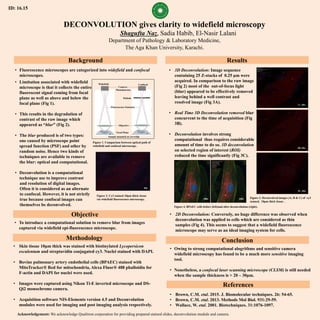
- 1. DECONVOLUTION gives clarity to widefield microscopy Shagufta Naz, Sadia Habib, El-Nasir Lalani Department of Pathology & Laboratory Medicine, The Aga Khan University, Karachi. Background Objective Methodology Results Conclusion • To introduce a computational solution to remove blur from images captured via widefield epi-fluorescence microscope. • Skin tissue 10μm thick was stained with biotinylated Lycopersicon esculentum and streptavidin conjugated cy3. Nuclei stained with DAPI. • Bovine pulmonary artery endothelial cells (BPAEC) stained with MitoTracker® Red for mitochondria, Alexa Fluor® 488 phalloidin for F-actin and DAPI for nuclei were used. • Images were captured using Nikon Ti-E inverted microscope and DS- Qi2 monochrome camera. • Acquisition software NIS-Elements version 4.5 and Deconvolution modules were used for imaging and post imaging analysis respectively. • Owing to strong computational alogrithms and sensitive camera widefield microscopy has found to be a much more sensitive imaging tool. • Nonetheless, a confocal laser scanning microscope (CLSM) is still needed when the sample thickness is > 20 – 30μm. Acknowledgement: We acknowledge Qualitron corporation for providing prepared stained slides, deconvolution module and camera. • Limitation associated with widefield microscope is that it collects the entire fluorescent signal coming from focal plane as well as above and below the focal plane (Fig 1). • This results in the degradation of contrast of the raw image which appeared as “blur” (Fig 2). • The blur produced is of two types: one caused by microscope point spread function (PSF) and other by random noise. Hence two kinds of techniques are available to remove the blur: optical and computational. • Deconvolution is a computational technique use to improve contrast and resolution of digital images. Often it is considered as an alternate to confocal. However, it is not strictly true because confocal images can themselves be deconvolved. Figure 1. Comparison between optical path of widefield and confocal microscope. Figure 2. Cy3 stained 10μm thick tissue via widefield fluorescence microscopy. • 3D Deconvolution: Image sequence containing 25 Z-stacks of 0.25 µm were acquired. In comparison to the raw image (Fig 2) most of the out-of-focus light (blur) appeared to be effectively removed leaving behind a well contrast and resolved image (Fig 3A). • Real Time 3D Deconvolution removed blur concurrent to the time of acquisition (Fig 3B). • Deconvolution involves strong computational thus requires considerable amount of time to do so. 3D deconvolution on selected region of interest (ROI) reduced the time significantly (Fig 3C). Figure 3. Deconvolved images (A, B & C) of cy3 stained, 10μm thick tissue . Figure 4. BPAEC cells before (left)and after deconvolution (right). • 2D Deconvolution: Conversely, no huge difference was observed when deconvolution was applied to cells which are considered as thin samples (Fig 4). This seems to suggest that a widefield fluorescence microscope may serve as an ideal imaging system for cells. • Fluorescence microscopes are categorized into widefield and confocal microscopes. 60x 3C, 60x 3A, 60x 3B, 60x 100x References • Brown, C.M. etal. 2015. J. Biomolecular techniques. 26: 54-65. • Brown, C.M. etal. 2013. Methods Mol Biol. 931:29-59. • Wallace, W. etal. 2001. Biotechniques. 31:1076-1097. 100x ID: 16.15 Pinhole Camera / Photodetector Widefield Confocal Objective Sample mounted on coverslip Focal Plane Fluorescence Emission