
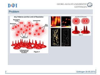
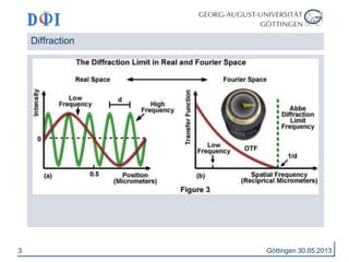
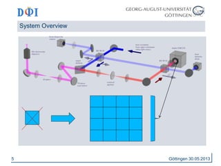
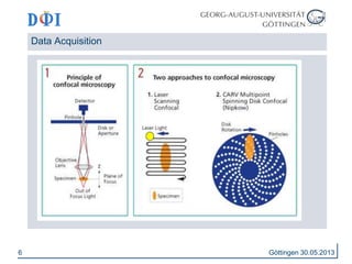
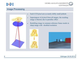
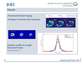
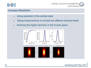
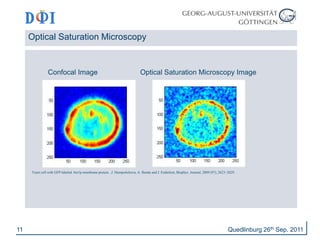
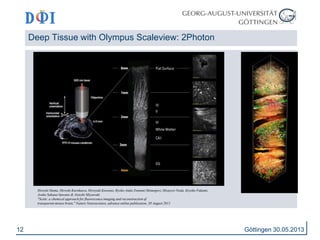
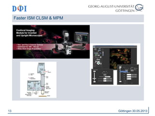
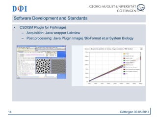


This document discusses image scanning microscopy (ISM), a technique that improves the resolution and contrast of fluorescence microscopy images. ISM works by acquiring multiple images of the sample from different positions and combining them to generate a single image with effectively doubled resolution. The document outlines the basic principles and components of ISM systems, provides examples of applications like imaging fluorescent beads and cell structures, and discusses related techniques for further enhancing resolution and depth of imaging. It concludes by emphasizing ISM's benefits like high resolution, low photodamage, and multicolor imaging capabilities.


![Getting Started with Apache Spark: Big Data Made Simple [Free Meetup]](https://cdn.slidesharecdn.com/ss_thumbnails/apachesparkgettingstarted-260203175547-8361bcc3-thumbnail.jpg?width=640&height=640&fit=bounds)


