Downloaded 53 times


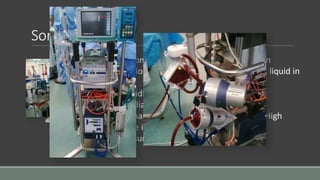
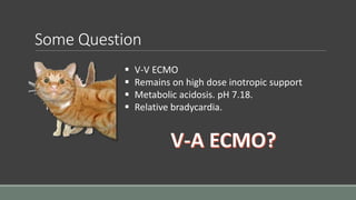
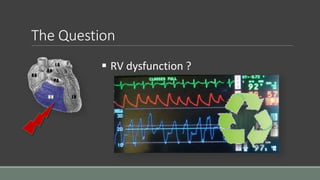
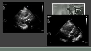
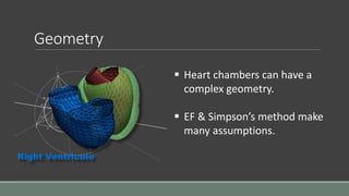
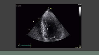
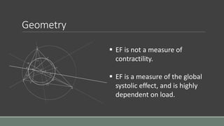

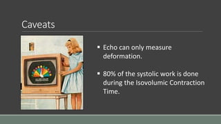
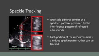

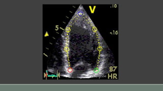
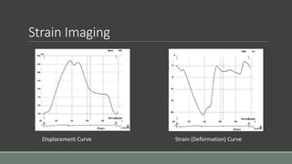
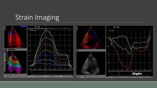
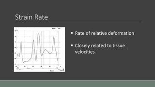
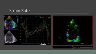
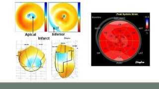
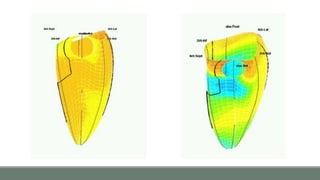
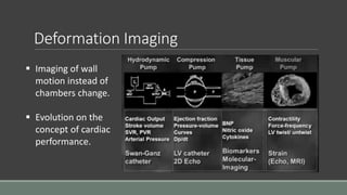
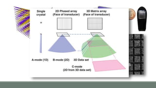
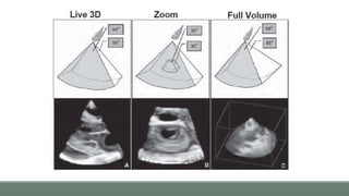
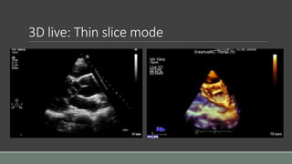
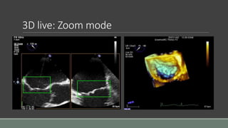
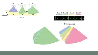
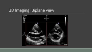
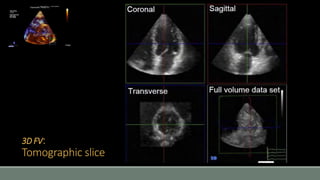
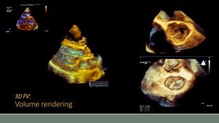
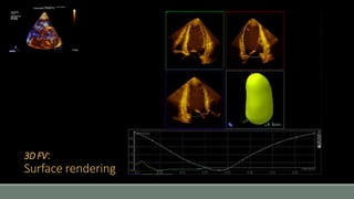
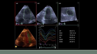
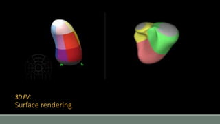
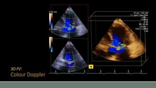
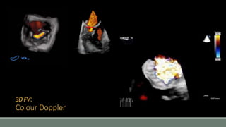
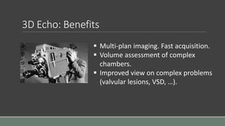
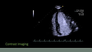
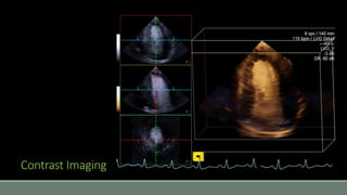


An obese male with a poorly documented medical history underwent a cystoscopy procedure under general anesthesia. During the procedure, he became hypoxic 15 minutes after induction and had a copious amount of frothy fluid in his endotracheal tube, making ventilation impossible. He went into cardiac arrest but was resuscitated. The patient remained hypoxic despite high levels of support. Strain imaging and 3D echocardiography were used to assess whether right ventricular dysfunction was contributing to the patient's condition, as 2D echocardiography has limitations in evaluating cardiac function and geometry. The document discusses the benefits and limitations of using newer echocardiography techniques like strain imaging and 3D imaging to evaluate cardiac performance and function.


