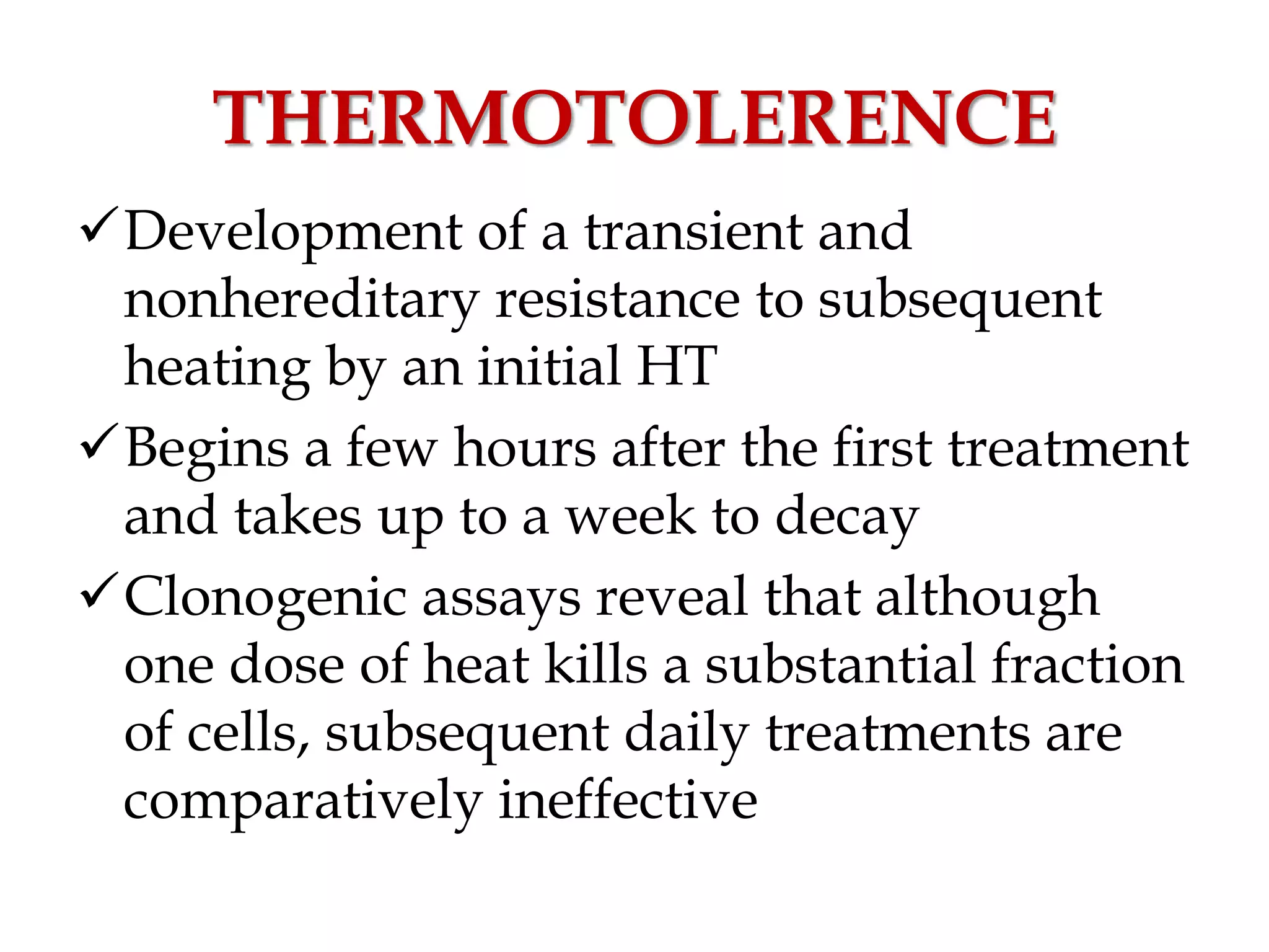
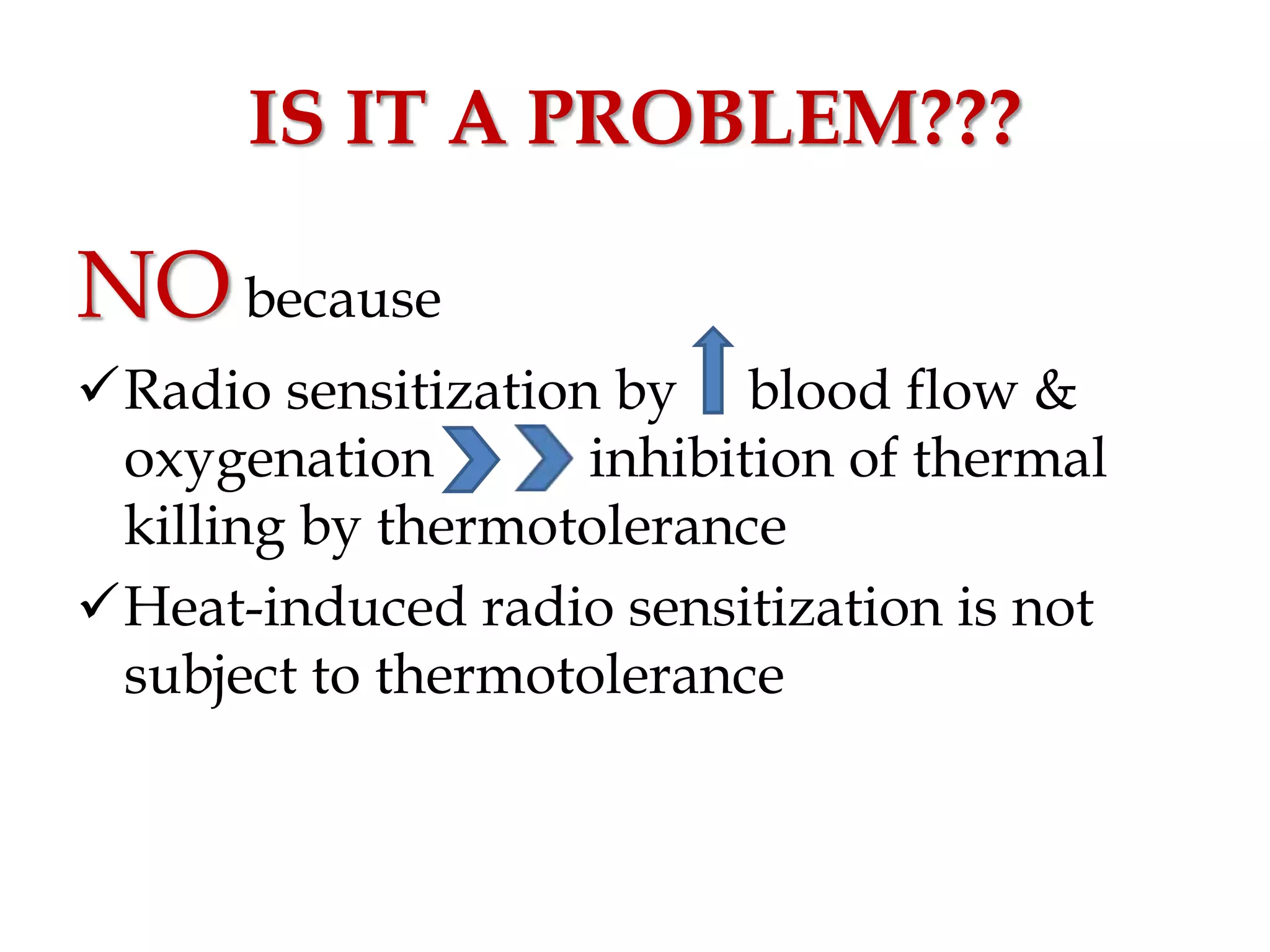
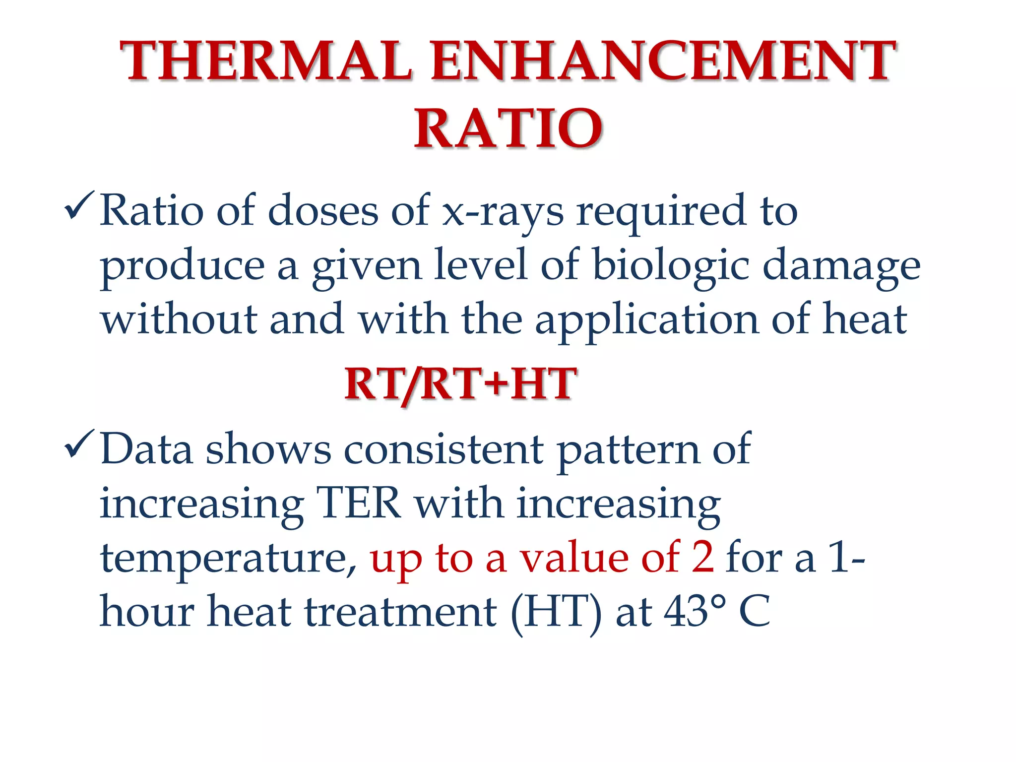
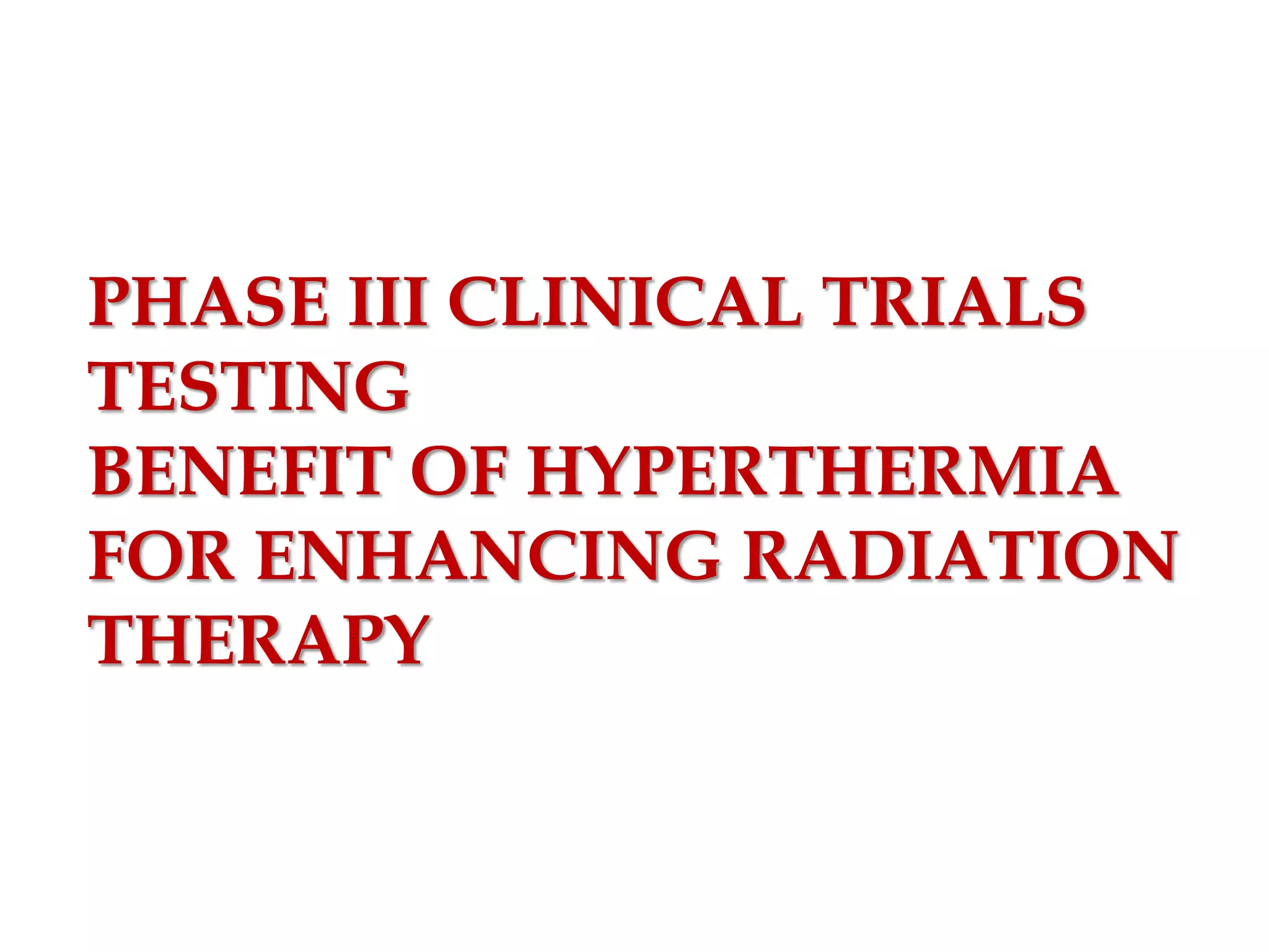
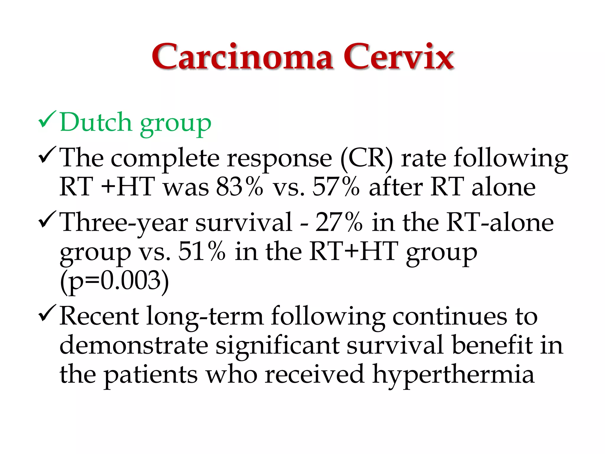
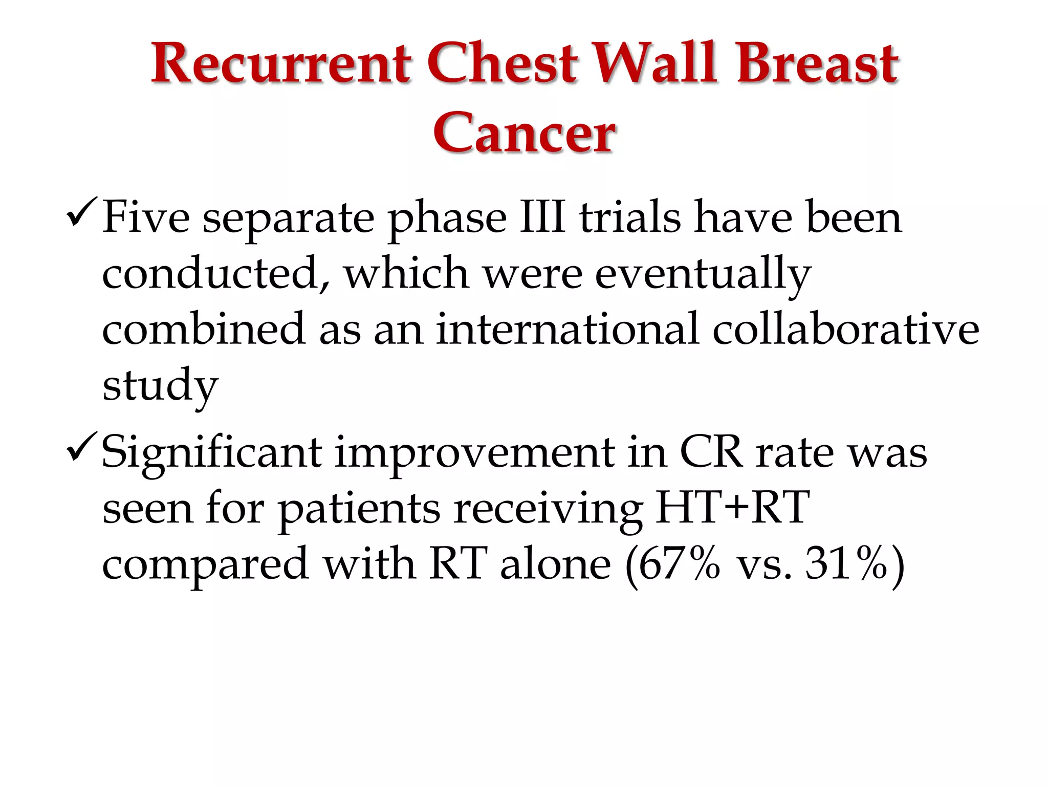
HYPERTHERMIA IN
RADIOTHERAPY
DR.JEWELL JOSEPH
INTRODUCTION
Elevation of temperature to a
supraphysiologic level
USA & Europe- 41.8–42 °C (107.2–107.6 °F)
Japan & Russia- 43–44 °C (109–111 °F)
Maintain the temperature about 1 hour
OVERVIEW
HISTORY OF MEDICAL HT
BIOLOGY OF HT
PHYSIOLOGICAL EFFECTS
PHYSICS
APPLICATIONS IN RADIOTHERAPY
700 - 600 BC
Western Zhou Dynasty – China
500 – 400 B.C
GREEKS
EGYPTIANS
400 – 300 B.C
"What medicines do not heal, the lance will;
what the lance does not heal, fire will"
100 A.D – 140 A.D
"If indeed any were so
good a physician as to be
able to produce fever, it
would not be necessary to
look for any other remedy
in sickness."
1400 – 1500 A.D
NATIVE AMERICANS
1600’S
“Fever is a mighty
engine, which nature
brings into the world
for the conquest of her
enemies”
THE MODERN ERA
Inhibition of tumor growth by high fever
caused by malaria - de Kizowitz (France) in
1779
Complete remission of histologically
confirmed face sarcoma after two erysipelas
infections with subsequent 2-year disease-
free survival - Busch (Germany) in 1866
George W. Crile Jr.
Long-lasting increase of the temperature of
some tumors to 42 – 50°C could selectively
destroy them without damaging the healthy
tissues.
James Fulton Percy
3–7-year survival of
inoperable cancer of the
uterus after local
hyperthermia above 45°C
The first International
Symposium on Cancer Therapy by
Hyperthermia and Radiation
Washington D.C in 1975
BIOLOGY OF HT
DIRECT EFFECTS INDIRECT EFFECTS
DIRECT EFFECTS
When cells are heated in vitro to a
sufficient temperature and for a long
enough duration, they die in a predictable,
exponential manner, and the rate of killing
increases with temperature
The cell survival curves for heat are
similar in shape to those obtained for x-
rays
Different cell lines differ in their
sensitivity to hyperthermia
There is NO consistent difference between
normal and malignant cells heated in
vitro.
Thermal Isoeffect Dose:
The Arrhenius Relationship
Temperature dependence of the rate of
cell killing by heat
Plots the log of the slope (1/Do) of cell
survival curves as a function of
temperature
Slope gives the activation energy of the
chemical process involved in the cell
killing
Arrhenius plots have a biphasic curve
Slope changes at the BREAK POINT(43
degrees for human cells)
Above this temperature, an increase of 1°C
doubles the rate of cell killing
Below the breakpoint, the rate of cell
killing by heat drops by a factor of 2 to 4
for each drop of 1° C
WHY DOES BREAK POINT
OCCUR?
Different mechanisms of cell killing above
and below BP
Development of thermotolerance within
cells
ARE PROTEINS THE TARGETS
FOR HEAT KILLING?
Activation energy for protein denaturation
to the activation energy for heat
cytotoxicity are similar
Heat of inactivation for cell killing and
thermal damage is similar to the energy
needed for protein denaturation (130–170
kcal/mol)
Increased expression of Heat Shock
Proteins
INDIRECT EFFECTS
Changes in the tumor microenvironment
42 to 44° C
Damage to tumor vascular endothelium
Decreased pO2,pH and nutrient status
Increased heat sensitivity but decreased
radiosensitivity
41 to 41.5° C
Damage to respiratory enzymes
Shift to anaerobic pathways
Increased oxygen tension in the tissues &
increased radiosensitivity
(Brezel et al., Jones et al)
PHYSIOLOGIC EFFECTS
EFFECT ON PERFUSION
Increased tissue perfusion (41 – 41.5°C)
Changes in vascular permeability
Vascular stasis and hemorrhage (42 - 44°C)
The increase in liposomal extravasation can
be exploited as a drug delivery vehicle
EFFECT ON OXYGENATION
EFFECT OF pH
Tumor cells
Decrease in extracellular pH
Cannot increase proton pumping
Increase in intracellular pH
APPLICATIONS
Reduction in pH by hyperglycemia,
glucose + metaiodobenzylguanidine
Vasoactive agents to reduce the blood
flow
IMMUNOLOGIC EFFECTS
Enhanced immunogenicity and HSP
expression seen after tumor cells heating
Thermally enhanced immune effector cell
activation and function
Thermally enhanced vascular perfusion
and delivery or trafficking of immune
effector cells to tumors
HYPERTHERMIA PHYSICS
CHALLENGES IN DELIVERING
HT
Anatomical variations
Varying tissue interfaces
Blood perfusion
MODALITIES
Thermal conduction
Nonionizing electromagnetic radiation
Ultrasound
THERMAL CONDUCTION
Circulating hot water in a needle, a
catheter, surface pad
Limited penetration of heat
Restricted to interstitial applications with
closely spaced implant arrays
ELECTROMAGNETIC HEATING
Heating from resistive losses as a result of
electric current in a resistive media (tissue)
At higher microwave frequencies, heat is
generated from mechanical interactions
between adjacent polar water molecules
aligning to the alternating EM field
Superficial applicators – penetration 1 – 4 cm
Deep HT devices – penetration more than 4
cm
ULTRASOUND HEATING
Energy transfer results from the
mechanical losses of viscous friction
Wavelength of US is orders of magnitude
smaller than that of an EM field of similar
penetration capabilities
So, US energy can be focused into small
tissue volumes
DOSIMETRY
Sapareto and Dewey - “cumulative
equivalent minutes” (CEM) at 43° C
Break point is set at 43° C
Above the break point,
t1/t2 = 2T1-T2
Below the break point,
t1/t2 = (4 to 6)T1-T2
CEM 43°C = tR(43-T)
R above BP – 0.5
below BP – 0.25
For a complex time–temperature history,
the heating profile is broken into short
intervals of time “t” length (typically 1 to 2
minutes), where the temperature remains
relatively constant
CEM 43° C = 𝒕R(43-avgT)
MEASURING LOCAL
TEMPERATURE
INVASIVE METHODS
Electrically conducting
Minimally conducting
Non conducting (optic)
NON-INVASIVE METHODS
Infrared thermography
Thermal monitoring sheet fiber optic
arrays
Electrical impedance tomography
Microwave tomography
Microwave radiometry
Ultrasonic temperature estimation
techniques
Magnetic resonance thermal imaging
(MRTI)
RATIONALE IN RT
Sensitizes the cells in S phase to RT
No difference in sensitivity to aerobic and
anaerobic cells
HT can lead to reoxygenation, which will
improve RT response
HT inhibits the repair of both sub lethal
and potentially lethal damage via its
effects in inactivating crucial DNA repair
pathways
THERMOTOLERENCE
Development of a transient and
nonhereditary resistance to subsequent
heating by an initial HT
Begins a few hours after the first treatment
and takes up to a week to decay
Clonogenic assays reveal that although
one dose of heat kills a substantial fraction
of cells, subsequent daily treatments are
comparatively ineffective
IS IT A PROBLEM???
NObecause
Radio sensitization by blood flow &
oxygenation inhibition of thermal
killing by thermotolerance
Heat-induced radio sensitization is not
subject to thermotolerance
THERMAL ENHANCEMENT
RATIO
Ratio of doses of x-rays required to
produce a given level of biologic damage
without and with the application of heat
RT/RT+HT
Data shows consistent pattern of
increasing TER with increasing
temperature, up to a value of 2 for a 1-
hour heat treatment (HT) at 43° C
Typical TER values
1.4 at 41° C
2.7 at 42.5° C
4.3 at 43° C
Canine oral squamous cell carcinomas –
1.15
Superficial human tumor types – 1.5
THERAPEUTIC GAIN FACTOR
Ratio of the TER in the tumor to the TER
in normal tissues
PHASE III CLINICAL TRIALS
TESTING
BENEFIT OF HYPERTHERMIA
FOR ENHANCING RADIATION
THERAPY
Carcinoma Cervix
Dutch group
The complete response (CR) rate following
RT +HT was 83% vs. 57% after RT alone
Three-year survival - 27% in the RT-alone
group vs. 51% in the RT+HT group
(p=0.003)
Recent long-term following continues to
demonstrate significant survival benefit in
the patients who received hyperthermia
Recurrent Chest Wall Breast
Cancer
Five separate phase III trials have been
conducted, which were eventually
combined as an international collaborative
study
Significant improvement in CR rate was
seen for patients receiving HT+RT
compared with RT alone (67% vs. 31%)
Superficial Malignancies
Single institution prospective randomized
trial conducted by Jones et al.
The complete response rate in the
hyperthermia/ radiation group was 66%
versus 42% in the radiation-alone group
Previously irradiated patients had the
greatest benefit, enjoying a 68.2% response
rate in the hyperthermia/radiation group
versus 23.5% in the radiation alone group
Head and neck cancer
Valdagni et al., showed patients in stage
III receiving RT+HT had a 58% CR
compared with 20% in the RT group
Patients with stage IV disease achieved a
CR of 38% compared with 7% for those
receiving RT alone
Glioblastoma multiforme
Sneed et al. – time to tumor progression
and 2-year survival were significantly
improved for patients who received
hyperthermia compared with those
treated with brachytherapy alone (31% vs.
15%)
ADVERSE EFFECTS
External application of heat may cause
surface burns
Whole body hyperthermia can cause
swelling, blood clots, and bleeding
Systemic shock
CHALLENGES IN
IMPLEMENATAION
It is difficult to heat tumor tissue volumes
with uniformity and precision
There is no standardized equipment to
effect loco regional HT
Techniques for measuring temperature
and the actual definition and calculation
of thermal dose remain significant
problems
Hyperthermia in radiotherapy

Hyperthermia in radiotherapy

  • 1.
  • 2.
    INTRODUCTION Elevation of temperatureto a supraphysiologic level USA & Europe- 41.8–42 °C (107.2–107.6 °F) Japan & Russia- 43–44 °C (109–111 °F) Maintain the temperature about 1 hour
  • 3.
    OVERVIEW HISTORY OF MEDICALHT BIOLOGY OF HT PHYSIOLOGICAL EFFECTS PHYSICS APPLICATIONS IN RADIOTHERAPY
  • 5.
    700 - 600BC Western Zhou Dynasty – China
  • 6.
    500 – 400B.C GREEKS EGYPTIANS
  • 7.
    400 – 300B.C "What medicines do not heal, the lance will; what the lance does not heal, fire will"
  • 8.
    100 A.D –140 A.D "If indeed any were so good a physician as to be able to produce fever, it would not be necessary to look for any other remedy in sickness."
  • 9.
    1400 – 1500A.D NATIVE AMERICANS
  • 10.
    1600’S “Fever is amighty engine, which nature brings into the world for the conquest of her enemies”
  • 11.
    THE MODERN ERA Inhibitionof tumor growth by high fever caused by malaria - de Kizowitz (France) in 1779
  • 12.
    Complete remission ofhistologically confirmed face sarcoma after two erysipelas infections with subsequent 2-year disease- free survival - Busch (Germany) in 1866
  • 13.
    George W. CrileJr. Long-lasting increase of the temperature of some tumors to 42 – 50°C could selectively destroy them without damaging the healthy tissues.
  • 14.
    James Fulton Percy 3–7-yearsurvival of inoperable cancer of the uterus after local hyperthermia above 45°C
  • 15.
    The first International Symposiumon Cancer Therapy by Hyperthermia and Radiation Washington D.C in 1975
  • 16.
    BIOLOGY OF HT DIRECTEFFECTS INDIRECT EFFECTS
  • 17.
    DIRECT EFFECTS When cellsare heated in vitro to a sufficient temperature and for a long enough duration, they die in a predictable, exponential manner, and the rate of killing increases with temperature The cell survival curves for heat are similar in shape to those obtained for x- rays
  • 18.
    Different cell linesdiffer in their sensitivity to hyperthermia There is NO consistent difference between normal and malignant cells heated in vitro.
  • 21.
    Thermal Isoeffect Dose: TheArrhenius Relationship
  • 22.
    Temperature dependence ofthe rate of cell killing by heat Plots the log of the slope (1/Do) of cell survival curves as a function of temperature Slope gives the activation energy of the chemical process involved in the cell killing Arrhenius plots have a biphasic curve Slope changes at the BREAK POINT(43 degrees for human cells)
  • 23.
    Above this temperature,an increase of 1°C doubles the rate of cell killing Below the breakpoint, the rate of cell killing by heat drops by a factor of 2 to 4 for each drop of 1° C
  • 24.
    WHY DOES BREAKPOINT OCCUR? Different mechanisms of cell killing above and below BP Development of thermotolerance within cells
  • 25.
    ARE PROTEINS THETARGETS FOR HEAT KILLING? Activation energy for protein denaturation to the activation energy for heat cytotoxicity are similar Heat of inactivation for cell killing and thermal damage is similar to the energy needed for protein denaturation (130–170 kcal/mol) Increased expression of Heat Shock Proteins
  • 26.
    INDIRECT EFFECTS Changes inthe tumor microenvironment 42 to 44° C Damage to tumor vascular endothelium Decreased pO2,pH and nutrient status Increased heat sensitivity but decreased radiosensitivity
  • 27.
    41 to 41.5°C Damage to respiratory enzymes Shift to anaerobic pathways Increased oxygen tension in the tissues & increased radiosensitivity (Brezel et al., Jones et al)
  • 28.
  • 29.
    EFFECT ON PERFUSION Increasedtissue perfusion (41 – 41.5°C) Changes in vascular permeability Vascular stasis and hemorrhage (42 - 44°C) The increase in liposomal extravasation can be exploited as a drug delivery vehicle
  • 30.
  • 31.
  • 32.
    Tumor cells Decrease inextracellular pH Cannot increase proton pumping Increase in intracellular pH
  • 33.
    APPLICATIONS Reduction in pHby hyperglycemia, glucose + metaiodobenzylguanidine Vasoactive agents to reduce the blood flow
  • 34.
  • 35.
    Enhanced immunogenicity andHSP expression seen after tumor cells heating Thermally enhanced immune effector cell activation and function Thermally enhanced vascular perfusion and delivery or trafficking of immune effector cells to tumors
  • 36.
  • 37.
    CHALLENGES IN DELIVERING HT Anatomicalvariations Varying tissue interfaces Blood perfusion
  • 38.
  • 39.
    THERMAL CONDUCTION Circulating hotwater in a needle, a catheter, surface pad Limited penetration of heat Restricted to interstitial applications with closely spaced implant arrays
  • 40.
    ELECTROMAGNETIC HEATING Heating fromresistive losses as a result of electric current in a resistive media (tissue) At higher microwave frequencies, heat is generated from mechanical interactions between adjacent polar water molecules aligning to the alternating EM field Superficial applicators – penetration 1 – 4 cm Deep HT devices – penetration more than 4 cm
  • 42.
    ULTRASOUND HEATING Energy transferresults from the mechanical losses of viscous friction Wavelength of US is orders of magnitude smaller than that of an EM field of similar penetration capabilities So, US energy can be focused into small tissue volumes
  • 44.
    DOSIMETRY Sapareto and Dewey- “cumulative equivalent minutes” (CEM) at 43° C Break point is set at 43° C Above the break point, t1/t2 = 2T1-T2 Below the break point, t1/t2 = (4 to 6)T1-T2 CEM 43°C = tR(43-T) R above BP – 0.5 below BP – 0.25
  • 45.
    For a complextime–temperature history, the heating profile is broken into short intervals of time “t” length (typically 1 to 2 minutes), where the temperature remains relatively constant CEM 43° C = 𝒕R(43-avgT)
  • 46.
    MEASURING LOCAL TEMPERATURE INVASIVE METHODS Electricallyconducting Minimally conducting Non conducting (optic)
  • 47.
    NON-INVASIVE METHODS Infrared thermography Thermalmonitoring sheet fiber optic arrays Electrical impedance tomography Microwave tomography Microwave radiometry Ultrasonic temperature estimation techniques Magnetic resonance thermal imaging (MRTI)
  • 48.
    RATIONALE IN RT Sensitizesthe cells in S phase to RT No difference in sensitivity to aerobic and anaerobic cells HT can lead to reoxygenation, which will improve RT response HT inhibits the repair of both sub lethal and potentially lethal damage via its effects in inactivating crucial DNA repair pathways
  • 49.
    THERMOTOLERENCE Development of atransient and nonhereditary resistance to subsequent heating by an initial HT Begins a few hours after the first treatment and takes up to a week to decay Clonogenic assays reveal that although one dose of heat kills a substantial fraction of cells, subsequent daily treatments are comparatively ineffective
  • 50.
    IS IT APROBLEM??? NObecause Radio sensitization by blood flow & oxygenation inhibition of thermal killing by thermotolerance Heat-induced radio sensitization is not subject to thermotolerance
  • 51.
    THERMAL ENHANCEMENT RATIO Ratio ofdoses of x-rays required to produce a given level of biologic damage without and with the application of heat RT/RT+HT Data shows consistent pattern of increasing TER with increasing temperature, up to a value of 2 for a 1- hour heat treatment (HT) at 43° C
  • 52.
    Typical TER values 1.4at 41° C 2.7 at 42.5° C 4.3 at 43° C Canine oral squamous cell carcinomas – 1.15 Superficial human tumor types – 1.5
  • 53.
    THERAPEUTIC GAIN FACTOR Ratioof the TER in the tumor to the TER in normal tissues
  • 54.
    PHASE III CLINICALTRIALS TESTING BENEFIT OF HYPERTHERMIA FOR ENHANCING RADIATION THERAPY
  • 55.
    Carcinoma Cervix Dutch group Thecomplete response (CR) rate following RT +HT was 83% vs. 57% after RT alone Three-year survival - 27% in the RT-alone group vs. 51% in the RT+HT group (p=0.003) Recent long-term following continues to demonstrate significant survival benefit in the patients who received hyperthermia
  • 56.
    Recurrent Chest WallBreast Cancer Five separate phase III trials have been conducted, which were eventually combined as an international collaborative study Significant improvement in CR rate was seen for patients receiving HT+RT compared with RT alone (67% vs. 31%)
  • 57.
    Superficial Malignancies Single institutionprospective randomized trial conducted by Jones et al. The complete response rate in the hyperthermia/ radiation group was 66% versus 42% in the radiation-alone group Previously irradiated patients had the greatest benefit, enjoying a 68.2% response rate in the hyperthermia/radiation group versus 23.5% in the radiation alone group
  • 58.
    Head and neckcancer Valdagni et al., showed patients in stage III receiving RT+HT had a 58% CR compared with 20% in the RT group Patients with stage IV disease achieved a CR of 38% compared with 7% for those receiving RT alone
  • 59.
    Glioblastoma multiforme Sneed etal. – time to tumor progression and 2-year survival were significantly improved for patients who received hyperthermia compared with those treated with brachytherapy alone (31% vs. 15%)
  • 60.
    ADVERSE EFFECTS External applicationof heat may cause surface burns Whole body hyperthermia can cause swelling, blood clots, and bleeding Systemic shock
  • 61.
    CHALLENGES IN IMPLEMENATAION It isdifficult to heat tumor tissue volumes with uniformity and precision There is no standardized equipment to effect loco regional HT Techniques for measuring temperature and the actual definition and calculation of thermal dose remain significant problems