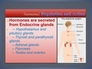
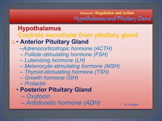
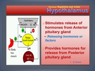
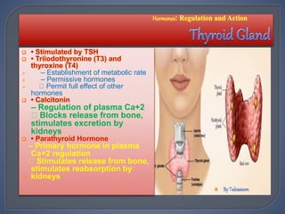
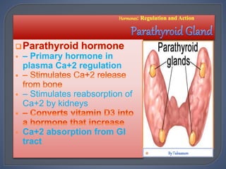
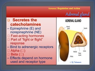
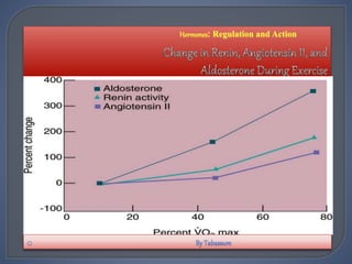
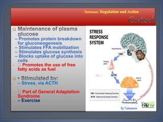
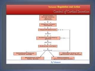
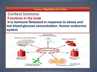
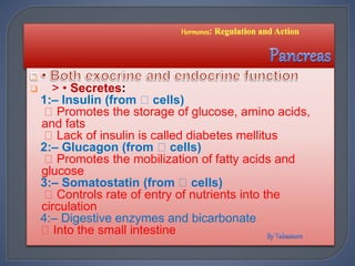
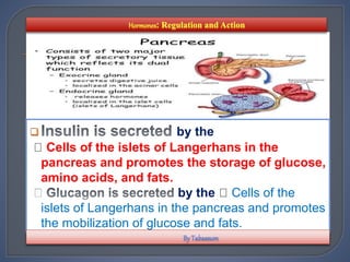
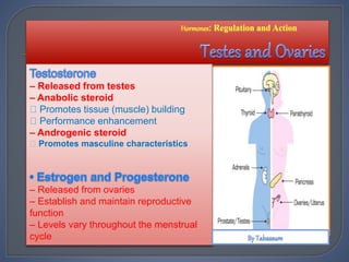
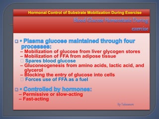
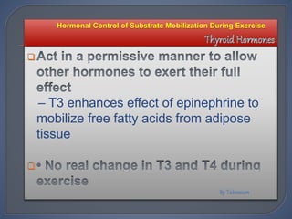
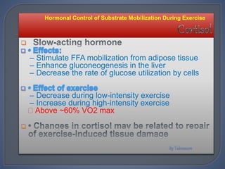
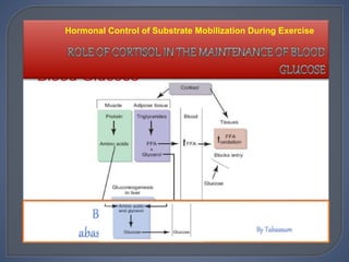
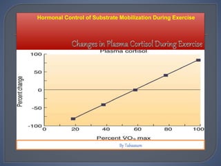
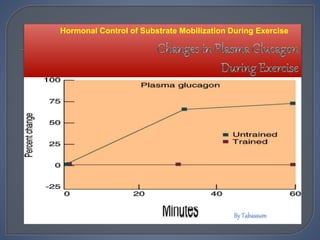
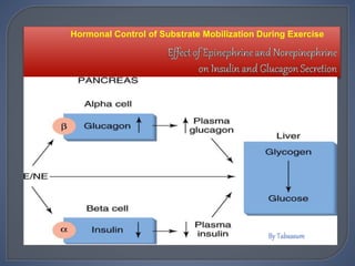
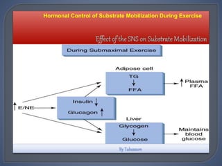
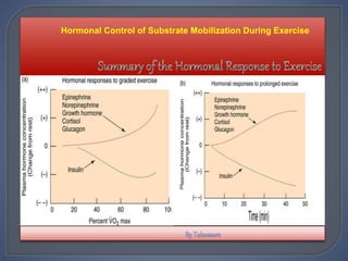
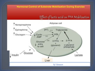
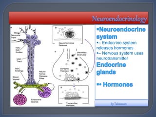
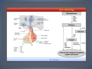
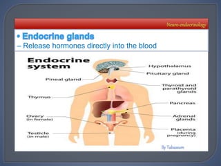
 Introduction
 Hormones: Regulation
and Action
 Hypothalamus and the
 Pituitary Gland
 Thyroid Gland
 Parathyroid Gland
 Adrenal Gland
 Pancreas
 Ovaries and Testes
 Neuroendocrinology
 Blood Hormone
 Concentration
 Hormone-Receptor
 Interaction
 Neuroendocrine aspects of
overtraining
 Hormonal Control of
Substrate Mobilization
During Exercise
 Muscle-Glycogen
 Utilization
 Blood Glucose
 Homeostasis During
 Exercise
 Hormone-Substrate
 Interaction
 Conclusions and
clinical relevance
References
 HORMNAL RESPONSE
Hormonal stimuli refers to the release of
a hormone in response to another hormone.
 A number of endocrine glands
release hormones when stimulated
by hormones released by other endocrine
glands.
 For example, the hypothalamus
produces hormones that stimulate the anterior
portion of the pituitary gland
 Hormones are necessary to help the body
make acute and chronic adaptations to
exercise
 Acute exercise:- single bout of exercise
 Exercise training or the “chronic exercise”
intervention can be defined as a repeated
amount of bouts of exercise during a short
or long-term period of time)
are necessary to help the body
make acute and chronic adaptations to
exercise
 single bout of exercise
 training or the “ ”
intervention can be defined as a repeated
amount of bouts of exercise during a short
or long-term period of time)

 It is, however, a very varied
and complex physiological
stimulus
.

.
 With the increasing
popularity of exercise
training for the
maintainence of good
health and even the
treatment of existing
diseases,
.



 or expressed as a percentage (measured directly
or calculated) of the individual subject’s
maximum work capacity.
 Work capacity, in turn, may be expressed as
either maximum tolerated work load in the
particular experimental system or as the
measured maximum aerobic capacity (V02
max)
 When assessing hormonal responses to
training reference should be made to the
relative intensity of the exercise stimulus,
expressed as VO2max ~ rather than absolute
work load, and this is indicated as appropriate

 Most exercise studies conclude that both high intensity
and endurance exercises cause an increase in plasma
ACTH ( Adreno cortico tropic hormone ) and cortisol in
humans
 However, the exercise-induced plasma cortisol increase
may not be directly related to the exercise-induced
ACTH increase,because both hormones show a
different exercise related response.
 ACTH increases curvilinear with exercise intensity, while
cortisol increases more with increasing exercise
duration.
 A significant increase in plasma cortisol concentration
was found following exercise. 
 However, there was no relationship with relative
work intensity or blood lactate concentration.
 The suggestion was made that the exercise test
was too short (total time test270 s) to induce a
consequent increase in cortisol concentration
 In general, for humans
 High intensity exercise results in a 2-3 fold
increase in cortisol,
 Peaking usually 15-30 minutes after exercise,
And
 Returning to pre-exercise levels within the hour
 Endurance exercise usually requires duration of
exercise of more than 20 minutes with


 High intensity exercise results in an increase in
ACTH, usually peaking the end of exercise and
returning to baseline within 60-120 minutes
 Prolonged sub maximal exercise or endurance
exercise shows a later peak in plasma cortisol and
a slower return to pre-exercise levels than high
intensity exercise
 Several factors influence the exercise-induced
cortisol response
EXAMPLE
 Circadian rhythm,
 Multiple exercise bouts
 High intensity versus
 Prolonged sub maximal exercise
 Nutrition
 Training. 
 A significant stimulatory effect of
exercise on Cortisol levels
 Only in the afternoon and
 Not in the early morning or during the night
after performing
 Low intensity exercise (3h at 40-60%
V02max
 However, the intensity of exercise
might have been too low to induce a
good response inthis study.

 Repeated bouts of high-intensity endurance exercise
 2 bouts of 65 min 70% V02 max, 3h rest in
between) resulted in increased plasma
concentrations of ACTH and cortisol during exercise
and early recovery
 Two bouts of high intensive exercise with a 4h rest
period in between showed an exaggerated response
for ACTH and cortis after the second bout
 In addition, the first bout of exercise was not able to
induce an increase in cortisol concentration

 Two bouts of moderate intensity exercise (30 min
50% V02 max, 45 minrest in between) did not
change the cortisol response to the second bout of
exercise
 Four bouts of moderate intensity (30 min, 50%
V02 max, 30min rest) progressively increased
concentrations of cortisol throughout the
series of exercise bouts
 However, cortisol concentrations were
still augmented during the rests in the
last study, which may partly account for
the progressive increase
Cortisol response after the second bout of
exercise(50% V02 max, 90-min, 3-h rest)
in men and an increased cortisol response
in women
 Comparison to other experiments is
difficult, because the athlete's plasma
glucose was maintained at euglycaemia
during the experiment by glucose
infusions 

 Some studies investigated the HPA axis during
Long-term recovery after exercise
 Studied the cortisol response after an incremental
exercise test till exhaustion and during a recovery
period of 32
hours in 4 geldings
 A significant increase of mean plasma cortisol
concentration after exercise was found, followed
by a significant decrease 4-8 hours post exercise
and a significant increase 20-24 hours post
exercise compared to basal levels, the same
alterations found in men 
 Most studies find elevated ACTH levels at rest
intrained subjects versus untrained subjects
 Mostly, basal cortisol levels are not elevated,
which might indicate that the adrenal gland is
less sensitive to ACTH due to training
decreased pituitary sensitivity to gluco-
corticoids in endurance-trained men (50-70 km
running per wk, completed marathon in less
than 3h30min) compared to sedentary
 men ( < lh physical activity per week)

 At rest, 3 out of 9 endurance-trained men
responded with an increase in ACTH and
cortisol after administration of CRH while at
the mean time dexa-methason was given to
suppress endogenous cortisol secretion
 The sedentary group as well as 5 out of
9 endurance-trained athletes did not respond
to an injection with oCRH
 Basal cortisol and ACTH levels were not
significantly different between both
groups
 Only the highly trained runners showed basal
elevated levels for ACTH and cortisol and a
blunted response of ACTH and cortisol to
administration of oCRH
 Some researchers tried to evaluate prolonged
periods of training by determination of the ratio
of catabolic to anabolic hormones using cortisol
as catabolic hormone and sex steroids or IGF-I as
anabolic hormone
 However, based on the current literature it is
difficult to conclude whether any androgen to
cortisol ratio is a useful indicator of training
status in endurance athletes

The responses of ACTH and cortisol
reflect the stress impact of the current
training load on the body, but it does
not necessarily indicate overtraining.

Hormones are secreted
from Endocrine glands
 – Hypothalamus and
pituitary glands
 – Thyroid and parathyroid
glands
 – Adrenal glands
 – Pancreas
 – Testes and ovaries

Hypothalamus
Controls secretions from pituitary gland
 • Anterior Pituitary Gland
–Adrenocorticotropic hormone (ACTH)
– Follicle-stimulating hormone (FSH)
– Luteinizing hormone (LH)
– Melanocyte-stimulating hormone (MSH)
– Thyroid-stimulating hormone (TSH)
– Growth hormone (GH)
– Prolactin
 • Posterior Pituitary Gland
– Oxytocin
– Antidiuretic hormone (ADH) 
 Stimulates release of
hormones from Anterior
pituitary gland
– Releasing hormones or
factors
 Provides hormones for
release from Posterior
pituitary gland

 While it’s very small, the hypothalamus
plays a crucial role in many important
functions, including:-
 Releasing hormones
 Regulating body temperature
 Maintaining daily physiological cycles
 Controlling appetite
 Managing of sexual behavior
 Regulating emotional responses

 • Adrenocorticotropic
hormone (ACTH)
– Stimulates cortisol release
from adrenal glands
 • Follicle-stimulating hormone
(FSH)
 • Luteinizing hormone (LH)
– Stimulates production of
testosterone and estrogen
 • Melanocyte-stimulating
hormone (MSH)
 • Thyroid-stimulating hormone
(TSH)
– Controls thyroid hormone
release from thyroid gland
 • Prolactin
 • Growth hormone (GH)

 Some of the most important hormones produced in
the anterior region include:
 Corticotropin-releasing hormone (CRH)
 CRH is involved in the body’s response to
both physical and emotional stress.
 It signals the pituitary gland to produce a
hormone called adreno-corticotropic
hormone (ACTH)
 ACTH triggers the production of cortisol, an
important stress hormone.
 Thyrotropin-releasing hormone (TRH). TRH
production stimulates the pituitary gland to
produce thyroid-stimulating hormone (TSH). TSH
plays an important role in the function of many
body parts, such as the heart, gastrointestinal
tract, and muscles.

 Gonadotropin-releasing hormone (GnRH):-
GnRH production causes the pituitary gland to produce
important reproductive hormones, such as follicle-
stimulating hormone (FSH) and luteinizing hormone
(LH). They act on the ovaries or testes to stimulate sex
hormone production, and egg and sperm maturity
 Oxytocin.:-This hormone controls many
important behaviors and emotions, such as
sexual arousal, trust, recognition, and maternal
behavior. It’s also involved in some functions of
the reproductive system, such as childbirth and
lactation. which stimulates uterine contractions
during labour and milk secretion during
breastfeeding.

 Vasopressin:- Also called antidiuretic hormone
(ADH), this hormone regulates water levels in the
body. When vasopressin is released, it signals the
kidneys to absorb water.
 Somatostatin:- Somatostatin works to stop the
pituitary gland from releasing certain hormones,
including growth hormones and thyroid-stimulating
hormones
 Growth hormone, which regulates growth, metabolism and
body composition
 The anterior region of the hypothalamus also helps regulate
body temperature through sweat. It also maintains circadian
rhythms. These are physical and behavioral changes that occur
on a daily cycle. For example, being awake during the day and
sleeping at nighttime is a circadian rhythm related to the
presence or absence of light.


– IGF-1 in muscle responsible for
muscle growth

– Amino acid uptake and protein
synthesis
– Long bone growth
-Reduces the use of plasma
glucose
-Increases gluconeogenesis
-Mobilizes fatty acids from
adipose tissue 

– Used to treat childhood
dwarfism
– Also used by athletes and
elderly

– Protein synthesis is collagen,
not contractile protein






 – Establishment of metabolic rate
 – Permissive hormones
 Permit full effect of other
hormones
– Regulation of plasma Ca+2
Blocks release from bone,
stimulates excretion by
kidneys
– Primary hormone in plasma
Ca+2 regulation
Stimulates release from bone,
stimulates reabsorption by
 kidneys

 Some of the important
functions of the thyroid
hormones include-
 :-Neural growth and
differentiation
 :-Myocardial contractility
:-Regulation of bone
formation and resorption,
 :- Development and
function of brown and
white adipose tissue
 :-Cholesterol metabolism
and synthesis
 :- In-utero they are
important for fetal growth
and differentiation

 – Primary hormone in
plasma Ca+2 regulation
 – Stimulates reabsorption of
Ca+2 by kidneys
 Ca+2 absorption from GI
tract

 The four parathyroid glands make more or less
parathyroid hormone (PTH) in response to the
level of calcium in the blood.
.
 Increased PTH causes the body to put more calcium
into the blood.
 Increased PTH causes the bones to release their
calcium into the blood.
– Epinephrine (E) and
norepinephrine (NE)
Fast-acting hormones
Part of “fight or flight”
response
– Bind to adrenergic receptors
Alpha ( )
Beta ( )
– Effects depend on hormone
used and receptor type









 Epinephrine and norepinephrine bind to -
and
 -adrenergic receptors and bring about
changes in
 Secretes steroid hormones
– Derived from cholesterol
 • Mineralcorticoids
– Aldosterone
– Maintenance of plasma
Na+ and K+
 • Glucocorticoids
– Cortisol
– Regulation of plasma
glucose
 • Sex steroids
– Androgens and estrogens
– Support prepubescent
growth
 The adrenal cortex secretes
 Aldosterone (mineralcorticoid)
 Cortisol (glucocorticoid)
 Estrogens and androgens (sex steroids).
 Aldosterone regulates Na+ reabsorption and K+ balance.
 Aldosterone secretion increases with strenuous exercise,
driven by the renin-angiotensin system
 . • Regulation of blood volume and blood pressure
 – Part of renin-angiotensin-aldosterone system
 – All three hormones increase during exercise
 • Stimulated by:
 – Increased K+ concentration
 – Decreased plasma volume

Exercise, to ensure that fuel (glucose and free
fatty acids) is available, and to make amino
acids available for tissue repair.

– Promotes protein breakdown
for gluconeogenesis
– Stimulates FFA mobilization
– Stimulates glucose synthesis
– Blocks uptake of glucose into
cells
 Promotes the use of free
fatty acids as fuel

 – Stress, via ACTH
 Part of General Adaptation
Syndrome
 – Exercise
Functions in the body
It is hormone Released in response to stress and
low blood-glucose concentration. Human endocrine
system
 – Leptin
 Influences appetite through the hypothalamus
 Enhances insulin sensitivity and fatty acid
oxidation
 – Adiponectin
 Increases insulin sensitivity and fatty acid
oxidation

 – Higher leptin levels and lower adiponectin
 – Leads to type 2 diabetes and low-grade
 inflammation
 > • Secretes:
1:– Insulin (from cells)
Promotes the storage of glucose, amino acids,
and fats
Lack of insulin is called diabetes mellitus
2:– Glucagon (from cells)
Promotes the mobilization of fatty acids and
glucose
3:– Somatostatin (from cells)
Controls rate of entry of nutrients into the
circulation
4:– Digestive enzymes and bicarbonate
Into the small intestine
 by the
Cells of the islets of Langerhans in the
pancreas and promotes the storage of glucose,
amino acids, and fats.
by the Cells of the
islets of Langerhans in the pancreas and promotes
the mobilization of glucose and fats.
– Released from testes
– Anabolic steroid
Promotes tissue (muscle) building
Performance enhancement
– Androgenic steroid
Promotes masculine characteristics
– Released from ovaries
– Establish and maintain reproductive
function
– Levels vary throughout the menstrual
cycle
is a steroid hormone that helps control and
guide female sexual development.
 Estrogen is responsible for stimulating the development
of female secondary sex characteristics.
 We know that secondary sex characteristics are defined
as characteristics specific to females or males, but not
directly related to reproduction.
 Therefore, in a female, we see estrogen helps with such
things as development of the breasts, widening of the
hips, and the growth of body hair.
 Estrogen works in harmony with , which is a
steroid hormone that acts to prepare the uterus to receive
the fertilized egg and maintain pregnancy.
 While it is correct to say that progesterone is secreted by
the ovaries, it's more correct to say that it is a hormone
produced by the corpus luteum of the ovaries.
 The corpus luteum is a structure that develops in an
 ovary after the egg has been discharged.
is a sex hormone that plays important
roles in the body
 In men, it’s thought to regulate sex drive (libido), bone
mass, fat distribution, muscle mass and strength, and
the production of red blood cells and sperm.
 A small amount of circulating testosterone is converted
to estradiol, a form of estrogen
and establish and maintain
reproductive function and determine secondary sex
characteristics
 Chronic exercise (training) can decrease
testosterone levels in males and estrogen levels in
females.
 The latter adaptation has potentially negative
consequences related to osteoporosis.
 Muscle mass
– In contrast to real-world reports
“Subjects” used 10 to 100 times the
recommended dosage
 • Also associated with negative side effects
– Revert to normal after discontinuation
 • Widespread use has led to testing of
competitive athletes
• Most users are not competitive athletes
– Take more than one steroid in megadoses
– High-intensity exercise results in greater
and more rapid glycogen depletion
- High-intensity exercise results in greater
increases in plasma epinephrin
Hormonal Control of Substrate Mobilization During Exercise
Plasma Ep
Hormonal Control of Substrate Mobilization During Exercise
TIME (MIN)
– Epinephrine-cyclic AMP
Via -adrenergic receptors
– Ca+2-calmodulin
Enhanced during exercise due to Ca+2 release
from sarcoplasmic reticulum
– Propranolol ( -receptor blocker) has no effect on
muscle glycogen utilization
Hormonal Control of Substrate Mobilization During Exercise
 Glycogen breakdown to glucose in
muscle is under the dual control of
epinephrine-cyclic AMP and Ca+2-
calmodulin
 The latter’s role is enhanced during
exercise due to the increase in Ca+2
from the sarcoplasmic reticulum
– Mobilization of glucose from liver glycogen stores
– Mobilization of FFA from adipose tissue
Spares blood glucose
– Gluconeogenesis from amino acids, lactic acid, and
glycerol
– Blocking the entry of glucose into cells
Forces use of FFA as a fuel
– Permissive or slow-acting
– Fast-acting
Hormonal Control of Substrate Mobilization During Exercise

– T3 enhances effect of epinephrine to
mobilize free fatty acids from adipose
tissue

Hormonal Control of Substrate Mobilization During Exercise

– Stimulate FFA mobilization from adipose tissue
– Enhance gluconeogenesis in the liver
– Decrease the rate of glucose utilization by cells
– Decrease during low-intensity exercise
– Increase during high-intensity exercise
Above ~60% VO2 max

Hormonal Control of Substrate Mobilization During Exercise

– Supports the action of cortisol
Decreases glucose uptake by tissues
Increases free fatty acid mobilization
Enhances gluconeogenesis in the liver
– Increase in plasma GH with increased
intensity
– Greater response in trained runners
Hormonal Control of Substrate Mobilization During Exercise

– Muscle glycogen mobilization
– Increasing liver glucose mobilization
– Increasing FFA mobilization
– Interfere with glucose uptake
– Also related to increased heart rate and blood
pressure during exercise
Hormonal Control of Substrate Mobilization During Exercise
 – Uptake and storage of glucose and FFA
 – Plasma concentration decreases during exercise
 – Decreased insulin response following training
 – Mobilization of glucose and FFA fuels
 – Plasma concentration increases during exercise
 – Decreased response following training
Hormonal Control of Substrate Mobilization During Exercise

Hormonal Control of Substrate Mobilization During Exercise

Glucose is taken up seven to twenty times
faster during exercise than at rest—even
with the decrease in plasma insulin
 The increases in intracellular Ca+2 and other
factors are associated with an increase in
the number of glucose transporters that
increase the membrane transport of glucose
Training causes a reduction in E, NE,
glucagon, and insulin responses to exercise

 – This occurs in spite of persisting hormonal
 stimulation for FFA mobilization
 – High levels of lactic acid
 Promotes resynthesis of triglycerides
 – Elevated H+ concentration inhibits HSL
 – Inadequate blood flow to adipose tissue
 – Insufficient albumin to transport FFA in plasma
Hormonal Control of Substrate Mobilization During Exercise

(a) the higher H+ concentration inhibiting hormone
sensitive lipase
 (b) the high levels of lactate during heavy exercise
promoting the resynthesis of triglycerides
 (c) an inadequate blood flow to adipose tissue, or
 (d) insufficient albumin needed to transport the FFA
 in the plasma
Hormonal Control of Substrate Mobilization During Exercise
– Endocrine system
releases hormones
– Nervous system uses
neurotransmitter
Neuro-endocrinology
– Release hormones directly into the blood
 – Alter the activity of tissues that possess
receptors to which the hormone can bind
 – Several classes based on chemical
makeup
 Amino acid derivatives
 Peptides/protein
 Steroids
Neuro-endocrinology
– Rate of secretion of hormone from endocrine
gland
Magnitude of input
Stimulatory versus inhibitory input
– Rate of metabolism or excretion of hormone
At the receptor and by the liver and kidneys
– Quantity of transport protein
Steroid hormones
– Changes in plasma volume
Neuro-endocrinology
– Concentration of the hormone
– Number of receptors on the cell
– Affinity of the receptor for the hormone
– Decrease in receptor number in response to
high concentration of hormone
– Increase in receptor number in response to
low concentration of hormone
Neuro-endocrinology
– Insulin
– Steroid hormones
– Cyclic AMP
– Ca+2
– Inositol triphosphate
– Diacylglycerol
– Insulin and growth hormone
Neuro-endocrinology
Hormones activate target cells by diffusing through the
plasma membrane of the target cells (lipid-soluble hormones)
to bind a receptor protein within the cytoplasm of the cell, or
by binding a specific receptor protein in the cell membrane of
the target cell (water-soluble proteins).
Neuro-endocrinology
Neuro-endocrinology
Neuro-endocrinology
Neuro-endocrinology
 The hormone-receptor interaction triggers events
at the cell; changing the concentration of the
hormone, the number of receptors on the cell, or the
affinity of the receptor for the hormone will all
influence the magnitude of the effect
 Hormones bring about their effects by modifying
membrane transport, activating/suppressing genes
to alter protein synthesis, and activating second
messengers (cyclic AMP, Ca++, inositol triphosphate,
and diacylglycerol)
Neuro-endocrinology
Hormonal response to exersize

Hormonal response to exersize

  • 3.
     Introduction  Hormones:Regulation and Action  Hypothalamus and the  Pituitary Gland  Thyroid Gland  Parathyroid Gland  Adrenal Gland  Pancreas  Ovaries and Testes  Neuroendocrinology  Blood Hormone  Concentration  Hormone-Receptor  Interaction  Neuroendocrine aspects of overtraining  Hormonal Control of Substrate Mobilization During Exercise  Muscle-Glycogen  Utilization  Blood Glucose  Homeostasis During  Exercise  Hormone-Substrate  Interaction  Conclusions and clinical relevance References
  • 4.
     HORMNAL RESPONSE Hormonalstimuli refers to the release of a hormone in response to another hormone.  A number of endocrine glands release hormones when stimulated by hormones released by other endocrine glands.  For example, the hypothalamus produces hormones that stimulate the anterior portion of the pituitary gland
  • 5.
     Hormones arenecessary to help the body make acute and chronic adaptations to exercise  Acute exercise:- single bout of exercise  Exercise training or the “chronic exercise” intervention can be defined as a repeated amount of bouts of exercise during a short or long-term period of time) are necessary to help the body make acute and chronic adaptations to exercise  single bout of exercise  training or the “ ” intervention can be defined as a repeated amount of bouts of exercise during a short or long-term period of time)
  • 6.
      It is,however, a very varied and complex physiological stimulus .  .  With the increasing popularity of exercise training for the maintainence of good health and even the treatment of existing diseases, . 
  • 7.
  • 10.
  • 11.
     or expressedas a percentage (measured directly or calculated) of the individual subject’s maximum work capacity.  Work capacity, in turn, may be expressed as either maximum tolerated work load in the particular experimental system or as the measured maximum aerobic capacity (V02 max)  When assessing hormonal responses to training reference should be made to the relative intensity of the exercise stimulus, expressed as VO2max ~ rather than absolute work load, and this is indicated as appropriate 
  • 12.
     Most exercisestudies conclude that both high intensity and endurance exercises cause an increase in plasma ACTH ( Adreno cortico tropic hormone ) and cortisol in humans  However, the exercise-induced plasma cortisol increase may not be directly related to the exercise-induced ACTH increase,because both hormones show a different exercise related response.  ACTH increases curvilinear with exercise intensity, while cortisol increases more with increasing exercise duration.  A significant increase in plasma cortisol concentration was found following exercise. 
  • 13.
     However, therewas no relationship with relative work intensity or blood lactate concentration.  The suggestion was made that the exercise test was too short (total time test270 s) to induce a consequent increase in cortisol concentration  In general, for humans  High intensity exercise results in a 2-3 fold increase in cortisol,  Peaking usually 15-30 minutes after exercise, And  Returning to pre-exercise levels within the hour  Endurance exercise usually requires duration of exercise of more than 20 minutes with  
  • 14.
     High intensityexercise results in an increase in ACTH, usually peaking the end of exercise and returning to baseline within 60-120 minutes  Prolonged sub maximal exercise or endurance exercise shows a later peak in plasma cortisol and a slower return to pre-exercise levels than high intensity exercise  Several factors influence the exercise-induced cortisol response EXAMPLE  Circadian rhythm,  Multiple exercise bouts  High intensity versus  Prolonged sub maximal exercise  Nutrition  Training. 
  • 15.
     A significantstimulatory effect of exercise on Cortisol levels  Only in the afternoon and  Not in the early morning or during the night after performing  Low intensity exercise (3h at 40-60% V02max  However, the intensity of exercise might have been too low to induce a good response inthis study. 
  • 16.
     Repeated boutsof high-intensity endurance exercise  2 bouts of 65 min 70% V02 max, 3h rest in between) resulted in increased plasma concentrations of ACTH and cortisol during exercise and early recovery  Two bouts of high intensive exercise with a 4h rest period in between showed an exaggerated response for ACTH and cortis after the second bout  In addition, the first bout of exercise was not able to induce an increase in cortisol concentration 
  • 17.
     Two boutsof moderate intensity exercise (30 min 50% V02 max, 45 minrest in between) did not change the cortisol response to the second bout of exercise  Four bouts of moderate intensity (30 min, 50% V02 max, 30min rest) progressively increased concentrations of cortisol throughout the series of exercise bouts  However, cortisol concentrations were still augmented during the rests in the last study, which may partly account for the progressive increase
  • 18.
    Cortisol response afterthe second bout of exercise(50% V02 max, 90-min, 3-h rest) in men and an increased cortisol response in women  Comparison to other experiments is difficult, because the athlete's plasma glucose was maintained at euglycaemia during the experiment by glucose infusions  
  • 19.
     Some studiesinvestigated the HPA axis during Long-term recovery after exercise  Studied the cortisol response after an incremental exercise test till exhaustion and during a recovery period of 32 hours in 4 geldings  A significant increase of mean plasma cortisol concentration after exercise was found, followed by a significant decrease 4-8 hours post exercise and a significant increase 20-24 hours post exercise compared to basal levels, the same alterations found in men 
  • 20.
     Most studiesfind elevated ACTH levels at rest intrained subjects versus untrained subjects  Mostly, basal cortisol levels are not elevated, which might indicate that the adrenal gland is less sensitive to ACTH due to training decreased pituitary sensitivity to gluco- corticoids in endurance-trained men (50-70 km running per wk, completed marathon in less than 3h30min) compared to sedentary  men ( < lh physical activity per week) 
  • 21.
     At rest,3 out of 9 endurance-trained men responded with an increase in ACTH and cortisol after administration of CRH while at the mean time dexa-methason was given to suppress endogenous cortisol secretion  The sedentary group as well as 5 out of 9 endurance-trained athletes did not respond to an injection with oCRH  Basal cortisol and ACTH levels were not significantly different between both groups
  • 22.
     Only thehighly trained runners showed basal elevated levels for ACTH and cortisol and a blunted response of ACTH and cortisol to administration of oCRH  Some researchers tried to evaluate prolonged periods of training by determination of the ratio of catabolic to anabolic hormones using cortisol as catabolic hormone and sex steroids or IGF-I as anabolic hormone  However, based on the current literature it is difficult to conclude whether any androgen to cortisol ratio is a useful indicator of training status in endurance athletes 
  • 23.
    The responses ofACTH and cortisol reflect the stress impact of the current training load on the body, but it does not necessarily indicate overtraining. 
  • 24.
    Hormones are secreted fromEndocrine glands  – Hypothalamus and pituitary glands  – Thyroid and parathyroid glands  – Adrenal glands  – Pancreas  – Testes and ovaries 
  • 25.
    Hypothalamus Controls secretions frompituitary gland  • Anterior Pituitary Gland –Adrenocorticotropic hormone (ACTH) – Follicle-stimulating hormone (FSH) – Luteinizing hormone (LH) – Melanocyte-stimulating hormone (MSH) – Thyroid-stimulating hormone (TSH) – Growth hormone (GH) – Prolactin  • Posterior Pituitary Gland – Oxytocin – Antidiuretic hormone (ADH) 
  • 26.
     Stimulates releaseof hormones from Anterior pituitary gland – Releasing hormones or factors  Provides hormones for release from Posterior pituitary gland 
  • 27.
     While it’svery small, the hypothalamus plays a crucial role in many important functions, including:-  Releasing hormones  Regulating body temperature  Maintaining daily physiological cycles  Controlling appetite  Managing of sexual behavior  Regulating emotional responses 
  • 28.
     • Adrenocorticotropic hormone(ACTH) – Stimulates cortisol release from adrenal glands  • Follicle-stimulating hormone (FSH)  • Luteinizing hormone (LH) – Stimulates production of testosterone and estrogen  • Melanocyte-stimulating hormone (MSH)  • Thyroid-stimulating hormone (TSH) – Controls thyroid hormone release from thyroid gland  • Prolactin  • Growth hormone (GH) 
  • 29.
     Some ofthe most important hormones produced in the anterior region include:  Corticotropin-releasing hormone (CRH)  CRH is involved in the body’s response to both physical and emotional stress.  It signals the pituitary gland to produce a hormone called adreno-corticotropic hormone (ACTH)  ACTH triggers the production of cortisol, an important stress hormone.  Thyrotropin-releasing hormone (TRH). TRH production stimulates the pituitary gland to produce thyroid-stimulating hormone (TSH). TSH plays an important role in the function of many body parts, such as the heart, gastrointestinal tract, and muscles. 
  • 30.
     Gonadotropin-releasing hormone(GnRH):- GnRH production causes the pituitary gland to produce important reproductive hormones, such as follicle- stimulating hormone (FSH) and luteinizing hormone (LH). They act on the ovaries or testes to stimulate sex hormone production, and egg and sperm maturity  Oxytocin.:-This hormone controls many important behaviors and emotions, such as sexual arousal, trust, recognition, and maternal behavior. It’s also involved in some functions of the reproductive system, such as childbirth and lactation. which stimulates uterine contractions during labour and milk secretion during breastfeeding. 
  • 31.
     Vasopressin:- Alsocalled antidiuretic hormone (ADH), this hormone regulates water levels in the body. When vasopressin is released, it signals the kidneys to absorb water.  Somatostatin:- Somatostatin works to stop the pituitary gland from releasing certain hormones, including growth hormones and thyroid-stimulating hormones  Growth hormone, which regulates growth, metabolism and body composition  The anterior region of the hypothalamus also helps regulate body temperature through sweat. It also maintains circadian rhythms. These are physical and behavioral changes that occur on a daily cycle. For example, being awake during the day and sleeping at nighttime is a circadian rhythm related to the presence or absence of light.  
  • 32.
    – IGF-1 inmuscle responsible for muscle growth  – Amino acid uptake and protein synthesis – Long bone growth -Reduces the use of plasma glucose -Increases gluconeogenesis -Mobilizes fatty acids from adipose tissue 
  • 33.
  • 34.
    – Used totreat childhood dwarfism – Also used by athletes and elderly  – Protein synthesis is collagen, not contractile protein 
  • 35.
  • 36.
  • 37.
  • 38.
  • 39.
     – Establishmentof metabolic rate  – Permissive hormones  Permit full effect of other hormones – Regulation of plasma Ca+2 Blocks release from bone, stimulates excretion by kidneys – Primary hormone in plasma Ca+2 regulation Stimulates release from bone, stimulates reabsorption by  kidneys 
  • 40.
     Some ofthe important functions of the thyroid hormones include-  :-Neural growth and differentiation  :-Myocardial contractility :-Regulation of bone formation and resorption,  :- Development and function of brown and white adipose tissue  :-Cholesterol metabolism and synthesis  :- In-utero they are important for fetal growth and differentiation 
  • 41.
     – Primaryhormone in plasma Ca+2 regulation  – Stimulates reabsorption of Ca+2 by kidneys  Ca+2 absorption from GI tract 
  • 42.
     The fourparathyroid glands make more or less parathyroid hormone (PTH) in response to the level of calcium in the blood. .  Increased PTH causes the body to put more calcium into the blood.  Increased PTH causes the bones to release their calcium into the blood.
  • 43.
    – Epinephrine (E)and norepinephrine (NE) Fast-acting hormones Part of “fight or flight” response – Bind to adrenergic receptors Alpha ( ) Beta ( ) – Effects depend on hormone used and receptor type
  • 44.
  • 45.
     Epinephrine andnorepinephrine bind to - and  -adrenergic receptors and bring about changes in
  • 47.
     Secretes steroidhormones – Derived from cholesterol  • Mineralcorticoids – Aldosterone – Maintenance of plasma Na+ and K+  • Glucocorticoids – Cortisol – Regulation of plasma glucose  • Sex steroids – Androgens and estrogens – Support prepubescent growth
  • 48.
     The adrenalcortex secretes  Aldosterone (mineralcorticoid)  Cortisol (glucocorticoid)  Estrogens and androgens (sex steroids).  Aldosterone regulates Na+ reabsorption and K+ balance.  Aldosterone secretion increases with strenuous exercise, driven by the renin-angiotensin system  . • Regulation of blood volume and blood pressure  – Part of renin-angiotensin-aldosterone system  – All three hormones increase during exercise  • Stimulated by:  – Increased K+ concentration  – Decreased plasma volume
  • 49.
     Exercise, to ensurethat fuel (glucose and free fatty acids) is available, and to make amino acids available for tissue repair.
  • 51.
     – Promotes proteinbreakdown for gluconeogenesis – Stimulates FFA mobilization – Stimulates glucose synthesis – Blocks uptake of glucose into cells  Promotes the use of free fatty acids as fuel   – Stress, via ACTH  Part of General Adaptation Syndrome  – Exercise
  • 53.
    Functions in thebody It is hormone Released in response to stress and low blood-glucose concentration. Human endocrine system
  • 56.
     – Leptin Influences appetite through the hypothalamus  Enhances insulin sensitivity and fatty acid oxidation  – Adiponectin  Increases insulin sensitivity and fatty acid oxidation   – Higher leptin levels and lower adiponectin  – Leads to type 2 diabetes and low-grade  inflammation
  • 57.
     > •Secretes: 1:– Insulin (from cells) Promotes the storage of glucose, amino acids, and fats Lack of insulin is called diabetes mellitus 2:– Glucagon (from cells) Promotes the mobilization of fatty acids and glucose 3:– Somatostatin (from cells) Controls rate of entry of nutrients into the circulation 4:– Digestive enzymes and bicarbonate Into the small intestine
  • 58.
     by the Cellsof the islets of Langerhans in the pancreas and promotes the storage of glucose, amino acids, and fats. by the Cells of the islets of Langerhans in the pancreas and promotes the mobilization of glucose and fats.
  • 59.
    – Released fromtestes – Anabolic steroid Promotes tissue (muscle) building Performance enhancement – Androgenic steroid Promotes masculine characteristics – Released from ovaries – Establish and maintain reproductive function – Levels vary throughout the menstrual cycle
  • 60.
    is a steroidhormone that helps control and guide female sexual development.  Estrogen is responsible for stimulating the development of female secondary sex characteristics.  We know that secondary sex characteristics are defined as characteristics specific to females or males, but not directly related to reproduction.  Therefore, in a female, we see estrogen helps with such things as development of the breasts, widening of the hips, and the growth of body hair.  Estrogen works in harmony with , which is a steroid hormone that acts to prepare the uterus to receive the fertilized egg and maintain pregnancy.  While it is correct to say that progesterone is secreted by the ovaries, it's more correct to say that it is a hormone produced by the corpus luteum of the ovaries.  The corpus luteum is a structure that develops in an  ovary after the egg has been discharged.
  • 61.
    is a sexhormone that plays important roles in the body  In men, it’s thought to regulate sex drive (libido), bone mass, fat distribution, muscle mass and strength, and the production of red blood cells and sperm.  A small amount of circulating testosterone is converted to estradiol, a form of estrogen and establish and maintain reproductive function and determine secondary sex characteristics  Chronic exercise (training) can decrease testosterone levels in males and estrogen levels in females.  The latter adaptation has potentially negative consequences related to osteoporosis.
  • 65.
     Muscle mass –In contrast to real-world reports “Subjects” used 10 to 100 times the recommended dosage  • Also associated with negative side effects – Revert to normal after discontinuation  • Widespread use has led to testing of competitive athletes • Most users are not competitive athletes – Take more than one steroid in megadoses
  • 66.
    – High-intensity exerciseresults in greater and more rapid glycogen depletion - High-intensity exercise results in greater increases in plasma epinephrin Hormonal Control of Substrate Mobilization During Exercise
  • 67.
    Hormonal Control ofSubstrate Mobilization During Exercise
  • 68.
    Plasma Ep Hormonal Controlof Substrate Mobilization During Exercise TIME (MIN)
  • 69.
    – Epinephrine-cyclic AMP Via-adrenergic receptors – Ca+2-calmodulin Enhanced during exercise due to Ca+2 release from sarcoplasmic reticulum – Propranolol ( -receptor blocker) has no effect on muscle glycogen utilization Hormonal Control of Substrate Mobilization During Exercise
  • 70.
    Hormonal Control ofSubstrate Mobilization During Exercise
  • 71.
    Hormonal Control ofSubstrate Mobilization During Exercise
  • 72.
     Glycogen breakdownto glucose in muscle is under the dual control of epinephrine-cyclic AMP and Ca+2- calmodulin  The latter’s role is enhanced during exercise due to the increase in Ca+2 from the sarcoplasmic reticulum
  • 73.
    – Mobilization ofglucose from liver glycogen stores – Mobilization of FFA from adipose tissue Spares blood glucose – Gluconeogenesis from amino acids, lactic acid, and glycerol – Blocking the entry of glucose into cells Forces use of FFA as a fuel – Permissive or slow-acting – Fast-acting Hormonal Control of Substrate Mobilization During Exercise
  • 74.
     – T3 enhanceseffect of epinephrine to mobilize free fatty acids from adipose tissue  Hormonal Control of Substrate Mobilization During Exercise
  • 75.
     – Stimulate FFAmobilization from adipose tissue – Enhance gluconeogenesis in the liver – Decrease the rate of glucose utilization by cells – Decrease during low-intensity exercise – Increase during high-intensity exercise Above ~60% VO2 max  Hormonal Control of Substrate Mobilization During Exercise
  • 76.
    Hormonal Control ofSubstrate Mobilization During Exercise
  • 77.
    Hormonal Control ofSubstrate Mobilization During Exercise
  • 78.
     – Supports theaction of cortisol Decreases glucose uptake by tissues Increases free fatty acid mobilization Enhances gluconeogenesis in the liver – Increase in plasma GH with increased intensity – Greater response in trained runners Hormonal Control of Substrate Mobilization During Exercise
  • 79.
    Hormonal Control ofSubstrate Mobilization During Exercise
  • 80.
    Hormonal Control ofSubstrate Mobilization During Exercise
  • 81.
    Hormonal Control ofSubstrate Mobilization During Exercise
  • 82.
     – Muscle glycogenmobilization – Increasing liver glucose mobilization – Increasing FFA mobilization – Interfere with glucose uptake – Also related to increased heart rate and blood pressure during exercise Hormonal Control of Substrate Mobilization During Exercise
  • 83.
    Hormonal Control ofSubstrate Mobilization During Exercise
  • 84.
    Hormonal Control ofSubstrate Mobilization During Exercise
  • 85.
    Hormonal Control ofSubstrate Mobilization During Exercise
  • 86.
     – Uptakeand storage of glucose and FFA  – Plasma concentration decreases during exercise  – Decreased insulin response following training  – Mobilization of glucose and FFA fuels  – Plasma concentration increases during exercise  – Decreased response following training Hormonal Control of Substrate Mobilization During Exercise
  • 87.
    Hormonal Control ofSubstrate Mobilization During Exercise
  • 88.
    Hormonal Control ofSubstrate Mobilization During Exercise
  • 89.
    Hormonal Control ofSubstrate Mobilization During Exercise
  • 90.
    Hormonal Control ofSubstrate Mobilization During Exercise
  • 91.
    Hormonal Control ofSubstrate Mobilization During Exercise
  • 92.
    Hormonal Control ofSubstrate Mobilization During Exercise
  • 93.
     Hormonal Control ofSubstrate Mobilization During Exercise
  • 94.
     Glucose is takenup seven to twenty times faster during exercise than at rest—even with the decrease in plasma insulin  The increases in intracellular Ca+2 and other factors are associated with an increase in the number of glucose transporters that increase the membrane transport of glucose Training causes a reduction in E, NE, glucagon, and insulin responses to exercise
  • 95.
      – Thisoccurs in spite of persisting hormonal  stimulation for FFA mobilization  – High levels of lactic acid  Promotes resynthesis of triglycerides  – Elevated H+ concentration inhibits HSL  – Inadequate blood flow to adipose tissue  – Insufficient albumin to transport FFA in plasma Hormonal Control of Substrate Mobilization During Exercise
  • 96.
    Hormonal Control ofSubstrate Mobilization During Exercise
  • 97.
    Hormonal Control ofSubstrate Mobilization During Exercise
  • 98.
     (a) the higherH+ concentration inhibiting hormone sensitive lipase  (b) the high levels of lactate during heavy exercise promoting the resynthesis of triglycerides  (c) an inadequate blood flow to adipose tissue, or  (d) insufficient albumin needed to transport the FFA  in the plasma Hormonal Control of Substrate Mobilization During Exercise
  • 99.
    – Endocrine system releaseshormones – Nervous system uses neurotransmitter
  • 100.
  • 101.
    – Release hormonesdirectly into the blood
  • 102.
     – Alterthe activity of tissues that possess receptors to which the hormone can bind  – Several classes based on chemical makeup  Amino acid derivatives  Peptides/protein  Steroids
  • 103.
  • 104.
    – Rate ofsecretion of hormone from endocrine gland Magnitude of input Stimulatory versus inhibitory input – Rate of metabolism or excretion of hormone At the receptor and by the liver and kidneys – Quantity of transport protein Steroid hormones – Changes in plasma volume
  • 105.
  • 106.
    – Concentration ofthe hormone – Number of receptors on the cell – Affinity of the receptor for the hormone – Decrease in receptor number in response to high concentration of hormone – Increase in receptor number in response to low concentration of hormone Neuro-endocrinology
  • 107.
    – Insulin – Steroidhormones – Cyclic AMP – Ca+2 – Inositol triphosphate – Diacylglycerol – Insulin and growth hormone Neuro-endocrinology
  • 108.
    Hormones activate targetcells by diffusing through the plasma membrane of the target cells (lipid-soluble hormones) to bind a receptor protein within the cytoplasm of the cell, or by binding a specific receptor protein in the cell membrane of the target cell (water-soluble proteins).
  • 109.
  • 110.
  • 111.
  • 112.
  • 113.
     The hormone-receptorinteraction triggers events at the cell; changing the concentration of the hormone, the number of receptors on the cell, or the affinity of the receptor for the hormone will all influence the magnitude of the effect  Hormones bring about their effects by modifying membrane transport, activating/suppressing genes to alter protein synthesis, and activating second messengers (cyclic AMP, Ca++, inositol triphosphate, and diacylglycerol) Neuro-endocrinology