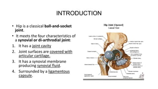
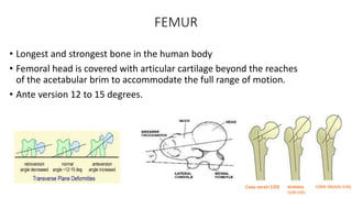
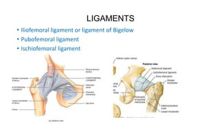
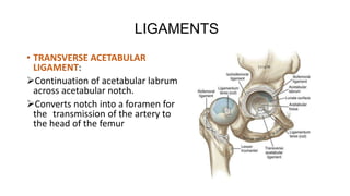
ANATOMY, BIOMECHANICS &
PRINCIPLES of HIP JOINT
Dr.M.KUMAR BHANU
Dept of orthopaedics
KIMS ,narketpally.
INTRODUCTION
• Hip is a classical ball-and-socket
joint.
• It meets the four characteristics of
a synovial or di-arthrodial joint:
1. It has a joint cavity
2. Joint surfaces are covered with
articular cartilage.
3. It has a synovial membrane
producing synovial fluid.
4. Surrounded by a ligamentous
capsule.
FEMUR
• Longest and strongest bone in the human body
• Femoral head is covered with articular cartilage beyond the reaches
of the acetabular brim to accommodate the full range of motion.
• Ante version 12 to 15 degrees.
Coxa vara(<120) NORMAL
(120-135)
COXA VALGA(>135)
LIGAMENTS
• Iliofemoral ligament or ligament of Bigelow
• Pubofemoral ligament
• Ischiofemoral ligament
LIGAMENTS
• TRANSVERSE ACETABULAR
LIGAMENT:
Continuation of acetabular labrum
across acetabular notch.
Converts notch into a foramen for
the transmission of the artery to
the head of the femur
HIP JOINT MUSCULATURE
• MOVEMENTS AT HIP JOINT: abduction/adduction flexion/extension
medial/lateral rotation
• HIP MUSCLES: Flexion 120
Extension 30
Abduction 45 to 50
Adductors 20 to 30
internal rotation 35
external rotation 45
HIP FLEXORS
Iliopsoas
Pectineus
Rectus femoris
sartorius
HIP EXTENSORS
Gluteus maximus
Semimembranosus
Semitendinosis
Biceps femoris
Add magnus(ischial part)
HIP ADDUCTORS
Adductor brevis
Adductor longus
Adductor magnus(ant part)
gracilis
HIP ABDUCTORS
Gluteus medius
Gluteus minimus
Tensor facia lata
EXTERNAL ROTATORS(SHORT)
Piriformis
Superior gemellus
Obturator internus
Inferior gemellus
Quadratus femoris
INTERNAL ROTATORS
Tensor fasciae lata
Gluteus medius
Gluteus minimus
BLOOD SUPPLY
Blood Supply to Head of Femur
• Child, obturator artery via ligamentum
teres supplies epiphysis
• Elderly, main supply via retinacular vessels from
trochanteric and cruciate anastamoses
• Medial and lateral circumflex femoral vessels
BLOOD SUPPLY
• Superior gluteal artery
• Inferior gluteal artery.
• Transverse and ascending
branches of lateral circumflex
femoral artery.
• Transverse and ascending
branch of medial circumflex
femoral
• Cruciate and trochanteric
anastomosis
LYMPHATICS
BIOMECHANICS OF HIP.
Science that deals with the study
of forces (internal or external )
acting on the living body
Forces acting on the hip
The abductor musculature ,acting
on the lever arm extending from
the lateral aspect of the greater
trochanter to the center of
femoral head ,must exert an equal
moment to the body weight arm to
hold the pelvis level
FORCES ACTING ON THE HIP
• Fig A. Lever arms acting on hip joint.
• Moment produced by body weight applied at body's center
of gravity, X, acting on lever arm, B-X,
must be counterbalanced by moment produced by
abductors, A, acting on shorter lever arm, A-B.
• Fig B. Lever arm A-B may be shorter than normal in arthritic
hip.
• Medialization of acetabulum shortens lever arm B-X,
• and use of high offset neck lengthens lever arm A-B. ,
• Fig c. Lateral and distal reattachment of osteotomized greater
trochanter lengthens lever arm A-B further and tightens
abductor musculature.
BIOMECHANICS OF HIP
• The Neck of Femur
• Angulated in relation to the shaft in
2 planes : sagittal & coronal
• Neck Shaft angle
– 140 deg at birth
– 120-135 deg in adult
• Anteversion
– Anteverted 40 deg at birth
– 12-15 deg in adults
AXIS OF LOWER LIMB
• Mechanical axis line passes
between center of hip joint and
center of ankle joint.
• Anatomic axis line is between
tip of greater trochanter to
center of knee joint.
• Angle formed between these
two is around 6 degrees.
BIOMECHANICS OF HIP.
• To maintain stable hip, torques produced by the body weight is
countered by abductor muscles pull.
• Abductor force X lever arm1 = weight X leverarm2
TRENDELENBURG GAIT
The factors influencing both the magnitude and the direction
of the compressive forces acting on the femoral head are
1) the position of the center of gravity
2) the abductor lever arm, which is a function of the neck-
shaft angle
3) the magnitude of body weight
STRATEGIES for reducing joint reaction force….
• This can be accomplished by limping, however the lateral movements
required take a considerable amount of energy and is a much less efficient
means of ambulation.
• Another strategy to reduce joint reaction force involves using a cane or
walking stick in the opposite hand.
• The moment produced from both the cane and abductor muscles
together produce a moment equal and opposite to that produced by the
effective body weight.
• The two-dimensional static analysis indicates that the joint reaction force
can be reduced by 50%(from 3 times body weight to 1.5 times body
weight)
PRINCIPLES OF THR
• Principle- to decrease joint reaction force
• Centralization of femoral head by deepening of Acetabulum
- decreases body weight lever arm
• Increase in neck length and Lateral reattachment of trochanter
- lengthens abductor lever arm
• This decreases abductor force, hence joint reaction force, & so the
wear of the implants
OFFSETS…..
• Vertical Ht (offset) Determined by the
Base length of the Prosthetic neck and
length gained by the head
• Horizontal Offset (Medial offset) center
of the head to the axis of the stem
• If medial offset is inadequate, it shortens
the moment arm ,produce limp, increase
bony impingement
• Excessive medial offset – dislocation,
increases stress on stem & cement stress
# or loosening
Thank you

Hip joint biomechanics

  • 1.
    ANATOMY, BIOMECHANICS & PRINCIPLESof HIP JOINT Dr.M.KUMAR BHANU Dept of orthopaedics KIMS ,narketpally.
  • 2.
    INTRODUCTION • Hip isa classical ball-and-socket joint. • It meets the four characteristics of a synovial or di-arthrodial joint: 1. It has a joint cavity 2. Joint surfaces are covered with articular cartilage. 3. It has a synovial membrane producing synovial fluid. 4. Surrounded by a ligamentous capsule.
  • 3.
    FEMUR • Longest andstrongest bone in the human body • Femoral head is covered with articular cartilage beyond the reaches of the acetabular brim to accommodate the full range of motion. • Ante version 12 to 15 degrees. Coxa vara(<120) NORMAL (120-135) COXA VALGA(>135)
  • 4.
    LIGAMENTS • Iliofemoral ligamentor ligament of Bigelow • Pubofemoral ligament • Ischiofemoral ligament
  • 5.
    LIGAMENTS • TRANSVERSE ACETABULAR LIGAMENT: Continuationof acetabular labrum across acetabular notch. Converts notch into a foramen for the transmission of the artery to the head of the femur
  • 6.
    HIP JOINT MUSCULATURE •MOVEMENTS AT HIP JOINT: abduction/adduction flexion/extension medial/lateral rotation • HIP MUSCLES: Flexion 120 Extension 30 Abduction 45 to 50 Adductors 20 to 30 internal rotation 35 external rotation 45
  • 7.
    HIP FLEXORS Iliopsoas Pectineus Rectus femoris sartorius HIPEXTENSORS Gluteus maximus Semimembranosus Semitendinosis Biceps femoris Add magnus(ischial part) HIP ADDUCTORS Adductor brevis Adductor longus Adductor magnus(ant part) gracilis
  • 8.
    HIP ABDUCTORS Gluteus medius Gluteusminimus Tensor facia lata EXTERNAL ROTATORS(SHORT) Piriformis Superior gemellus Obturator internus Inferior gemellus Quadratus femoris INTERNAL ROTATORS Tensor fasciae lata Gluteus medius Gluteus minimus
  • 9.
    BLOOD SUPPLY Blood Supplyto Head of Femur • Child, obturator artery via ligamentum teres supplies epiphysis • Elderly, main supply via retinacular vessels from trochanteric and cruciate anastamoses • Medial and lateral circumflex femoral vessels
  • 10.
    BLOOD SUPPLY • Superiorgluteal artery • Inferior gluteal artery. • Transverse and ascending branches of lateral circumflex femoral artery. • Transverse and ascending branch of medial circumflex femoral • Cruciate and trochanteric anastomosis
  • 11.
  • 12.
    BIOMECHANICS OF HIP. Sciencethat deals with the study of forces (internal or external ) acting on the living body Forces acting on the hip The abductor musculature ,acting on the lever arm extending from the lateral aspect of the greater trochanter to the center of femoral head ,must exert an equal moment to the body weight arm to hold the pelvis level
  • 13.
    FORCES ACTING ONTHE HIP • Fig A. Lever arms acting on hip joint. • Moment produced by body weight applied at body's center of gravity, X, acting on lever arm, B-X, must be counterbalanced by moment produced by abductors, A, acting on shorter lever arm, A-B. • Fig B. Lever arm A-B may be shorter than normal in arthritic hip. • Medialization of acetabulum shortens lever arm B-X, • and use of high offset neck lengthens lever arm A-B. , • Fig c. Lateral and distal reattachment of osteotomized greater trochanter lengthens lever arm A-B further and tightens abductor musculature.
  • 14.
    BIOMECHANICS OF HIP •The Neck of Femur • Angulated in relation to the shaft in 2 planes : sagittal & coronal • Neck Shaft angle – 140 deg at birth – 120-135 deg in adult • Anteversion – Anteverted 40 deg at birth – 12-15 deg in adults
  • 15.
    AXIS OF LOWERLIMB • Mechanical axis line passes between center of hip joint and center of ankle joint. • Anatomic axis line is between tip of greater trochanter to center of knee joint. • Angle formed between these two is around 6 degrees.
  • 16.
    BIOMECHANICS OF HIP. •To maintain stable hip, torques produced by the body weight is countered by abductor muscles pull. • Abductor force X lever arm1 = weight X leverarm2
  • 17.
    TRENDELENBURG GAIT The factorsinfluencing both the magnitude and the direction of the compressive forces acting on the femoral head are 1) the position of the center of gravity 2) the abductor lever arm, which is a function of the neck- shaft angle 3) the magnitude of body weight
  • 18.
    STRATEGIES for reducingjoint reaction force…. • This can be accomplished by limping, however the lateral movements required take a considerable amount of energy and is a much less efficient means of ambulation. • Another strategy to reduce joint reaction force involves using a cane or walking stick in the opposite hand. • The moment produced from both the cane and abductor muscles together produce a moment equal and opposite to that produced by the effective body weight. • The two-dimensional static analysis indicates that the joint reaction force can be reduced by 50%(from 3 times body weight to 1.5 times body weight)
  • 19.
    PRINCIPLES OF THR •Principle- to decrease joint reaction force • Centralization of femoral head by deepening of Acetabulum - decreases body weight lever arm • Increase in neck length and Lateral reattachment of trochanter - lengthens abductor lever arm • This decreases abductor force, hence joint reaction force, & so the wear of the implants
  • 20.
    OFFSETS….. • Vertical Ht(offset) Determined by the Base length of the Prosthetic neck and length gained by the head • Horizontal Offset (Medial offset) center of the head to the axis of the stem • If medial offset is inadequate, it shortens the moment arm ,produce limp, increase bony impingement • Excessive medial offset – dislocation, increases stress on stem & cement stress # or loosening
  • 21.