• The word‘Pathology’ is derived from two Greek words
—pathos meaning suffering, and logos meaning
study. Pathology is, thus, scientific study of structure
and function of the body in disease; or in other words,
pathology consists of the abnormalities that occur in
normal anatomy (including histology) and physiology
owing to disease. Another commonly used term with
reference to study of diseases is ‘pathophysiology’
comprised by two words: patho=suffering;
physiology=study of normal function. Pathophysiology,
thus, includes study of disordered function or
breakdown of homeostasis in diseases.
3.
• Pathologists arethe diagnosticians of disease.
Therefore, knowledge and understanding of
pathology is essential for all would-be doctors,
general medical practitioners and specialists since
unless they know the causes, mechanisms, nature
and type of disease, and understand the language
spoken by the pathologist in the form of laboratory
reports, they would not be able to institute
appropriate treatment or suggest preventive
measures to the patient
4.
SUBDIVISIONS OF PATHOLOGY
•Depending upon the species studied:
• human pathology
• Plant pathology
• Animal pathology
• Veterinary pathology
• Poultry pathology
• Comparative pathology: studies of diseases in
animals in comparison with those found in man
5.
• Human pathologydivided into
• General pathology-general principles of disease
• Systemic pathology –diseases pertaining specific
systems
6.
Subspecialities:
• A. Histopathology: Anatomic pathology,
pathologic anatomy, morbid anatomy. Structural
changes observed by naked eye exam referred to
as gross or macroscopic changes and the changes
detected by light and electron microscopy:
surgical pathology, forensic pathology,
cytopathology (exfoliative cytology, fine needle
aspiration cytology
7.
• B. Haematologydiseases of blood
• C. Chemical pathology: Analysis of biochemical
constituents of blood , urine, semen, CSF and other
body fluids
• D. Immunology
• E. Experimental pathology . Production of disease in
the experimental animal and its study
• F. Geographic pathology. Study of differences in
distribution of frequency and type of diseases in
populations in different parts of the world
8.
• G. MedicalGenetics: Studies relationship between
heredity and disease
• H. Molecular pathology: Detection and diagnosis of
abnormalities at the level of DNA
9.
TECHNIQUES FOR
THE STUDYOF PATHOLOGY
Autopsy pathology
• Block extraction of abdominal and thoracic organs
• Insitu organ by organ dissection
• Mini autopsy or limited autopsy where a particular
organ specific disease is suspected
10.
• Purpose ofautopsy.
• 1. Quality assurance of patient care by:
• i) confirming the cause of death;
• ii) establishing the final diagnosis; and
• iii) study of therapeutic response to treatment.
11.
• 2. Educationof the entire team involved in patient
care by:
• i) making autopsy diagnosis of conditions which are
often missed clinically e.g. pneumonia, pulmonary
• embolism, acute pancreatitis, carcinoma prostate;
• ii) discovery of newer diseases made at autopsy
e.g. Reye’s syndrome, Legionnaire’s disease, severe
acute respiratory syndrome (SARS);
12.
• Declining autopsyrate throughout world in the
recent times is owing to the following reasons:
1. Higher diagnostic confidence made possible by
advances in imaging techniques e.g. CT, MRI,
angiography etc.
2. Physician’s fear of legal liability on being wrong.
13.
• SURGICAL PATHOLOGY
•Request forms filling : Patient ID, history, physical
and operative findings , results of other relevant
investigations
• TISSUE ACCESSION. The laboratory staff receiving
the biopsy specimen must always match the ID of
the patient on the request form with that on the
specimen container
14.
• GROSS ROOM.Gross examination of the specimen
received in the laboratory is the next most
important step. Proper gross tissue cutting, gross
description and selection of representative tissue
sample in larger specimens
15.
• HISTOPATHOLOGY LABORATORY.Tissue cassettes
along with unique number given in the gross room
to the tissue sample is carried throughout laboratory
procedures. Staining and observation under
microscope.
16.
• SURGICAL PATHOLOGYREPORT. The final and the
most important task of pathology laboratory is
issuance of a prompt, accurate, brief, and
prognostically significant report. The ideal report
must contain five aspects:
• i) History (as available to the pathologist including
patient’s identity).
ii) Precise gross description.
iii) Brief microscopic findings.
• iv) Morphologic diagnosis
v) Additional comments in some cases.
17.
SPECIAL STAINS
• InH & E staining, haematoxylin stains nuclei and
eosin is used as counterstain for cytoplasm and
various extracellular material.
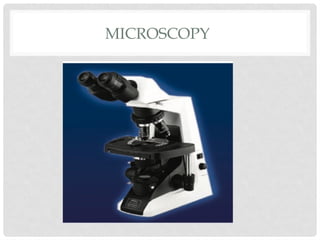
• LIGHT MICROSCOPY.The usual type of microscope
used in clinical laboratories is called light
microscope. In general, there are two types of light
microscopes:
• Simple microscope- a simple hand magnifying lens
• Compound microscope –has a battery of lenses
20.
IMMUNOFLORESCENCE
• Technique employedto localise antigenic
molecules on the cells by microscopic examination.
• This is done by using specific antibody against the
antigenic molecule forming antigen-antibody
complex at the specific antigenic site
CYTOGENETICS
• Human somaticcell are diploid containing 46
chromosomes
• 22 pair autosomes
• 1 pair sex chromosome XX,XY
• Gametes : sperm and ova , are haploid as they
contain 23 chromosomes
23.
DIAGNOSTIC MOLECULAR PATHOLOGY
•IN SITU HYBRIDISATION. In situ hybridisation (ISH) is a
molecular hybridisation technique which allows
localisation of nucleic acid sequence directly in the
intact cell (i.e. in situ) without DNA extraction
• FILTER HYBRIDISATION. In this method, target DNA or
RNA is extracted from the tissue, which may either
be fresh, frozen and unfixed tissue, or formalin-fixed
paraffin- embedded tissue


![CASE_PRESENTATION_ON_subdural_hematoma(SDH)[1 FINAL PPT]-1.pptx](https://cdn.slidesharecdn.com/ss_thumbnails/casepresentationonsubduralhematomasdh1finalppt-1-260129172522-d405d375-thumbnail.jpg?width=640&height=640&fit=bounds)
