EDEMA
• 60% ofbody weight is
composed of water
• 2/3 --> intracellular.
• Most remaining is found as
interstitial fluid
• 5% of the body’s water is in
blood plasma.
•Accumulation of fluid
within the cells, interstitial
tissue, and body cavities.
5.
• Fluid movementbetween the vascular and interstitial spaces is
governed by two opposing forces
• The vascular hydrostatic pressure and the colloid osmotic pressure
• Only a small fluid remains in the interstitial space.
• This is drained by lymphatic vessels.
6.
Extravascular fluid
(little proteinor pressure)
NORMAL REGULATION OF MICROCIRCULATION
Hydrostatic pressure at the arteriolar end
is high, at the venular end it is low.
Osmotic pressure > hydrostatic pressure
in the venular end.
Therefore, only a small amount of fluid is
retained in the interstitium (drained by
lymphatics).
7.
- Subcutaneous edemais important to recognize because it
signals potential underlying cardiac or renal disease.
8.
Two factors determinethe
movement of fluid between the
vessel and the tissue:
1) Intravascular hydrostatic
pressure
2) Plasma colloid pressure
Fluid will accumulate in the
interstitium,
1) If hydrostatic pressure is
increased.
2) If intravascular colloid oncotic
pressure decreases.
Mechanisms of edema
9.
Important concepts andterms
■ Transudate: Protein and cell-poor fluid that has a specific gravity < 1.012.
• Cardiac failure or decreased protein levels cause a transudate.
■ Exudate: Protein and cell-rich fluid that has a specific gravity > 1.020.
• Inflammation causes an exudate.
■ Pitting edema: When the skin and underlying soft tissues of a leg with
edema are compressed with fingers, the impressions remain.
• Most commonly associated with heart failure.
■ Anasarca: Generalized edema of the entire body that is most commonly
seen in glomerular diseases (caused by protein loss)
10.
Pitting edema: fingerpressure over edematous tissue displaces the
interstitial fluid and leaves a finger-shaped depression.
12.
Causes of
increased
vascular
hydrostatic
pressure
■ Heartfailure:
• If heart cannot pump blood as effectively
as it should.
• There will be pooling of blood in the veins.
■ Cirrhosis:
• Fibrous scarring impairs return of blood
through the portal vein
This causes fluid to leak into the peritoneal
cavity.
■ Venous obstruction:
• Tumor pushing on a vein will cause back up
of blood
• This will cause leakage of fluid into
the interstitium.
Causes of
decreased
oncotic
pressure
■ Decreasedproduction of albumin by the liver (e.g., in
cirrhosis /or liver damage).
• Plasma osmotic pressure (=oncotic pressure) will decrease.
■ Increased loss of protein by the kidney (glomerular
diseases) or in the gut (protein-losing
gastroenteropathy).
■ Malnutrition.
15.
OTHER CAUSES
OF EDEMA
3.Obstruction in lymphatics
• 1.Inflamation
• 2.Neoplasia
• 3. After surgery
• 4. After radiotherapy
4. Sodium retention
• Kidney failure and excessive salt
intake
• Increased sodium reabsorption in
tubules
5. Inflammation
• 1. Acute
• 2. Chronic
16.
????
What isedema?
Which pressure changes are responsible of edema?
What is transuda, exudate, pitting edema, anasarca?
What are the causes of edema?
What are the disease examples to increased
hydrostatic pressure and decreased oncotic pressure?
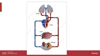
HYPEREMIA
AND
CONGESTION
Hyperemia and congestionmeans an
increase in local blood flow in the tissue.
Active accumulation of blood within vessels,
(as seen in vasodilation due to acute inflammation)
hyperemia
Passive accumulation of blood within vessels,
( such as seen in the lungs due to left-sided heart
failure,) or (in the liver due to right-sided heart
failure) congestion
20.
In hyperemia, sincethe
increased inflow is well
oxygenated erythema
occur.
In congestion, venous outflow
decreases and venous blood is
deoxygenated cyanosis occur.
Chronic passive congestion-lung
•Cause: Left-sided
heart failure, which
causes blood to back
up into the lungs
• Left ventricle cannot
pump the blood out
efficiently
• Grossly; darkly pigmented,
heavy and firm lungs.
• Hemosiderin in macrophages
(“heart failure cells”) &
• fibrosis of the alveolar septae
Examples to conjestion
24.
Chronic passive congestion-
liver
Cause:Right-sided heart
failure
Right ventricle cannot collect
the blood in efficiently
Macroscopic examination:
• Red, brown, depressed
(from cell loss as a result of
ischemic necrosis)
• It is called "Nutmeg (small
coconut) Liver"
Examples to conjestion
25.
Right heart failure:Chronic passive congestion in the central
parts of liver lobules.
26.
• What arethe reasons of hyperemia and
conjestion? What is the difference?
• What are the disease examples to conjestion
and hyperemia?
?????
Bleeding
• Bleeding isthe outflow of blood as a result of a rupture of the blood
vessel.
• Causes are ; trauma, atherosclerosis, inflammation of the vascular
wall, neoplastic erosion etc.
• Accumulation of bleeding in the tissue is called a hematoma.
• Small bleeding in the skin, mucosa and serosae are called petechiae, some
large ones are purpura, and those larger than 1-2 cm are called
ecchymosis.
• Bleeding can result in hemorrhagic shock !!
30.
What is bleeding?
Whatis heematoma, purpura, petechia,
ecchymosis?
What may be the complication of bleeding?
?????
HEMOSTASIS
AND
TROMBOSIS
Hemostasis:
• Physiologic coagulationof
blood with the purpose of
preventing bleeding.
Thrombosis:
• Pathologic coagulation of blood
resulting in the formation of a
solid mass within a chamber of
the heart or within a blood vessel.
33.
NORMAL
HEMOSTASIS
In case ofbleeding, the
following steps occur
respectively;
• Short-term vasoconstriction
(spasm) occurs.
• Primary hemostasis
• Secondary hemostasis
• Fibrinolysis
34.
NORMAL
HEMOSTASIS
In case ofbleeding, the following
steps occur respectively;
• If endothelium is injured or
ruptured somehow
• A short-term vasoconstriction
(spasm) occurs.
35.
NORMAL HEMOSTASIS
• Primaryhemostasis: When ECM is
exposed to platelets, they are
activated and aggregate to form a
hemostatic plug.
• Secondary hemostasis: Tissue factor is
released in the area of damage. The
coagulation cascade begins, eventually
fibrin is formed and accumulates.
• The opposite regulatory mechanisms
act and fibrin is destroyed
(fibrinolysis) to prevent thrombus
formation and occlusion of the
vessel.
Factors predisposing tothrombus formation
(Virchow triad)
■ Stasis of blood (e.g., due to congestive heart failure, obesity,
immobilization).
Stasis is a particularly common predisposing condition in patients
who develop venous thrombi.
■ Hypercoagulability: Hypercoagulable states are important in the
development of thrombi in any location ( hereditary or acquired).
■ Endothelial damage: Endothelial damage plays a major role in
many arterial thrombi.
38.
TROMBUS
• It canbe arterial or venous.
• Arterial thrombosis;
• coronary, cerebral arteries and femoral arteries.
They mostly sit on the atheroma plate.
• Venous thrombosis (phlebotrombosis);
• is often in the lower limbs.
• If the thrombus breaks, embolism may develop.
39.
What are thefactors causing abnormal
blood flow;
• Hypertension
• Ulcerous atherosclerotic plaques
• Aneurysms the aorta and other artery
• Myocardial infarction
• Mitral valve stenosis-atrial fibrillation
• Hyperviscosity syndromes (polistemia vera)
• Sickle cell anemia
40.
Hereditary conditions predisposingto thrombosis
■ Factor V Leiden mutation:
A mutation in the factor V gene is responsible for hypercoagulability. (protein C
cannot cleave and activate factor V).
• The incidence of factor V Leiden mutations is 2–15% of the Caucasian population.
■ Prothrombin gene mutation:
Causes an elevated level of prothrombin.
• Patients who have this mutation have a threefold risk of having venous
thromboses.
• The incidence is 1–2% of the general population.
Fates and complicationsof thrombi
• Fates of thrombi:
• Organization
• Recanalization,
• Dissolution
• Embolization.
• Complications of thrombi:
• Occlusion of the blood vessel ischemia.
• Ischemia causes cell injury and cell death
(necrosis).
43.
EMBOLISM
• Embolus isa free solid, liquid or gaseous mass that is
carried away from the source by blood flow in the vein.
• Most of the time (99%) a “thrombus” is the causative
factor.
• Trombus may leave from where it develops, carried out in
blood flow and is is stucked in a vessel lumen.
• Rare sources are oil droplets, nitrogen bubbles,
atherosclerotic residues, tumor crumbs, foreign bodies
such as bullets.
• They can block a narrow vein, leading to ischemic necrosis.
44.
PULMONARY EMBOLISM
• Themost common form of thromboembolic disease.
• 95% of venous emboli originate from deep leg vein thrombus.
• It travels to the right heart and occludes pulmonary artery branches.
• If > 60% pulmonary circulation is blocked
• sudden death, right heart failure (cor pulmonale), cardiovascular collapse may
occur.
• Multiple embolus leads to pulmonary hypertension and right heart
failure.
45.
PULMONARY EMBOLUS
Emboli thatis derived from a lower
extremity deep venous thrombosis
travels to the right side of the heart.
Afterwards, it goes to the pulmonary
artery branches and occludes them.
46.
SYSTEMIC THROMBOEMBOLISM
• Embolitraveling within systemic arterial
circulation.
• Most (80%) arise from cardiac mural thrombi.
• Major sites for arteriolar embolization are:
• 1) Lower extremities (75%)
• 2) Brain (10%)
47.
????
• What isthe difference between hemostasis and thrombosis?
• What are the steps of normal hemostasis?
• What is the reason of coagulation cascade?
• What are the predisposing factors for thrombosis? What is
Virchow triad?
• Types of thrombus?
• What is embolus? What are the sources? What is the main
complication?
• What is the examples of thromboembolus?
INFARCT
•A localized areaof dead (necrotic) cells within an organ. (infarct =
the pathologic finding; infarction =the process) .
• Mechanisms of infarct formation:
• Hypoxia and ischemia are two main mechanisms.
• Hypoxia = lack of oxygen to an organ
• Ischemia = lack of blood flow to an organ.
• Important point: «Ischemia is more damaging than hypoxia»
• In ischemia, both oxygen and nutrient delivery is impaired.
• In addition, toxic metabolites cannot be taken out.
50.
Causes of infarcts
■Obstruction of vessel:
Due to atherosclerosis, thrombi, emboli
Damage to the vessels
(e.g., trauma, neoplasms and cytomegalovirus infection)
External compression of an artery or vein (e.g., torsion of organ).
■ Generalized hypotension: As occurs in forms of shock.
51.
RED INFACT- HEMORAGIC
INFARCT
Red(hemorrhagic) infarct occurs in
•Organs drained by a single vein (over-testis),
•Loose organs (lung),
•Dual circulating organs (lung, small intestines),
•Tissues are congested due to previous venous insufficiency,
•If the ischemic area is reperfused.
52.
PALE (WHITE) INFARCT:
Causedby the occlusion of arterial
circulation in solid organs such as;
•pancreas,
•heart,
•spleen
•kidney
53.
• White splenicinfarct. This spleen
has a white (“anemic”) infarct
(arrowhead).
• White infarcts often occur in solid
organs with a single blood supply,
such as the spleen
54.
INFARCTION
• The dominanthistological
feature of infarction is
coagulation necrosis.
• There is melting
necrosis (liquefaction)
in the brain.
55.
????
• What isinfarct? Infarction?
• What is the mechanism of formation and etiology
(cause) of infarct?
• What is red and white infarct? Examples?
SHOCK (CARDIOVASCULAR COLLAPSE)
•Shock is the final common pathway for several
potentially lethal clinical events.
• Generalized hypoperfusion
of the body !!
sudden decrease in the amount of
body circulation
insufficiency of systemic blood supply
(ISCHEMIA)
İnefficient O2 supply to tissues
HYPOXIA
SHOCK
Types of shockEtiology Main mechanism
Cardiogenic Myocardial infarct
Ventricular rupture
Arrythmia
Cardiac tamponade
Pulmonary embolism
Heart can not pump blood
• myocard is damaged
• external pressure
• blockage in the outlet
Hypovolemic • Excessive bleeding
• Fluid loss, diarrhare, vomiting,
burns, travma
Loss of blood or plasma occurs
Septic • Severe microbic enfections,
endotoxic shock, gram (-) sepsis
Peripheral vasodilatation,
endothelial damage by leukocytes
activation of thrombotic &
fibrinolytic cascades DIC
(disseminated intravascular
coagulation)
61.
Types of shockClinic Main mechanism
Neurogenic Spinal damage Blood is pooled in peripheral vessels
Anaflactic Allergens Type I hypersensitivity reaction,
Systemic vasodilation
62.
CARDIOGENIC SHOCK
• Basicdescription: Failure of the heart as a pump.
Examples of causes of cardiogenic shock
■ A large myocardial infarct—damages the myocardium & heart cannot
pump effectively.
■ Acute mitral regurgitation—the heart pumps enough blood, but
much of it leaks back into the left atrium.
Clinical presentation:
*Blood pressure is
low, skin is cool
63.
HYPOVOLEMIC SHOCK
Basic description:Lack of enough blood (due to loss) to properly perfuse the
body—most commonly due to trauma.
• Clinical presentation of hypovolemic shock
• ■ If less than 20% of the body’s total blood volume is lost:
• Cool and clammy skin with increased heart rate.
• ■ If 20–40% of the body’s total blood volume is lost:
• Increased respiratory rate, possibly confusion.
• ■ If more than 40% of the body’s total blood volume is lost:
• Hypotension, oliguria.
• Preexisting heart disease may exacerbate the effects of hypovolemic shock!
64.
SEPTIC SHOCK
• Basicdescription: Generalized vascular dilation caused by an infectious
organism,
• usually due to lipopolysaccharides (LPS) in the cell wall of gram-negative bacterial
organisms such as «Escherichia coli, Pseudomonas, and Klebsiella».
• Blood pools in the venous system and peripheral vessels.
• Not enough blood returns to the heart to be pumped out.
• The mortality rate is between 25–50%.
• Risk is high in patients that have diabetes and immunodeficiency !!
65.
FEATURES AND COMPLICATIONSOF SHOCK
• Generalized hypoperfusion of organs leads to cell injury and death.
• ■ In the brain: «Global hypoxic-ischemic encephalopathy» may occur.
• Microscopy; “red” neurons (i.e., dead neurons that have red
cytoplasm and pyknotic nuclei)
• Red neurons occur in areas most prone to ischemic injury.
• Some areas in the adult brain are more vulnerable to injury (the area
between the distribution of two major cerebral arteries), hippocampus,
and cerebellum.
66.
• Red neurons.This section of the
hippocampus demonstrates “red” neurons
(arrowhead).
• Indicative of ischemic injury, such as
occurs in shock,
• these neurons have a shrunken pyknotic
nucleus, an eosinophilic cytoplasm, and a
rounded cellular outline.
67.
FEATURES AND COMPLICATIONSOF SHOCK
■ In the heart: «Subendocardial contraction band necrosis»
• myocytes traversed by darkly eosinophilic bands.
■ In the lungs: «Diffuse alveolar damage»
• proteinaceous exudates in the alveoli and hyaline membranes (i.e.,
eosinophilic “membranes” composed of protein and cellular debris)
■ In the liver: «Centrilobular necrosis»
• Centrilobular hepatocytes are last to receive oxygenated blood
• That is why they are most prone to injury from shock.
• Nutmeg liver (appearance similar to chronic passive congestion)
68.
Centrilobular necrosis.
This sectiondemonstrates the
changes of centrilobular necrosis,
Preservation of hepatocytes around
the portal tracts (arrowheads),
The centrilobular hepatocytes are
the last hepatocytes to receive
oxygenated blood;
Thus, these cells are at most risk for
injury.
69.
FEATURES AND COMPLICATIONSOF SHOCK
■ In the kidney: «Acute tubular necrosis»
• coagulative necrosis of tubular epithelial cells and dilation of tubules.
■ In the adrenal gland: «Corticomedullary hemorrhage.»
■ In the gastrointestinal system «Acute gastric hemorrhages and ulcers»
• «intestinal ischemia» occurs at borderzone areas between the distribution of
major vessels,
• commonly in the regions of the cecum and splenic flexure.
70.
• Shock-induced injuryof the stomach.
• The gastric mucosa contains
innumerable punctate hemorrhages
(red-black spots).
71.
STAGES OF SHOCK
■Compensated: Although the organs are hypoperfused, they are still able to
maintain homeostasis without injury.
■ Progressive: Organs can no longer maintain homeostasis and organ
damage begins to occur.
■ Irreversible: Irreversible organ damage has occurred. Even if the source of
the shock is eliminated (e.g., a transfusion to correct blood loss secondary
to trauma), the organs cannot repair themselves.
72.
???
What isshock?
What are the causes ? Types? Etiology? Pathogenesis?
Stages of shock?