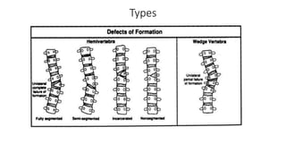
Hemivertebra
Dr.Simranjeet Kaur (PT)
• Segmentation and formation defects in vertebrae cause congenital
deformities of the spine: scoliosis, kyphosis, and kyphoscoliosis ,
Hemivertebrae
• Incidence : about 0.1–1.0% in live births
•Hemivertebrae (HV) is a condition where one half of the vertebra
completely fails to form.
•It is the most common etiology of congenital scoliosis, lordosis or
kyphosis ( fully segmented, partially segmented, or unsegmented and
are more common in females.
•Exact etiology is unclear and involves both genetic and environmental
factors
•
•Hemivertebrae are the most frequent cases of congenital deformity, and
their growth potential is similar to a normal vertebra, creating a wedge-
shaped deformity on both the sagittal and frontal planes, which can
progress during spine growth
• Fully segmented no incarcerated hemivertebrae with normal growth
plate are considered a more likely cause of progressing deformity,
particularly when associated with contra lateral bone bar. However,
even semi-segmented hemivertebrae can cause significant deformity.
Hemivertebrae location is significant in deformity progression risk, as
in congenital scoliosis by hemivertebrae of the mobile spine at the
lumbar, thoracolumbar, cervical, and thoracocervical level levels, and
it is more likely to progress than hemivertebrae at the thoracic level,
where the thoracic cage could limit deformity progression. In
congenital kyphosis, high thoracic location is associated with the
worst progression risk
Types
• The type and location of HV can affect the likelihood and the rate of
progression . The degree of spine deformity tends to worsen as the
child grows ( 5 ). The rate at which scoliosis or spine deformity
develops depends on many factors including the type of HV, number
and position of HV in the spine, ipsilateral v/s bilat eral in case of
multiple HV and age of the patient
• This occurs very early, from the fifth to the eighth week of gestation,
and it is frequently associated with other pathological conditions, like
congenital kidney disorders, congenital heart disease, and spinal cord
dysraphism
• Antenatal 3D ultrasound is potentially a useful modality to detect the
exact location, type, number of affected vertebra and associated
anomalies and hence determine
• Even though most HV occur in isolation, a significant number could be
associated with GIT, CNS, renal, other vertebral and cardiovascular
anomalies. Segmentation anomalies may be associated with syrinx,
diastematomyelia and te thered cord ( They can also be part of
various genetic syndromes: Jarcho10 Levin syndrome (AR, fused
vertebrae, scoliosis, abnormal rib alignment), Klippel). Fiel syndrome
(AR/ AD, fused cervical vertebrae), VACTERL (sporadic) and OEIS
complex (sporadic; omphalocele, cloacal exstrophy, imperforate anus
and spinal defects) . The mainstay of treatment remains early
diagnosis and intervention before severe curvature and deformity
occur.
Clinical Features
• Tilted pelvis
• Difficulty walking
• Difficulty breathing
• Abnormal curvature or twisting in the back, left or right, forward or
backward
• Uneven shoulders, hips, waist or legs
oManagement of a Neonate with Antenatal / Postnatal Diagnosis of HV
Neonate with antenatal or postnatal diagnosis of HV 1. Thorough
review of antenatal scans, foetal MRI (if available) and family history
Careful general and systemic examination Normal examination (no
dysmorphism, systemic exam normal)
• Abnormal examination (dysmorphism or abnormal systemic
examination) Complete spine x ray (AP and lateral) Consider spinal
and renal US Genetic referral and chromosomal studies Complete
spine x ray (AP and lateral views) Further investigations based on
abnormal system (Echo, cranial US, spinal US, renal US) MRI spine if
abnormal spine USS 1. Orthopaedic referral before discharge 2.
*Parental counselling by consultant or SR about possibility of long
term follow up and scoliosis depending on location of HV and other
anomalies
• Bracing is ineffective in limiting deformity progression and with
potential risks of neurologic impairment in the case of Kyhosis and
kyphoscoliosis,4–6 surgery is proposed to treat progressive deformity.
• Surgery :Hemivertebra Resection and Spinal Arthrodesis
• For treating congenital scoliosis caused by hemivertebra posterior
hemivertebra, resection and monosegmental fusion appears to be
effective. This treatment results in an excellent correction in both the
frontal and sagittal planes. Early surgery is typically prescribed as a
treatment for children with congenital scoliosis, even though there is
little evidence for its long term results.

Hemivertebra.pptx

  • 1.
  • 2.
    • Segmentation andformation defects in vertebrae cause congenital deformities of the spine: scoliosis, kyphosis, and kyphoscoliosis , Hemivertebrae • Incidence : about 0.1–1.0% in live births
  • 3.
    •Hemivertebrae (HV) isa condition where one half of the vertebra completely fails to form. •It is the most common etiology of congenital scoliosis, lordosis or kyphosis ( fully segmented, partially segmented, or unsegmented and are more common in females. •Exact etiology is unclear and involves both genetic and environmental factors • •Hemivertebrae are the most frequent cases of congenital deformity, and their growth potential is similar to a normal vertebra, creating a wedge- shaped deformity on both the sagittal and frontal planes, which can progress during spine growth
  • 4.
    • Fully segmentedno incarcerated hemivertebrae with normal growth plate are considered a more likely cause of progressing deformity, particularly when associated with contra lateral bone bar. However, even semi-segmented hemivertebrae can cause significant deformity. Hemivertebrae location is significant in deformity progression risk, as in congenital scoliosis by hemivertebrae of the mobile spine at the lumbar, thoracolumbar, cervical, and thoracocervical level levels, and it is more likely to progress than hemivertebrae at the thoracic level, where the thoracic cage could limit deformity progression. In congenital kyphosis, high thoracic location is associated with the worst progression risk
  • 5.
  • 6.
    • The typeand location of HV can affect the likelihood and the rate of progression . The degree of spine deformity tends to worsen as the child grows ( 5 ). The rate at which scoliosis or spine deformity develops depends on many factors including the type of HV, number and position of HV in the spine, ipsilateral v/s bilat eral in case of multiple HV and age of the patient
  • 7.
    • This occursvery early, from the fifth to the eighth week of gestation, and it is frequently associated with other pathological conditions, like congenital kidney disorders, congenital heart disease, and spinal cord dysraphism • Antenatal 3D ultrasound is potentially a useful modality to detect the exact location, type, number of affected vertebra and associated anomalies and hence determine
  • 10.
    • Even thoughmost HV occur in isolation, a significant number could be associated with GIT, CNS, renal, other vertebral and cardiovascular anomalies. Segmentation anomalies may be associated with syrinx, diastematomyelia and te thered cord ( They can also be part of various genetic syndromes: Jarcho10 Levin syndrome (AR, fused vertebrae, scoliosis, abnormal rib alignment), Klippel). Fiel syndrome (AR/ AD, fused cervical vertebrae), VACTERL (sporadic) and OEIS complex (sporadic; omphalocele, cloacal exstrophy, imperforate anus and spinal defects) . The mainstay of treatment remains early diagnosis and intervention before severe curvature and deformity occur.
  • 11.
    Clinical Features • Tiltedpelvis • Difficulty walking • Difficulty breathing • Abnormal curvature or twisting in the back, left or right, forward or backward • Uneven shoulders, hips, waist or legs
  • 12.
    oManagement of aNeonate with Antenatal / Postnatal Diagnosis of HV Neonate with antenatal or postnatal diagnosis of HV 1. Thorough review of antenatal scans, foetal MRI (if available) and family history Careful general and systemic examination Normal examination (no dysmorphism, systemic exam normal) • Abnormal examination (dysmorphism or abnormal systemic examination) Complete spine x ray (AP and lateral) Consider spinal and renal US Genetic referral and chromosomal studies Complete spine x ray (AP and lateral views) Further investigations based on abnormal system (Echo, cranial US, spinal US, renal US) MRI spine if abnormal spine USS 1. Orthopaedic referral before discharge 2. *Parental counselling by consultant or SR about possibility of long term follow up and scoliosis depending on location of HV and other anomalies
  • 13.
    • Bracing isineffective in limiting deformity progression and with potential risks of neurologic impairment in the case of Kyhosis and kyphoscoliosis,4–6 surgery is proposed to treat progressive deformity. • Surgery :Hemivertebra Resection and Spinal Arthrodesis • For treating congenital scoliosis caused by hemivertebra posterior hemivertebra, resection and monosegmental fusion appears to be effective. This treatment results in an excellent correction in both the frontal and sagittal planes. Early surgery is typically prescribed as a treatment for children with congenital scoliosis, even though there is little evidence for its long term results.