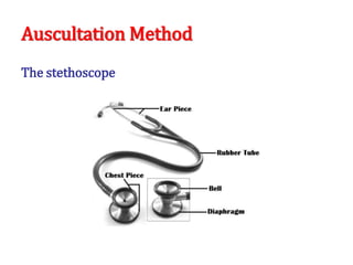
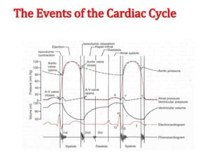
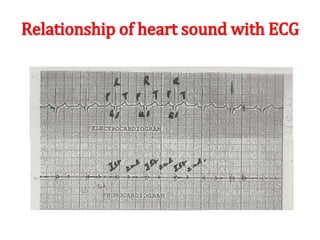
HEART SOUNDS
Objectives
• To understand why the different heart sounds are
produced.
• To know the sites at which heart sounds are best
recorded.
• To recognize the value of phonocardiography.
Auscultation Method
The stethoscope
The position of the patient
• The heart should be auscultated when the patient
is in the following positions:
• Supine.
• Left lateral.
• Sitting.
Areas of auscultation
• The mitral area (apex): This is found in the left 5th
intercostal space, approximately 1 cm medial to
the mid-clavicular line.
• The tricuspid area: This is found just to the left of
the lower border of the sternum.
• The pulmonary area: This is found in the left 2nd
intercostal space at the sternal border.
• The aortic area: This is found in the right 2nd
intercostal space at the sternal border.
Phonocardiography
Phonocardiography is the sensitive technique, by
which a recording can be made of all four heart
sounds by placing a transducer on specific areas of
auscultation.
First heart sound (S1)
• It is always normal. It sounds as “lub”. It is also called
S1.
• It is usually prolonged, but dull in nature.
• It is caused by the closure of AV valves.
• It is best heard when auscultated at mitral and
tricuspid areas.
• It occurs at the beginning of ventricular systole in
relation to cardiac cycle.
• It occurs just after QRS complex if we relate it to ECG
• Frequency: 50-60 Htz
• Time: 0.15 sec
Second heart sound (S2)
• It is always normal. It sounds as “dub”. It is also called
S2.
• It is usually short and sharp in nature.
• It is caused by the closure of semi-lunar valves.
• It is best heard when auscultated at aortic and
pulmonary areas.
• It occurs at the beginning of ventricular diastole in
relation to cardiac cycle.
• It occurs just after T wave if we relate it to ECG.
• Frequency:80-90 Htz
• Time: 0.11 sec
Third heart sound (S3)
• It may be heard normally in children, thin adults, and
pregnant women or after exercise. It is also called S3.
• It is caused by the striking of the blood to the wall of
ventricles during rapid filling phase of ventricular
diastole.
• It occurs in the early diastole in relation to cardiac
cycle.
• Frequency: 20-30 Htz
• Time: 0.1 sec
Fourth heart sound (S4)
• It may be heard normally in older people. It is also
called S4.
• It is caused by the forceful contraction of atria.
• It occurs just before the first heart sound during late
diastole in relation to cardiac cycle.
• Frequency: < 20 Htz
Heart sounds using Phonocardiography
The Events of the Cardiac Cycle
Relationship of heart sound with ECG
Splitting of second heart sound A2-P2
• Physiologic splitting of the 2nd heart sound occurs
during deep inspiration when the A2 component
splits from the P2 component by more than 0.2
seconds.
• It is auscultated as “dub, dub” over the aortic or
pulmonary areas
Heart Murmurs
Murmurs are abnormal sounds produced due to
abnormal flow of blood through abnormal heart
valves e.g. stenosis or regurgitation.
Function of papillary muscle & Chordae tendineae
Thank you

heart sounds, auscultation , points basics

  • 1.
  • 2.
    Objectives • To understandwhy the different heart sounds are produced. • To know the sites at which heart sounds are best recorded. • To recognize the value of phonocardiography.
  • 3.
  • 4.
    The position ofthe patient • The heart should be auscultated when the patient is in the following positions: • Supine. • Left lateral. • Sitting.
  • 5.
  • 6.
  • 7.
    • The mitralarea (apex): This is found in the left 5th intercostal space, approximately 1 cm medial to the mid-clavicular line. • The tricuspid area: This is found just to the left of the lower border of the sternum. • The pulmonary area: This is found in the left 2nd intercostal space at the sternal border. • The aortic area: This is found in the right 2nd intercostal space at the sternal border.
  • 8.
    Phonocardiography Phonocardiography is thesensitive technique, by which a recording can be made of all four heart sounds by placing a transducer on specific areas of auscultation.
  • 9.
    First heart sound(S1) • It is always normal. It sounds as “lub”. It is also called S1. • It is usually prolonged, but dull in nature. • It is caused by the closure of AV valves. • It is best heard when auscultated at mitral and tricuspid areas. • It occurs at the beginning of ventricular systole in relation to cardiac cycle. • It occurs just after QRS complex if we relate it to ECG • Frequency: 50-60 Htz • Time: 0.15 sec
  • 10.
    Second heart sound(S2) • It is always normal. It sounds as “dub”. It is also called S2. • It is usually short and sharp in nature. • It is caused by the closure of semi-lunar valves. • It is best heard when auscultated at aortic and pulmonary areas. • It occurs at the beginning of ventricular diastole in relation to cardiac cycle. • It occurs just after T wave if we relate it to ECG. • Frequency:80-90 Htz • Time: 0.11 sec
  • 11.
    Third heart sound(S3) • It may be heard normally in children, thin adults, and pregnant women or after exercise. It is also called S3. • It is caused by the striking of the blood to the wall of ventricles during rapid filling phase of ventricular diastole. • It occurs in the early diastole in relation to cardiac cycle. • Frequency: 20-30 Htz • Time: 0.1 sec
  • 12.
    Fourth heart sound(S4) • It may be heard normally in older people. It is also called S4. • It is caused by the forceful contraction of atria. • It occurs just before the first heart sound during late diastole in relation to cardiac cycle. • Frequency: < 20 Htz
  • 13.
    Heart sounds usingPhonocardiography
  • 14.
    The Events ofthe Cardiac Cycle
  • 15.
    Relationship of heartsound with ECG
  • 16.
    Splitting of secondheart sound A2-P2 • Physiologic splitting of the 2nd heart sound occurs during deep inspiration when the A2 component splits from the P2 component by more than 0.2 seconds. • It is auscultated as “dub, dub” over the aortic or pulmonary areas
  • 17.
    Heart Murmurs Murmurs areabnormal sounds produced due to abnormal flow of blood through abnormal heart valves e.g. stenosis or regurgitation.
  • 18.
    Function of papillarymuscle & Chordae tendineae
  • 19.