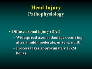
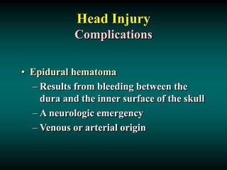
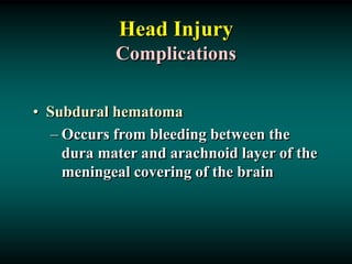
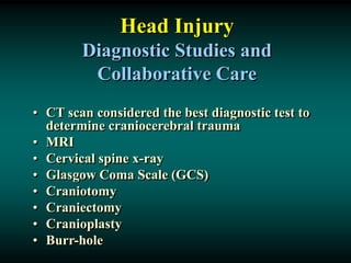
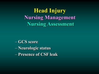
Head Injury
Head Injury
• Any trauma to the scalp, skull, or brain
• Head trauma includes an alteration in
consciousness no matter how brief
Head Injury
• Causes
– Motor vehicle accidents
– Firearm-related injuries
– Falls
– Assaults
– Sports-related injuries
– Recreational accidents
Head Injury
• High potential for poor outcome
• Deaths occur at three points in time after
injury:
– Immediately after the injury
– Within 2 hours after injury
– 3 weeks after injury
Head Injury
Types of Head Injuries
• Scalp lacerations
– The most minor type of head trauma
– Scalp is highly vascular  profuse
bleeding
– Major complication is infection
Head Injury
Types of Head Injuries
• Skull fractures
– Linear or depressed
– Simple, comminuted, or compound
– Closed or open
– Direct & Indirect
– Coup & Contrecoup
Head Injury
Types of Head Injuries
• Skull fractures
– Location of fracture alters the
presentation of the manifestations
– Facial paralysis
– Conjugate deviation of gaze
– Battle’s sign
Head Injury
Types of Head Injuries
• Basal Skull fractures
– CSF leak (extravasation) into ear (Otorrhea)
or nose (Rhinorrhea)
– High risk infection or meningitis
– “HALO Sign (Battle Sign)” on clothes of
linen
– Possible injury to Internal carotid artery
– Permanent CSF leaks possible
Battle’s Sign
Fig. 55-13
Nursing Care of Skull
Fractures
• Minimize CSF leak
– Bed flat
– Never suction orally; never insert NG tube; never use Q-Tips
in nose/ears; caution patient not to blow nose
• Place sterile gauze/cotton ball around area
• Verify CSK leak:
– DEXTROSTIX: positive for glucose
• Monitor closely: Respiratory status+++
Head Injury
Types of Head Injuries
• Minor head trauma
– Concussion
• A sudden transient mechanical head
injury with disruption of neural activity
and a change in LOC
• Brief disruption in LOC
• Amnesia
• Headache
• Short duration
Head Injury
Types of Head Injuries
• Minor head trauma
– Postconcussion syndrome
• 2 weeks to 2 months
• Persistent headache
• Lethargy
• Personality and behavior changes
Head Injury
Types of Head Injuries
• Major head trauma
– Includes cerebral contusions and
lacerations
– Both injuries represent severe trauma
to the brain
Head Injury
Types of Head Injuries
• Major head trauma
– Contusion
• The bruising of brain tissue within a focal
area that maintains the integrity of the pia
mater and arachnoid layers
– Lacerations
• Involve actual tearing of the brain tissue
• Intracerebral hemorrhage is generally
associated with cerebral laceration
Head Injury
Pathophysiology
• Diffuse axonal injury (DAI)
– Widespread axonal damage occurring
after a mild, moderate, or severe TBI
– Process takes approximately 12-24
hours
Head Injury
Pathophysiology
• Diffuse axonal injury (DAI)
– Clinical signs:
•  LOC
•  ICP
• Decerebration or decortication
• Global cerebral edema
Head Injury
Complications
• Epidural hematoma
– Results from bleeding between the
dura and the inner surface of the skull
– A neurologic emergency
– Venous or arterial origin
Head Injury
Complications
• Subdural hematoma
– Occurs from bleeding between the
dura mater and arachnoid layer of the
meningeal covering of the brain
Epidural and Subdural Hematomas
Fig. 55-15
Epidural Hematoma
Subdural Hematoma
Head Injury
Complications
• Subdural hematoma
– Usually venous in origin
– Much slower to develop into a mass
large enough to produce symptoms
– May be caused by an arterial
hemorrhage
Head Injury
Complications
• Subdural hematoma
– Acute subdural hematoma
• High mortality
• Signs within 48 hours of the injury
• Associated with major trauma (Shearing
Forces)
• Patient appears drowsy and confused
• Pupils dilate and become fixed
Head Injury
Complications
• Subdural hematoma
– Subacute subdural hematoma
• Occurs within 2-14 days of the
injury
• Failure to regain consciousness may
be an indicator
Head Injury
Complications
• Subdural hematoma
– Chronic subdural hematoma
• Develops over weeks or months after
a seemingly minor head injury
Head Injury
Diagnostic Studies and
Collaborative Care
• CT scan considered the best diagnostic test to
determine craniocerebral trauma
• MRI
• Cervical spine x-ray
• Glasgow Coma Scale (GCS)
• Craniotomy
• Craniectomy
• Cranioplasty
• Burr-hole
Head Injury
Nursing Management
Nursing Assessment
– GCS score
– Neurologic status
– Presence of CSF leak
Head Injury
Nursing Management
Nursing Diagnoses
– Ineffective tissue perfusion
– Hyperthermia
– Acute pain
– Anxiety
– Impaired physical mobility
Head Injury
Nursing Management
Planning
– Overall goals:
• Maintain adequate cerebral perfusion
• Remain normothermic
• Be free from pain, discomfort, and
infection
• Attain maximal cognitive, motor, and
sensory function
Head Injury
Nursing Management
Nursing implementation
Health Promotion
• Prevent car and motorcycle accidents
• Wear safety helmets
Head Injury
Nursing Management
Nursing implementation
Acute Intervention
• Maintain cerebral perfusion and
prevent secondary cerebral ischemia
• Monitor for changes in neurologic
status
Head Injury
Nursing Management
Nursing implementation
Ambulatory and Home Care
• Nutrition
• Bowel and bladder management
• Spasticity
• Dysphagia
• Seizure disorders
• Family participation and education
Head Injury
Nursing Management
Evaluation
Expected Outcomes
• Maintain normal cerebral perfusion
pressure
• Achieve maximal cognitive, motor, and
sensory function
• Experience no infection, hyperthermia,
or pain

Head Injury.ppt

  • 1.
  • 2.
    Head Injury • Anytrauma to the scalp, skull, or brain • Head trauma includes an alteration in consciousness no matter how brief
  • 3.
    Head Injury • Causes –Motor vehicle accidents – Firearm-related injuries – Falls – Assaults – Sports-related injuries – Recreational accidents
  • 4.
    Head Injury • Highpotential for poor outcome • Deaths occur at three points in time after injury: – Immediately after the injury – Within 2 hours after injury – 3 weeks after injury
  • 5.
    Head Injury Types ofHead Injuries • Scalp lacerations – The most minor type of head trauma – Scalp is highly vascular  profuse bleeding – Major complication is infection
  • 6.
    Head Injury Types ofHead Injuries • Skull fractures – Linear or depressed – Simple, comminuted, or compound – Closed or open – Direct & Indirect – Coup & Contrecoup
  • 7.
    Head Injury Types ofHead Injuries • Skull fractures – Location of fracture alters the presentation of the manifestations – Facial paralysis – Conjugate deviation of gaze – Battle’s sign
  • 8.
    Head Injury Types ofHead Injuries • Basal Skull fractures – CSF leak (extravasation) into ear (Otorrhea) or nose (Rhinorrhea) – High risk infection or meningitis – “HALO Sign (Battle Sign)” on clothes of linen – Possible injury to Internal carotid artery – Permanent CSF leaks possible
  • 9.
  • 10.
    Nursing Care ofSkull Fractures • Minimize CSF leak – Bed flat – Never suction orally; never insert NG tube; never use Q-Tips in nose/ears; caution patient not to blow nose • Place sterile gauze/cotton ball around area • Verify CSK leak: – DEXTROSTIX: positive for glucose • Monitor closely: Respiratory status+++
  • 11.
    Head Injury Types ofHead Injuries • Minor head trauma – Concussion • A sudden transient mechanical head injury with disruption of neural activity and a change in LOC • Brief disruption in LOC • Amnesia • Headache • Short duration
  • 12.
    Head Injury Types ofHead Injuries • Minor head trauma – Postconcussion syndrome • 2 weeks to 2 months • Persistent headache • Lethargy • Personality and behavior changes
  • 13.
    Head Injury Types ofHead Injuries • Major head trauma – Includes cerebral contusions and lacerations – Both injuries represent severe trauma to the brain
  • 14.
    Head Injury Types ofHead Injuries • Major head trauma – Contusion • The bruising of brain tissue within a focal area that maintains the integrity of the pia mater and arachnoid layers – Lacerations • Involve actual tearing of the brain tissue • Intracerebral hemorrhage is generally associated with cerebral laceration
  • 15.
    Head Injury Pathophysiology • Diffuseaxonal injury (DAI) – Widespread axonal damage occurring after a mild, moderate, or severe TBI – Process takes approximately 12-24 hours
  • 16.
    Head Injury Pathophysiology • Diffuseaxonal injury (DAI) – Clinical signs: •  LOC •  ICP • Decerebration or decortication • Global cerebral edema
  • 17.
    Head Injury Complications • Epiduralhematoma – Results from bleeding between the dura and the inner surface of the skull – A neurologic emergency – Venous or arterial origin
  • 18.
    Head Injury Complications • Subduralhematoma – Occurs from bleeding between the dura mater and arachnoid layer of the meningeal covering of the brain
  • 19.
    Epidural and SubduralHematomas Fig. 55-15 Epidural Hematoma Subdural Hematoma
  • 20.
    Head Injury Complications • Subduralhematoma – Usually venous in origin – Much slower to develop into a mass large enough to produce symptoms – May be caused by an arterial hemorrhage
  • 21.
    Head Injury Complications • Subduralhematoma – Acute subdural hematoma • High mortality • Signs within 48 hours of the injury • Associated with major trauma (Shearing Forces) • Patient appears drowsy and confused • Pupils dilate and become fixed
  • 22.
    Head Injury Complications • Subduralhematoma – Subacute subdural hematoma • Occurs within 2-14 days of the injury • Failure to regain consciousness may be an indicator
  • 23.
    Head Injury Complications • Subduralhematoma – Chronic subdural hematoma • Develops over weeks or months after a seemingly minor head injury
  • 24.
    Head Injury Diagnostic Studiesand Collaborative Care • CT scan considered the best diagnostic test to determine craniocerebral trauma • MRI • Cervical spine x-ray • Glasgow Coma Scale (GCS) • Craniotomy • Craniectomy • Cranioplasty • Burr-hole
  • 25.
    Head Injury Nursing Management NursingAssessment – GCS score – Neurologic status – Presence of CSF leak
  • 26.
    Head Injury Nursing Management NursingDiagnoses – Ineffective tissue perfusion – Hyperthermia – Acute pain – Anxiety – Impaired physical mobility
  • 27.
    Head Injury Nursing Management Planning –Overall goals: • Maintain adequate cerebral perfusion • Remain normothermic • Be free from pain, discomfort, and infection • Attain maximal cognitive, motor, and sensory function
  • 28.
    Head Injury Nursing Management Nursingimplementation Health Promotion • Prevent car and motorcycle accidents • Wear safety helmets
  • 29.
    Head Injury Nursing Management Nursingimplementation Acute Intervention • Maintain cerebral perfusion and prevent secondary cerebral ischemia • Monitor for changes in neurologic status
  • 30.
    Head Injury Nursing Management Nursingimplementation Ambulatory and Home Care • Nutrition • Bowel and bladder management • Spasticity • Dysphagia • Seizure disorders • Family participation and education
  • 31.
    Head Injury Nursing Management Evaluation ExpectedOutcomes • Maintain normal cerebral perfusion pressure • Achieve maximal cognitive, motor, and sensory function • Experience no infection, hyperthermia, or pain