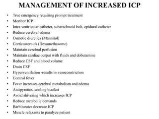
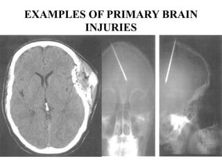
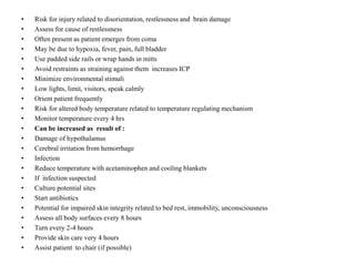
NEUROLOGICAL NURSING:
Increased ICP and Head Injury
This unit will assist learners in applying nursing knowledge to the
care of a patient with increased ICP and head injury, in an
acute care setting
OBJECTIVES
By the end of this unit, the learners will be able to:
1. Review anatomy and physiology of brain and its protective
structures.
2. Differentiate between primary and secondary head injuries
and demonstrate an understanding of the related patho
physiology.
3. Anticipate major complications that may result from head
injuries.
4. Utilize nursing process in caring for a patient, with head
injury.
INTRODUCTION:
Brain is divided into four main parts:
• Brain stem (medulla oblongata, midbrain and the Pons,
) controls breathing, heartbeat rates and reactions to
auditory and visual stimuli
• Diencephalon controls homeostasis
The diencephalon is a complex of structures within the
brain, whose major divisions are the thalamus and
hypothalamus. It functions as a relay system between
sensory input neurons and other parts of the brain, as an
interactive site for the central nervous and endocrine
systems, and works in tandem with the limbic system.
• Cerebrum controls intellectual processes and emotions
• Cerebellum- maintains body posture and balance
THE PRINCIPAL PARTS OF THE BRAIN
• Main parts: brain stem, diencephalon,
cerebrum and cerebellum
• Protection(Cranial bones, Meninges, The
meninges are three layers of connective tissue
that surround the brain and spinal cord. They
are known as: Dura mater, Arachnoid mater
and Pia mater and cerebrospinal fluid
• Ventricles: Intraventricular foramen (CSF
secretes from it, CSF acts as protection, shock
absorber and contain nutrient)
THE PRINCIPAL PARTS OF BRAIN
HEAD INJURIES

• Head injuries are one of the most common
causes of disability and death in children
• The Centers of Disease Control and Prevention
(CDC) estimates that more than 10,000
children become disabled from a brain injury
each year
• Head injuries can be defined as mild as bump
to severe in nature
PREVALENCE OF PEDIATRIC TRAUMA
• Trauma is the leading cause of death in infants and children
• Trauma is the cause of 50% of deaths in people between 5
and 34 years of age
• Motor vehicle related accidents accounts for 50% of
pediatric trauma cases
• $ 16 billion is spent annually caring for injuries to children
less than 16 years of age
• Other causes include: falls, assaults, sports injuries
• Two thirds of patients are under 30 years, most are males.
• Head injuries often cause damage to the brain from bleeding
or swelling which results in increased intracranial pressure
INTRACRANIAL PRESSURE
• The cranial skull contains three components: brain, blood,
and cerebrospinal fluid (CSF)
• The cranial skull is a closed system, and if one of the three
components increases in volume, at least one of the other
two must decrease in volume or the pressure increases. Any
bleeding or swelling within the skull increases the volume
of contents within the skull and therefore causes increases
intracranial pressure.
• Normal ICP is 10-20 mm Hg
PATHOPHYSIOLOGY
• Increased ICP- Pathophysiology the pressure increases
enough, it can cause displacement of the brain through or
against the rigid structures of the skull. This causes
restriction of blood flow to the brain, decreasing oxygen
delivery and waste removal. Cells in the brain become
anoxic and cannot metabolize properly, producing ischemia,
infarction, irreversible brain damage and eventually brain
death.
CLINICAL MANIFESTATIONS
• Depends on the severity and anatomic location of the
underlying brain injury. Localized pain usually suggests that
a fracture is present. Fracture of the cranial skull may or
may not produce swelling in the region of the fracture. But
frequently produce hemorrhage. Therefore, an x-ray is
needed for diagnosis.
EARLY SIGNS OF INCREASED ICP
Earliest sign is change in LOC:
• Slowing of speech, delay in responding to questions
• Restlessness, confusion
• Papillary changes
• Weakness in one extremity
• Headache
• Constant, aggravated by movement, increasing in intensity
LATE SIGNS OF INCREASED ICP:
• LOC, deteriorates to comatose
• Bradycardia, fluctuating to tachycardia
• Decreased respiratory rate
• Altered respiratory pattern (Cheyne-stokes)
• BP and temperature rises
• Widening pulse pressure (difference between systolic and
diastolic pressure)
• Projectile vomiting
• Decorticate or decerebrate posturing, followed by bilateral
flaccidity
• Loss of brainstem reflexes(pupils, corneal, gag, swallowing)
are continuous signs
MANAGEMENT OF INCREASED ICP
•
•
•
•
•
•
•
•
•
•
•
•
•
•
•
•
•
•

True emergency requiring prompt treatment
Monitor ICP
Intra ventricular catheter, subarachnoid bolt, epidural catheter
Reduce cerebral edema
Osmotic diuretics (Mannitol)
Corticosteroids (Dexamethasome)
Maintain cerebral perfusion
Maintain cardiac output with fluids and dobutamine
Reduce CSF and blood volume
Drain CSF
Hyperventilation -results in vasoconstriction
Control fever
Fever increases cerebral metabolism and edema
Antipyretics, cooling blanket
Avoid shivering which increases ICP
Reduce metabolic demands
Barbiturates decrease ICP
Muscle relaxants to paralyze patient
HEAD INJURY CLASSIFICATON
•
•
•
•
•
•
•
•
•

Scalp
Skull
Brain
Scalp- Trauma causes abrasion, contusion, laceration or
hematoma beneath the layers of tissue of the scalp.
Skull fracture- breaks in the continuity of the skull caused
by forceful trauma
Classified as linear, depressed or basilar
Skull fracture may be open or closed
Open- tear in the dura
Closed- dura is intact
BRAIN INJURY
• Closed- damage to brain tissue, but no opening through
skull and dura
• Open- occurs when object penetrates the skull, enters the
brain-opens the scalp, skull, dura to enter the brain.
COMMON REACTIONS:
• Minor Injury- Client rapidly regains mental function
• Concussion- temporary loss of neurologic function with brief
loss of consciousness few seconds to few minutes
• Contusion- bruising and hemorrhaging at brain surfaceunconsciousness for more than a few seconds to few minutes
• Loss of neurologic function- paralysis, speech and visual
disturbances
• Increases ICP- Brain compression
• Intracranial hemorrhage –hematomas (collection of blood) that
develops within the cranial vault- most serious of brain injuries
• Hematoma- epidural (above the dura )
• Subdural(below the dura)
• Intracerebral (within the dura)
TRAUMATIC BRAIN INJURY:

Primary brain injury: Results Secondary brain injury:
from what has occurred to the physiologic and biochemical
brain at the time of injury
events which follow the
primary injury

HEMATOMA:
• Most serious brain injury
• Collection of blood
TYPES OF HEMATOMA
• Epidural (between the skull and the dura) this can result
from a skull fracture that causes a rupture or laceration in
the artery between the skull and the dura
• Subdural (between the dura and the brain) this can cause
from the trauma, but can also occur as a result of rupture of
an aneurysm.
• Intracerebral (into the substance of the brain) result from:
systemic hypertension rupture of aneurysm- intracranial
tumors- when force is exerted to the head e.g. bullet wound
EXAMPLES OF PRIMARY BRAIN
INJURIES
FACTORS THAT EFFECT SECONDARY
BRAIN INJURIES
•
•
•
•
•
•

Blood Pressure
Oxygenation
Temperature
Control of Blood Glucose
Fluid volume status
Increased Intracranial Pressure
TRAUMATIC BRAIN INJURY
• Complications: several complications can occur
immediately or soon after a traumatic brain injury. Severe
injuries increase the risk of a greater number of
complications and more- severe complications
COMPLICATIONS
•
•
•
•
•
•
•
•
•
•
•
•
•
•

Altered consciousness
Coma
Vegetative state
Minimally conscious state.
Locked-in syndrome
Seizures
Infections
Sensory problems
Degenerative brain diseases
Nerve damage
Cognitive problems
Communication problems
Behavioral changes
Emotional changes
MANAGEMENT
•
•
•
•
•
•
•
•
•
•

Based on physical and neurological examination
X-ray
CT scan
MRI
Treatment of increased ICP
supportive measures
Ventilatory support
Fluid and electrolyte maintenance
Nutritional support
Pain and anxiety management
NURSING ASSESSMENT
• History of trauma- time, cause, direction and force of the
blow
• Loss of consciousness, duration
• Assess LOC, Glasgow coma scale
• Response to verbal commands or tactile stimuli
• Pupillary response to light
• Motor function
• Vital signs
• Monitor for signs of increased ICP
• Motor function
• Move extremities, hand grasp, pedal push, speech
NURSING DIAGNOSIS AND
INTERVENTIONS
• Ineffective airway clearance related to accumulation of
secretions and decresed LOC
• Maintain patient airway
• Suction carefully
• Discourage coughing(causes increase in ICP)
• Elevate HOB 30 degrees.
• Guard against aspiration
• Monitor ABGs to assess ventilation
• Ineffective breathing pattern related to neurological
dysfunction
• Monitor constantly for respiratory irregularities
• Cheyne stokes, hyperventilation
• Effective suctioning
• HOB 30 degrees
• Position patient lateral or semi prone
•
•
•
•
•
•
•
•
•
•
•
•
•
•
•
•

Altered cerebral tissue perfusion related to increased intracranial pressure
Position patient to reduce ICP
Head in midline position to promote venous drainage
Elevate HOB 30 degree
Avoid extreme rotation or flexion of neck
Avoid extreme hip flexion
Prevent straining
Stool softeners
High fibre diet space nursing activities
Main calm atmosphere, reduce stimuli
Risk for fluid volume deficit related to dehydration procedures and
decreased LOC
Monitor electrolytes
Brain damage can produce metabolic and hormonal dysfunctions
Monitor intake and output
Monitor IV fluids carefully
Record daily weights
• Altered nutrition related to metabolic changes,
inadequate intake
• Start enteral feedings when patient stabilized
• NG feeding unless CSF rhinorrhea
• Elevate HOB 30 degrees
• Aspirate for residual before feeding to prevent
distention and aspiration
• Use pump to regulate feeds
•
•
•
•
•
•
•
•
•
•
•
•
•
•
•
•
•
•
•
•
•
•
•
•

Risk for injury related to disorientation, restlessness and brain damage
Assess for cause of restlessness
Often present as patient emerges from coma
May be due to hypoxia, fever, pain, full bladder
Use padded side rails or wrap hands in mitts
Avoid restraints as straining against them increases ICP
Minimize environmental stimuli
Low lights, limit, visitors, speak calmly
Orient patient frequently
Risk for altered body temperature related to temperature regulating mechanism
Monitor temperature every 4 hrs
Can be increased as result of :
Damage of hypothalamus
Cerebral irritation from hemorrhage
Infection
Reduce temperature with acetaminophen and cooling blankets
If infection suspected
Culture potential sites
Start antibiotics
Potential for impaired skin integrity related to bed rest, immobility, unconsciousness
Assess all body surfaces every 8 hours
Turn every 2-4 hours
Provide skin care very 4 hours
Assist patient to chair (if possible)
GLASCOW COMA SCALE

Icp

  • 1.
    NEUROLOGICAL NURSING: Increased ICPand Head Injury This unit will assist learners in applying nursing knowledge to the care of a patient with increased ICP and head injury, in an acute care setting
  • 2.
    OBJECTIVES By the endof this unit, the learners will be able to: 1. Review anatomy and physiology of brain and its protective structures. 2. Differentiate between primary and secondary head injuries and demonstrate an understanding of the related patho physiology. 3. Anticipate major complications that may result from head injuries. 4. Utilize nursing process in caring for a patient, with head injury.
  • 3.
    INTRODUCTION: Brain is dividedinto four main parts: • Brain stem (medulla oblongata, midbrain and the Pons, ) controls breathing, heartbeat rates and reactions to auditory and visual stimuli • Diencephalon controls homeostasis The diencephalon is a complex of structures within the brain, whose major divisions are the thalamus and hypothalamus. It functions as a relay system between sensory input neurons and other parts of the brain, as an interactive site for the central nervous and endocrine systems, and works in tandem with the limbic system. • Cerebrum controls intellectual processes and emotions • Cerebellum- maintains body posture and balance
  • 4.
    THE PRINCIPAL PARTSOF THE BRAIN • Main parts: brain stem, diencephalon, cerebrum and cerebellum • Protection(Cranial bones, Meninges, The meninges are three layers of connective tissue that surround the brain and spinal cord. They are known as: Dura mater, Arachnoid mater and Pia mater and cerebrospinal fluid • Ventricles: Intraventricular foramen (CSF secretes from it, CSF acts as protection, shock absorber and contain nutrient)
  • 5.
  • 6.
    HEAD INJURIES • Headinjuries are one of the most common causes of disability and death in children • The Centers of Disease Control and Prevention (CDC) estimates that more than 10,000 children become disabled from a brain injury each year • Head injuries can be defined as mild as bump to severe in nature
  • 7.
    PREVALENCE OF PEDIATRICTRAUMA • Trauma is the leading cause of death in infants and children • Trauma is the cause of 50% of deaths in people between 5 and 34 years of age • Motor vehicle related accidents accounts for 50% of pediatric trauma cases • $ 16 billion is spent annually caring for injuries to children less than 16 years of age • Other causes include: falls, assaults, sports injuries • Two thirds of patients are under 30 years, most are males. • Head injuries often cause damage to the brain from bleeding or swelling which results in increased intracranial pressure
  • 8.
    INTRACRANIAL PRESSURE • Thecranial skull contains three components: brain, blood, and cerebrospinal fluid (CSF) • The cranial skull is a closed system, and if one of the three components increases in volume, at least one of the other two must decrease in volume or the pressure increases. Any bleeding or swelling within the skull increases the volume of contents within the skull and therefore causes increases intracranial pressure. • Normal ICP is 10-20 mm Hg
  • 9.
    PATHOPHYSIOLOGY • Increased ICP-Pathophysiology the pressure increases enough, it can cause displacement of the brain through or against the rigid structures of the skull. This causes restriction of blood flow to the brain, decreasing oxygen delivery and waste removal. Cells in the brain become anoxic and cannot metabolize properly, producing ischemia, infarction, irreversible brain damage and eventually brain death.
  • 10.
    CLINICAL MANIFESTATIONS • Dependson the severity and anatomic location of the underlying brain injury. Localized pain usually suggests that a fracture is present. Fracture of the cranial skull may or may not produce swelling in the region of the fracture. But frequently produce hemorrhage. Therefore, an x-ray is needed for diagnosis.
  • 11.
    EARLY SIGNS OFINCREASED ICP Earliest sign is change in LOC: • Slowing of speech, delay in responding to questions • Restlessness, confusion • Papillary changes • Weakness in one extremity • Headache • Constant, aggravated by movement, increasing in intensity
  • 12.
    LATE SIGNS OFINCREASED ICP: • LOC, deteriorates to comatose • Bradycardia, fluctuating to tachycardia • Decreased respiratory rate • Altered respiratory pattern (Cheyne-stokes) • BP and temperature rises • Widening pulse pressure (difference between systolic and diastolic pressure) • Projectile vomiting • Decorticate or decerebrate posturing, followed by bilateral flaccidity • Loss of brainstem reflexes(pupils, corneal, gag, swallowing) are continuous signs
  • 13.
    MANAGEMENT OF INCREASEDICP • • • • • • • • • • • • • • • • • • True emergency requiring prompt treatment Monitor ICP Intra ventricular catheter, subarachnoid bolt, epidural catheter Reduce cerebral edema Osmotic diuretics (Mannitol) Corticosteroids (Dexamethasome) Maintain cerebral perfusion Maintain cardiac output with fluids and dobutamine Reduce CSF and blood volume Drain CSF Hyperventilation -results in vasoconstriction Control fever Fever increases cerebral metabolism and edema Antipyretics, cooling blanket Avoid shivering which increases ICP Reduce metabolic demands Barbiturates decrease ICP Muscle relaxants to paralyze patient
  • 14.
    HEAD INJURY CLASSIFICATON • • • • • • • • • Scalp Skull Brain Scalp-Trauma causes abrasion, contusion, laceration or hematoma beneath the layers of tissue of the scalp. Skull fracture- breaks in the continuity of the skull caused by forceful trauma Classified as linear, depressed or basilar Skull fracture may be open or closed Open- tear in the dura Closed- dura is intact
  • 15.
    BRAIN INJURY • Closed-damage to brain tissue, but no opening through skull and dura • Open- occurs when object penetrates the skull, enters the brain-opens the scalp, skull, dura to enter the brain.
  • 16.
    COMMON REACTIONS: • MinorInjury- Client rapidly regains mental function • Concussion- temporary loss of neurologic function with brief loss of consciousness few seconds to few minutes • Contusion- bruising and hemorrhaging at brain surfaceunconsciousness for more than a few seconds to few minutes • Loss of neurologic function- paralysis, speech and visual disturbances • Increases ICP- Brain compression • Intracranial hemorrhage –hematomas (collection of blood) that develops within the cranial vault- most serious of brain injuries • Hematoma- epidural (above the dura ) • Subdural(below the dura) • Intracerebral (within the dura)
  • 17.
    TRAUMATIC BRAIN INJURY: Primarybrain injury: Results Secondary brain injury: from what has occurred to the physiologic and biochemical brain at the time of injury events which follow the primary injury HEMATOMA: • Most serious brain injury • Collection of blood
  • 18.
    TYPES OF HEMATOMA •Epidural (between the skull and the dura) this can result from a skull fracture that causes a rupture or laceration in the artery between the skull and the dura • Subdural (between the dura and the brain) this can cause from the trauma, but can also occur as a result of rupture of an aneurysm. • Intracerebral (into the substance of the brain) result from: systemic hypertension rupture of aneurysm- intracranial tumors- when force is exerted to the head e.g. bullet wound
  • 20.
    EXAMPLES OF PRIMARYBRAIN INJURIES
  • 21.
    FACTORS THAT EFFECTSECONDARY BRAIN INJURIES • • • • • • Blood Pressure Oxygenation Temperature Control of Blood Glucose Fluid volume status Increased Intracranial Pressure
  • 22.
    TRAUMATIC BRAIN INJURY •Complications: several complications can occur immediately or soon after a traumatic brain injury. Severe injuries increase the risk of a greater number of complications and more- severe complications
  • 23.
    COMPLICATIONS • • • • • • • • • • • • • • Altered consciousness Coma Vegetative state Minimallyconscious state. Locked-in syndrome Seizures Infections Sensory problems Degenerative brain diseases Nerve damage Cognitive problems Communication problems Behavioral changes Emotional changes
  • 24.
    MANAGEMENT • • • • • • • • • • Based on physicaland neurological examination X-ray CT scan MRI Treatment of increased ICP supportive measures Ventilatory support Fluid and electrolyte maintenance Nutritional support Pain and anxiety management
  • 25.
    NURSING ASSESSMENT • Historyof trauma- time, cause, direction and force of the blow • Loss of consciousness, duration • Assess LOC, Glasgow coma scale • Response to verbal commands or tactile stimuli • Pupillary response to light • Motor function • Vital signs • Monitor for signs of increased ICP • Motor function • Move extremities, hand grasp, pedal push, speech
  • 26.
    NURSING DIAGNOSIS AND INTERVENTIONS •Ineffective airway clearance related to accumulation of secretions and decresed LOC • Maintain patient airway • Suction carefully • Discourage coughing(causes increase in ICP) • Elevate HOB 30 degrees. • Guard against aspiration • Monitor ABGs to assess ventilation
  • 27.
    • Ineffective breathingpattern related to neurological dysfunction • Monitor constantly for respiratory irregularities • Cheyne stokes, hyperventilation • Effective suctioning • HOB 30 degrees • Position patient lateral or semi prone
  • 28.
    • • • • • • • • • • • • • • • • Altered cerebral tissueperfusion related to increased intracranial pressure Position patient to reduce ICP Head in midline position to promote venous drainage Elevate HOB 30 degree Avoid extreme rotation or flexion of neck Avoid extreme hip flexion Prevent straining Stool softeners High fibre diet space nursing activities Main calm atmosphere, reduce stimuli Risk for fluid volume deficit related to dehydration procedures and decreased LOC Monitor electrolytes Brain damage can produce metabolic and hormonal dysfunctions Monitor intake and output Monitor IV fluids carefully Record daily weights
  • 29.
    • Altered nutritionrelated to metabolic changes, inadequate intake • Start enteral feedings when patient stabilized • NG feeding unless CSF rhinorrhea • Elevate HOB 30 degrees • Aspirate for residual before feeding to prevent distention and aspiration • Use pump to regulate feeds
  • 30.
    • • • • • • • • • • • • • • • • • • • • • • • • Risk for injuryrelated to disorientation, restlessness and brain damage Assess for cause of restlessness Often present as patient emerges from coma May be due to hypoxia, fever, pain, full bladder Use padded side rails or wrap hands in mitts Avoid restraints as straining against them increases ICP Minimize environmental stimuli Low lights, limit, visitors, speak calmly Orient patient frequently Risk for altered body temperature related to temperature regulating mechanism Monitor temperature every 4 hrs Can be increased as result of : Damage of hypothalamus Cerebral irritation from hemorrhage Infection Reduce temperature with acetaminophen and cooling blankets If infection suspected Culture potential sites Start antibiotics Potential for impaired skin integrity related to bed rest, immobility, unconsciousness Assess all body surfaces every 8 hours Turn every 2-4 hours Provide skin care very 4 hours Assist patient to chair (if possible)
  • 31.