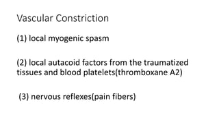
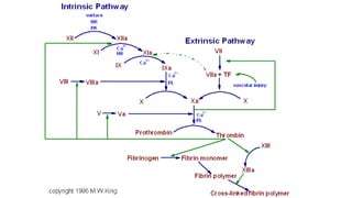
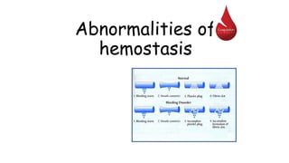
Haemostasis
by
Dr.APARNA.A
CONTENTS
• What is haemostasis
• Mechanism of haemostasis- vasoconstriction , platelet plug
formation,
clot formation, clot retraction, closure of ruptured area by growth of
fibroblasts.
-Vasoconstriction- local myospasm, local autocoid factors from
ruptured vessel and platelets, sympathetic reflex
-Temporary platelet plug
-Blood clotting mechanism- intrinsic and extrinsic mechanisms leading
to common pathway.
Hemostasis („hemo”=blood; sta=„remain”)
is the stoppage of bleeding, which is vitally
important when blood vessels are damaged.
Following an injury to blood vessels several
actions may help prevent blood loss, including:
Formation of a clot
Vascular Constriction
(1) local myogenic spasm
(2) local autacoid factors from the traumatized
tissues and blood platelets(thromboxane A2)
(3) nervous reflexes(pain fibers)
Local vasoconstriction
• is due to local spasm
of the smooth
muscle (symp.
reflex)
• can be maintained
by platelet
vasoconstrictors
Formation of
platelet aggregate
• Injured blood vessel
releases ADP, which
attracts platelets (PLT)
• PLT comming in contact
with exposed collagen
release: serotonin, ADP,
TXA2, which accelerate
vasoconstriction and
causes PLT to swell and
become more sticky
The micrograph shows activated
platelets adhering to some
damaged cells
Extrinsic pathway
• Release of tissue factor. Traumatized tissue releases a complex of
several factors called tissue factor or tissue thromboplastin.
• This factor is composed especially of phospholipids from the
membranes of the tissue plus a lipoprotein complex that functions
mainly as a proteolytic enzyme.
COMMON PATHWAY
Calcium ions
• Are required for promotion and acceleration
of almost all blood clotting reactions
• Except: activation of XII and XI (intrinsic
mechanism)
Ca2+
http://www.mhhe.com/biosci/esp/2002_general/Esp/folder_structure/tr/m1/s7/trm1s7_3.htm
Antihaemostatic mechanisms
1.Factors preventing platelet aggregation-
-Endothelial Surface Factors- sialic acid
-prostacyclin- inhibits TxA2 (aspirin - similar action -
used in MI )
2.Circulatory anticoagulants
3.Fibrinolytic system
Fibrinolysis
Clot Dissolution (extrinsic PA)
1. Plasmin is formed from plasminogen - enzyme called
activator (e.g. enzymes from urine, tears, saliva or
bacterial enzyme streptokinase)
2. Plasmin as an enzyme is involved in breaking down fibrin
into soluble fragments (fibrinolysis)
Plasminogen Plasmin
Activator (e.g. t-PA)
Fibrin soluble fragments
Plasminogen may be produced
by eosinophils
Fibrinolytic system- intrinsic PA system
Anticoagulants
Hirudo medicinalis produce
Hirudin that inhibits Thrombin
Anticoagulants
• Although tissue breakdown and platelets
destruction are normal events in the absence
of trauma, intravascular clotting does not
usually occur because:
- the amounts of procoagulants released are
very small
- natural anticoagulants are present (Antithrombin
III, Heparin, Antithromboplastin, Protein C and S,
fibrin fibers)
Natural anticoagulants
• Antithrombin III – inhibits factor X and thrombin
• Heparin from basophils and mast cells potentiates effects
of antithrombin III (together they inhibit IX, X, XI, XII
and thrombin)
• Antithromboplastin (inhibits „tissue factors” – tissue
thromboplastins)
• Protein C and S – activated by thrombin; degrade factor
Va and VIIIa
Synthetic Anticoagulants
• Heparin-
• Warfarin- vit k antagonist-inhibits synthesis of factors 2,7, 9, 10
• Decalcifying agents/calcium sequestors-
- Sodium oxolates and citrates- form insoluble salts with ca
- EDTA-ethylene diamine tetraacetic acid- chealates ca
• Defibrination substances- malaysian pit viper venom- breakdown
fibrin products by stimulating fibrinolytic system.
• Cold(5-10 degrees slows coagulation)
Abnormalities of
hemostasis
Thrombocytopenia
 Severe reduction in the
number of PLTs -
thrombocytopenia
 this causes spontaneous
bleeding as a reaction to
minor trauma
 in the skin - reddish-
purple blotchy rash
 it may result from:
 decreased production (toxins,
radiation, infection, leukemias)
 increased destruction (autoimmune
processes)
 increased PLTs consumption (DIC)
Hemorrhagic spots (petechiae)
Thrombocytopenia
• Lethal when
PLTs<10000/Cumm
• Bleeding occurs when
PLTs<50000/cumm
• Norm: 1,50,000-
400000/cumm
Hepatic failure
• Most of the clotting
factors are formed
in the liver
Subconjunctival hemorrhage
Disseminated intravascular
coagulation (DIC)
• Widespread coagulation 
thrombosis in small blood vessels
 increased fibrinolysis,
and depletion of coagulating
factors  generalized bleeding
• It may result from:
- bacterial infections
(endothelial damage)
- disseminated cancers
(release of procoagulants)
- complications of pregnancy
Disseminated cervical
cancer metastases (PET
imaging)
Hemophilia A
(lack of F VIII; 85%)
• Spontaneous or
traumatic
subcutaneous bleeding
• Blood in the urine
• Bleeding in the mouth,
lips, tongue
• Bleeding to the joints,
CNS, gastrointestinal
tract
Mild hemophilia after
injection in buttock
Haemophilia B
VON WILLEBRAND DISEASE
• defect in platelety adhesion- prolonged bleeding time.
• defect in factor 8- clotting time prolonged.
"Intrinsic" and "extrinsic"
coagulation pathways
N: 9.9 – 13 sec
Activated Partial
Thromboplastin Time
N: 25-35 sec
Prothrombin Time
Selected causes of abnormal
coagulation tests
Partial
Thromboplastin
Time (aPTT)
Prothrombin
Time (PT)
Thrombin Time
(TT)
Bleeding Time
(BT)
Factor deficiency
(except VII)
VII, X, V, II,
fibrinogen
deficiency
Low or absent
fibrinogen
Thrombocytopenia
Antibodies to
clotting factors
Antibodies Dysfibrinogenemia,
hypofibrinogenemia
Von Willebrand’s
disease
Heparin Warfarin; Vit K
defficiency (mild to
severe)
Heparin Drugs (Aspirin,
NSAIDs, high dose
penicillins, etc.)
Excessive Warfarin Excessive Heparin Cirrhosis, Uremia,
PLTs dysfunction
Haemostasis.ppt

Haemostasis.ppt

  • 1.
  • 2.
    CONTENTS • What ishaemostasis • Mechanism of haemostasis- vasoconstriction , platelet plug formation, clot formation, clot retraction, closure of ruptured area by growth of fibroblasts. -Vasoconstriction- local myospasm, local autocoid factors from ruptured vessel and platelets, sympathetic reflex -Temporary platelet plug -Blood clotting mechanism- intrinsic and extrinsic mechanisms leading to common pathway.
  • 3.
    Hemostasis („hemo”=blood; sta=„remain”) isthe stoppage of bleeding, which is vitally important when blood vessels are damaged. Following an injury to blood vessels several actions may help prevent blood loss, including: Formation of a clot
  • 4.
    Vascular Constriction (1) localmyogenic spasm (2) local autacoid factors from the traumatized tissues and blood platelets(thromboxane A2) (3) nervous reflexes(pain fibers)
  • 5.
    Local vasoconstriction • isdue to local spasm of the smooth muscle (symp. reflex) • can be maintained by platelet vasoconstrictors
  • 6.
    Formation of platelet aggregate •Injured blood vessel releases ADP, which attracts platelets (PLT) • PLT comming in contact with exposed collagen release: serotonin, ADP, TXA2, which accelerate vasoconstriction and causes PLT to swell and become more sticky
  • 7.
    The micrograph showsactivated platelets adhering to some damaged cells
  • 9.
  • 10.
    • Release oftissue factor. Traumatized tissue releases a complex of several factors called tissue factor or tissue thromboplastin. • This factor is composed especially of phospholipids from the membranes of the tissue plus a lipoprotein complex that functions mainly as a proteolytic enzyme.
  • 12.
  • 14.
    Calcium ions • Arerequired for promotion and acceleration of almost all blood clotting reactions • Except: activation of XII and XI (intrinsic mechanism) Ca2+ http://www.mhhe.com/biosci/esp/2002_general/Esp/folder_structure/tr/m1/s7/trm1s7_3.htm
  • 15.
    Antihaemostatic mechanisms 1.Factors preventingplatelet aggregation- -Endothelial Surface Factors- sialic acid -prostacyclin- inhibits TxA2 (aspirin - similar action - used in MI ) 2.Circulatory anticoagulants 3.Fibrinolytic system
  • 16.
  • 17.
    Clot Dissolution (extrinsicPA) 1. Plasmin is formed from plasminogen - enzyme called activator (e.g. enzymes from urine, tears, saliva or bacterial enzyme streptokinase) 2. Plasmin as an enzyme is involved in breaking down fibrin into soluble fragments (fibrinolysis) Plasminogen Plasmin Activator (e.g. t-PA) Fibrin soluble fragments Plasminogen may be produced by eosinophils
  • 18.
  • 19.
  • 20.
    Anticoagulants • Although tissuebreakdown and platelets destruction are normal events in the absence of trauma, intravascular clotting does not usually occur because: - the amounts of procoagulants released are very small - natural anticoagulants are present (Antithrombin III, Heparin, Antithromboplastin, Protein C and S, fibrin fibers)
  • 21.
    Natural anticoagulants • AntithrombinIII – inhibits factor X and thrombin • Heparin from basophils and mast cells potentiates effects of antithrombin III (together they inhibit IX, X, XI, XII and thrombin) • Antithromboplastin (inhibits „tissue factors” – tissue thromboplastins) • Protein C and S – activated by thrombin; degrade factor Va and VIIIa
  • 23.
    Synthetic Anticoagulants • Heparin- •Warfarin- vit k antagonist-inhibits synthesis of factors 2,7, 9, 10 • Decalcifying agents/calcium sequestors- - Sodium oxolates and citrates- form insoluble salts with ca - EDTA-ethylene diamine tetraacetic acid- chealates ca • Defibrination substances- malaysian pit viper venom- breakdown fibrin products by stimulating fibrinolytic system. • Cold(5-10 degrees slows coagulation)
  • 24.
  • 25.
    Thrombocytopenia  Severe reductionin the number of PLTs - thrombocytopenia  this causes spontaneous bleeding as a reaction to minor trauma  in the skin - reddish- purple blotchy rash  it may result from:  decreased production (toxins, radiation, infection, leukemias)  increased destruction (autoimmune processes)  increased PLTs consumption (DIC) Hemorrhagic spots (petechiae)
  • 26.
    Thrombocytopenia • Lethal when PLTs<10000/Cumm •Bleeding occurs when PLTs<50000/cumm • Norm: 1,50,000- 400000/cumm
  • 27.
    Hepatic failure • Mostof the clotting factors are formed in the liver Subconjunctival hemorrhage
  • 28.
    Disseminated intravascular coagulation (DIC) •Widespread coagulation  thrombosis in small blood vessels  increased fibrinolysis, and depletion of coagulating factors  generalized bleeding • It may result from: - bacterial infections (endothelial damage) - disseminated cancers (release of procoagulants) - complications of pregnancy Disseminated cervical cancer metastases (PET imaging)
  • 29.
    Hemophilia A (lack ofF VIII; 85%) • Spontaneous or traumatic subcutaneous bleeding • Blood in the urine • Bleeding in the mouth, lips, tongue • Bleeding to the joints, CNS, gastrointestinal tract Mild hemophilia after injection in buttock
  • 30.
  • 31.
    VON WILLEBRAND DISEASE •defect in platelety adhesion- prolonged bleeding time. • defect in factor 8- clotting time prolonged.
  • 32.
    "Intrinsic" and "extrinsic" coagulationpathways N: 9.9 – 13 sec Activated Partial Thromboplastin Time N: 25-35 sec Prothrombin Time
  • 33.
    Selected causes ofabnormal coagulation tests Partial Thromboplastin Time (aPTT) Prothrombin Time (PT) Thrombin Time (TT) Bleeding Time (BT) Factor deficiency (except VII) VII, X, V, II, fibrinogen deficiency Low or absent fibrinogen Thrombocytopenia Antibodies to clotting factors Antibodies Dysfibrinogenemia, hypofibrinogenemia Von Willebrand’s disease Heparin Warfarin; Vit K defficiency (mild to severe) Heparin Drugs (Aspirin, NSAIDs, high dose penicillins, etc.) Excessive Warfarin Excessive Heparin Cirrhosis, Uremia, PLTs dysfunction