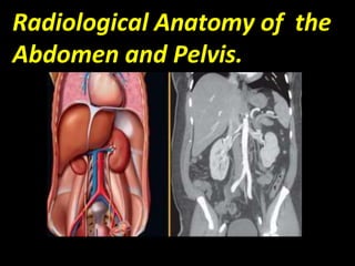
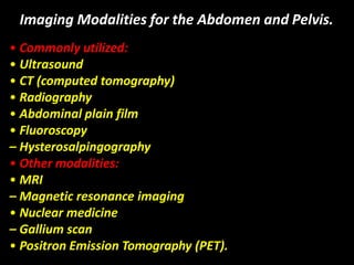
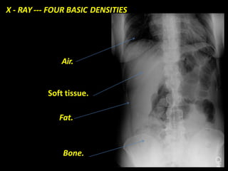
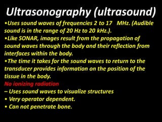
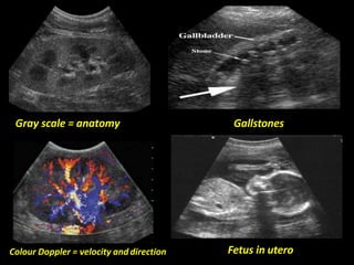
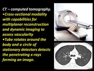
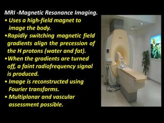
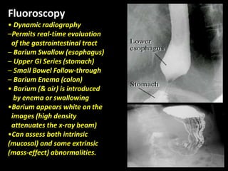
Radiological Anatomy of the
Abdomen and Pelvis.
Imaging Modalities for the Abdomen and Pelvis.
• Commonly utilized:
• Ultrasound
• CT (computed tomography)
• Radiography
• Abdominal plain film
• Fluoroscopy
– Hysterosalpingography
• Other modalities:
• MRI
– Magnetic resonance imaging
• Nuclear medicine
– Gallium scan
• Positron Emission Tomography (PET).
X - RAY --- FOUR BASIC DENSITIES
Air.
Soft tissue.
Fat.
Bone.
Ultrasonography (ultrasound)
•Uses sound waves of frequencies 2 to 17 MHz. (Audible
sound is in the range of 20 Hz to 20 kHz.).
•Like SONAR, images result from the propagation of
sound waves through the body and their reflection from
interfaces within the body.
•The time it takes for the sound waves to return to the
transducer provides information on the position of the
tissue in the body.
No ionizing radiation
– Uses sound waves to visualize structures
• Very operator dependent.
• Can not penetrate bone.
Gray scale = anatomy Gallstones
Fetus in uteroColour Doppler = velocity anddirection
CT – computed tomography.
•Cross-sectional modality
with capabilities for
multiplanar reconstruction
and dynamic imaging to
assess vascularity
•Tube rotates around the
body and a circle of
stationary detectors detects
the penetrating x-rays
forming an image.
MRI -Magnetic Resonance Imaging.
• Uses a high-field magnet to
image the body.
•Rapidly switching magnetic field
gradients align the precession of
the H protons (water and fat).
•When the gradients are turned
off, a faint radiofrequency signal
is produced.
• Image is reconstructed using
Fourier transforms.
• Multiplanar and vascular
assessment possible.
Fluoroscopy
• Dynamic radiography
–Permits real-time evaluation
of the gastrointestinal tract
– Barium Swallow (esophagus)
– Upper GI Series (stomach)
– Small Bowel Follow-through
– Barium Enema (colon)
• Barium (& air) is introduced
by enema or swallowing
•Barium appears white on the
images (high density
attenuates the x-ray beam)
•Can assess both intrinsic
(mucosal) and some extrinsic
(mass-effect) abnormalities.
Nuclear Medicine - GI Bleeding Scan
• Evaluates bleeding, particularly from the lower GI tract.
• Radiopharmaceutical = Tc99m in vitro labelled RBCs.
• Sequential 5 minute images acquired over an hour.
• Looking for progressive accumulation of tracer.
Bleeding on the cecum.
Introduction.
• The primary imaging modalities for the abdomen
and pelvis are plain film, ultrasound, and CT.
• Most common indications for imaging include
pain, trauma, distention, nausea, vomiting,
and/or change in bowel habits.
•Choice of modality depends upon clinical
symptoms, patient age & gender, and findings
on physical exam.
• Mastery of the anatomy within each quadrant
can help explain particular symptoms, clinical
presentations, and/or imaging findings.
Reading the Abdominal Plain Film.
• Also known as the
“KUB” (kidney, ureter, &
bladder).
•Use a systematic
approach to
Interpretation.
– Lung bases & diaphragms.
– Bones.
– Soft tissues.
• Abnormal calcifications.
• Organs.
Stomach
AP SUPINE ABDOMEN X-RAY GAS PATTERN.
•Colon has sacculations called
haustra as teniae coli are
shorter than the colonic wall
•Colon is relatively peripheral
but can be very mobile
Plain Film Soft tissues : Liver, Spleen, & Kidney.
Soft Tissue Structures: Subtle on KUB.
What’s Up on an Abdominal Film?
• Always check the lung bases for an infiltrate.
•Look for free air on the upright film: commonly beneath
the right hemidiaphragm.
Free air under right hemidiaphragm
due to perforated duodenal ulcer
Diaphragm
Liver edge
WITHOUT CONTRAST
STOMACH
COLON
UPPER GI ORAL BARIUM CONTRAST. BARIUM ENEMA.
UPPER GASTRIC STUDY
BARIUM FILLED
STOMACH.
SMALL BOWEL
Calcifications, Metallic Surgical and Foreign Bodies
AP ABDOMEN
(MALE).
AP ABDOMEN
FEMALE.
Common bile duct
Gallbladder stones.
Right common iliac vein.
MR Angiography.
Right pelvic renal transplant
as seen on MRA.
CT cross sectional anatomy.
MRI anatomy images of the abdomen.
BILIARY TRACT SCAN
HIDA SCAN.
Hepato-biliary scan.
MRA.
AORTOGRAM
INFERIOR
VENA CAVAGRAM
Conclusions
• The primary imaging modalities for the abdomen
and pelvis are plain film, ultrasound, and CT.
• Basic anatomic knowledge can improve the
diagnostic value of the radiological imaging.
• Correct use of anatomic terms facilitates
communication with referring clinicians.
•Choice of modality depends upon clinical symptoms,
patient age & gender, and findings on physical exam.
•Mastery of the anatomy within each quadrant
can help explain particular symptoms, clinical
presentations, and/or imaging findings.
Thank You.

Git

  • 1.
    Radiological Anatomy ofthe Abdomen and Pelvis.
  • 2.
    Imaging Modalities forthe Abdomen and Pelvis. • Commonly utilized: • Ultrasound • CT (computed tomography) • Radiography • Abdominal plain film • Fluoroscopy – Hysterosalpingography • Other modalities: • MRI – Magnetic resonance imaging • Nuclear medicine – Gallium scan • Positron Emission Tomography (PET).
  • 3.
    X - RAY--- FOUR BASIC DENSITIES Air. Soft tissue. Fat. Bone.
  • 4.
    Ultrasonography (ultrasound) •Uses soundwaves of frequencies 2 to 17 MHz. (Audible sound is in the range of 20 Hz to 20 kHz.). •Like SONAR, images result from the propagation of sound waves through the body and their reflection from interfaces within the body. •The time it takes for the sound waves to return to the transducer provides information on the position of the tissue in the body. No ionizing radiation – Uses sound waves to visualize structures • Very operator dependent. • Can not penetrate bone.
  • 5.
    Gray scale =anatomy Gallstones Fetus in uteroColour Doppler = velocity anddirection
  • 6.
    CT – computedtomography. •Cross-sectional modality with capabilities for multiplanar reconstruction and dynamic imaging to assess vascularity •Tube rotates around the body and a circle of stationary detectors detects the penetrating x-rays forming an image.
  • 7.
    MRI -Magnetic ResonanceImaging. • Uses a high-field magnet to image the body. •Rapidly switching magnetic field gradients align the precession of the H protons (water and fat). •When the gradients are turned off, a faint radiofrequency signal is produced. • Image is reconstructed using Fourier transforms. • Multiplanar and vascular assessment possible.
  • 8.
    Fluoroscopy • Dynamic radiography –Permitsreal-time evaluation of the gastrointestinal tract – Barium Swallow (esophagus) – Upper GI Series (stomach) – Small Bowel Follow-through – Barium Enema (colon) • Barium (& air) is introduced by enema or swallowing •Barium appears white on the images (high density attenuates the x-ray beam) •Can assess both intrinsic (mucosal) and some extrinsic (mass-effect) abnormalities.
  • 9.
    Nuclear Medicine -GI Bleeding Scan • Evaluates bleeding, particularly from the lower GI tract. • Radiopharmaceutical = Tc99m in vitro labelled RBCs. • Sequential 5 minute images acquired over an hour. • Looking for progressive accumulation of tracer. Bleeding on the cecum.
  • 10.
    Introduction. • The primaryimaging modalities for the abdomen and pelvis are plain film, ultrasound, and CT. • Most common indications for imaging include pain, trauma, distention, nausea, vomiting, and/or change in bowel habits. •Choice of modality depends upon clinical symptoms, patient age & gender, and findings on physical exam. • Mastery of the anatomy within each quadrant can help explain particular symptoms, clinical presentations, and/or imaging findings.
  • 11.
    Reading the AbdominalPlain Film. • Also known as the “KUB” (kidney, ureter, & bladder). •Use a systematic approach to Interpretation. – Lung bases & diaphragms. – Bones. – Soft tissues. • Abnormal calcifications. • Organs. Stomach
  • 12.
    AP SUPINE ABDOMENX-RAY GAS PATTERN.
  • 13.
    •Colon has sacculationscalled haustra as teniae coli are shorter than the colonic wall •Colon is relatively peripheral but can be very mobile
  • 14.
    Plain Film Softtissues : Liver, Spleen, & Kidney.
  • 15.
  • 16.
    What’s Up onan Abdominal Film? • Always check the lung bases for an infiltrate. •Look for free air on the upright film: commonly beneath the right hemidiaphragm. Free air under right hemidiaphragm due to perforated duodenal ulcer Diaphragm Liver edge
  • 17.
    WITHOUT CONTRAST STOMACH COLON UPPER GIORAL BARIUM CONTRAST. BARIUM ENEMA.
  • 19.
  • 20.
  • 22.
  • 25.
  • 26.
  • 62.
  • 87.
  • 95.
    MR Angiography. Right pelvicrenal transplant as seen on MRA.
  • 111.
  • 143.
    MRI anatomy imagesof the abdomen.
  • 151.
  • 152.
  • 155.
  • 156.
  • 158.
  • 159.
    Conclusions • The primaryimaging modalities for the abdomen and pelvis are plain film, ultrasound, and CT. • Basic anatomic knowledge can improve the diagnostic value of the radiological imaging. • Correct use of anatomic terms facilitates communication with referring clinicians. •Choice of modality depends upon clinical symptoms, patient age & gender, and findings on physical exam. •Mastery of the anatomy within each quadrant can help explain particular symptoms, clinical presentations, and/or imaging findings.
  • 160.