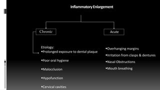
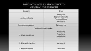
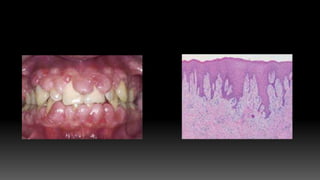
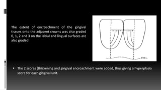
GINGIVAL ENLARGEMENT
CONTENTS:
 Introduction
 Classification
 Inflammatory Enlargement
 Drug-induced Enlargement
 Enlargements associated with systemic diseases and conditions
 Neoplastic Enlargement
 False Enlargement
 Treatment of Gingival Enlargement
 Conclusion
INTRODUCTION:
 Increase in the size of gingiva is a common feature of gingival disease.
Accepted current terminology for this condition is Gingival Enlargement or
Gingival Overgrowth.
 Hyperplasia- The actual increase in number of cells.
 Hypertrophy- The increase in size of cells.
CLASSIFICATION:
 INFLAMMATORY ENLARGEMENT
A. Chronic
B. Acute
 II.DRUG INDUCED ENLARGEMENT
A. General information
B. Anticonvulsants
C. Immunosuppressant’s
D. Calcium channel blockers
IV. NEOPLASTIC ENLARGEMENT
A. Benign Tumors
III. ENLARGEMENTS ASSOCIATED WITH SYSTEMIC DISEASES OR
CONDITIONS
A. Conditioned enlargement
1.Pregnancy
2.Puberty
3.Vitamin C deficiency
4.Plasma cell gingivitis
5.Non-neoplastic conditioned enlargement
A. Conditioned enlargement
B.SYSTEMIC DISEASES CAUSING GINGIVAL ENLARGEMENT
1. Leukemia
2. Granulomatous diseases (Wegener’s granulomatosis, Sarcoidosis)
IV. NEOPLASTIC ENLARGEMENT
A. Benign Tumors
B. Malignant Tumors
V.FALSE ENLARGEMENT
2.Using the criteria of location and distribution, gingival
enlargement is classified as follows
 Localized: Limited to gingiva adjacent to single tooth or group of teeth
 Generalized: Involving gingiva throughout the mouth
 Marginal: Confined to marginal gingiva
 Papillary: Confined to interdental papilla
 Diffuse: Involving marginal; attached gingiva and papilla
 Discrete: An isolated sessile or pedunculated,tumorlike enlargement
DEGREE OF GINGIVAL ENLARGEMENT (Bokenkamp and
Bohnhorst 1994)
 Grade 0: No signs of gingival enlargement
 Grade I: Enlargement confined to interdental papilla
 Grade II: Enlargement involves papilla& marginal gingiva
 Grade III: Enlargement covers three quarters or more of the crown.
Angelopoulous and Goaz (1972)
Grade 0: None
Grade I: No more than one third of the clinical crown covered.
Grade II: Any part of the middle third of the crown covered.
Grade III: Greater than two third of the crown covered.
Photographic analysis –Ellis & Seymour
 Criteria for assessing gingival encroachment/overgrowth on adjacent tooth
surfaces for a gingival unit (where a discrepancy exists between adjacent teeth,
the highest score is awarded).
 0= No encroachment of interdental papilla onto tooth surface.
 1= Mild encroachment of interdental papilla, producing a blunted appearance to
papilla tip.
 2= Moderate encroachment, involving lateral spread of papilla across buccal
tooth surface of less than one quarter tooth width.
 3= Marked encroachment of papilla, i.e., more than 1/4 tooth width. Loss of
normal papilla form.
InflammatoryEnlargement
Acute
Chronic
Overhanging margins
Irritation from clasps & dentures
Nasal Obstructions
Mouth breathing
Etiology:
Prolonged exposure to dental plaque
Poor oral hygiene
Malocclusion
Hypofunction
Cervical cavities
CHRONIC INFLAMMATORY ENLARGEMENT
CLINICAL FEATURES:
Originates as a slight ballooning of the
interdental papilla or marginal gingiva.
In early stages, it produces a life-saver like
bulge around the involved tooth.
Progresses slowly and painlessly unless it is
complicated by acute infection.
Discrete sessile or pedunculated mass
resembling a tumor.
HISTOPATHOLOGY
Shows exudative and proliferative features of chronic
inflammation.
Lesions are clinically deep red or bluish red and are
soft, friable with smooth, shiny surface and bleed
easily.
These lesions have preponderance of inflammatory
cells and fluid, vascular engorgement, new capillary
formation and associated degenerative changes.
Whereas, lesions which are firm and resilient show
greater fibrotic component with abundance of
fibroblasts and collagen fibers.
GINGIVAL CHANGES ASSOCIATED WITH MOUTH
BREATHING
 Gingiva appears red and edematous, with
a diffuse surface shininess of the exposed
area.
 Maxillary anterior region is the most
commonly affected site.
 The altered gingiva is clearly demarcates
from adjacent unexposed normal gingiva.
 The harmful effects are attributed to
irritation from surface dehydration.
ACUTE INFLAMMATORY ENLARGEMENT
GINGIVAL ABSCESS
Localized,painful,rapidly expanding lesion of sudden
onset.
Limited to marginal gingiva or interdental papilla.
The lesion becomes fluctuant and pointed with a
surface orifice from which purulent exudate may be
expressed.
HISTOPATHOLOGY
Consistsofpurulentfocusintheconnectivetissue,surroundedbydiffuseinfiltrationofPMN’s,
edematoustissueandvascularengorgement.
ETIOLOGY
Resultsfrombacteriacarrieddeepinsidethetissuewhenforeignsubstance(eg.Tooth-brushbristle,
pieceofapplecoreorlobstershellfragment)isforcefullyembeddedingingiva.
PERIODONTAL (LATERAL) ABSCESS
 It is a localized purulent inflammation in
periodontal tissues.
 Also known as lateral abscess or parietal
abscess.
 Involves the supporting periodontal tissues and
generally produces enlargement of gingiva.
DRUG INDUCED GINGIVAL INLARGEMENT
Category Drugs
Anticonvulsants
Phenytoin
Sodium valproate
Phenobarbitone
Vigabatrin
Immunosuppressant Cyclosporine
Calcium channel blockers
1. Dihydropyridines
Nifedipine
Felodipine
Amlodipine
2. Phenylalkylamine Verapamil
3. Benzodiazepine Diltiazem
DRUGS COMMONLY ASSOCIATED WITH
GINGIVAL OVERGROWTH
ANTICONVULSANTS
CLINICAL FEATURES:
First reported in 1939 by Kimball associated with chronic usage of the anti-epileptic drug
Phenytoin.
 Overgrowth usually becomes apparent in first three months of phenytoin usage.
 Starts as painless, beadlike enlargement of facial and lingual gingival margins and
interdental papilla.
 As the condition progresses, the marginal and papillary enlargements unite and develop
into massive tissue fold, covering a considerable portion of crown and may interfere with
occlusion.
 Hyperplasia is usually generalized but severe in maxillary and mandibular anterior region.
 Secondary inflammatory process may complicate gingival hyperplasia.
HISTOPATHOLOGY
 Consists of pronounce hyperplasia of epithelium and connective tissue.
 There is acanthosis of the epithelium, and elongated rete pegs extend deep
into the connective tissue, which exhibits densely arranged collagen bundles
with an increase in number of fibroblasts and new blood vessels.
 Usual therapeutic plasma level of phenytoin - 10-20μg/ml
Classification of gingival hyperplasia caused by Dilantin
James R. Babocks(1974)
R. Babocks1974)
Grade-I Minimal: No hyperplasia
Grade-II Moderate: Definite hyperplasia of gingiva, with encroachment on the
clinical crown of the teeth, but no interference of function.
Grade-III Severe: Interference with function due to overgrowth of tissue .
Rees TD (1993)
Histological Classification [Barak in 1987]987)]
 Grade I – Normal Gingiva - Width of epithelium - 0.3-0.5 mm.
 Grade II – Slight Overgrowth - Width of epithelium 0.5-1.5 mm.
 Grade III- Moderate Overgrowth –Width of epithelium 1.5-3 mm.
 Grade IV – Severe Overgrowth- Width of epithelium 3- 4 mm.
Seymour, Smith and Turnbill 1985
 The degree of gingival thickening on both labial and lingual aspects
was graded as follows:
 0 = Normal
 1 = Thickening from the normal up to 2 mm;
 2 = Thickening from the normal greater than 2 mm
 The 2 scores (thickening and gingival encroachment were added, thus giving a hyperplasia
score for each gingival unit.
The extent of encroachment of the gingival
tissues onto the adjacent crowns was also graded
0, 1, 2 and 3 on the labial and lingual surfaces are
also graded
Hassell 1981
 Different subpopulations of fibroblasts.
 Some of which are capable of high protein and collagen synthesis.
 In presence of Phenytoin, the high activity fibroblasts react and produce a significant
increase in collagen production.
 Higher concentration in GCF than in peripheral or systemic circulation.
Sorrell et al 1971
 Long-term phenytoin causes immunosuppression.
Tissues more susceptible to inflammation.
 But other anti-epileptic drugs though they have immunosupressive action don’t
cause gingival overgrowth.
Staple 1951,1952
 Phenytoin therapy is reported to cause an alteration in the metabolism of adrenal
gland secretions.
 Supression of ACTH and alteration in pituitary gland activity.
 Reduced glucocorticoid synthesis and this has been suggested as an explanation for
the gingival overgrowth.
Waxman 1970
 Patients on phenytoin have low serum level of folic acid.
 Drug may reduce the absorption from GIT or block it’s transport across intestinal
epithelium.
 Also inhibit folate reductase.
 A deficiency results in impaired maturation of epithelium, rendering C.T. more
susceptible to inflammation.
IMMUNOSUPPRESANTS
 Cyclosporine is a potent immunosuppressant used to prevent organ transplant
rejection and to treat various autoimmune diseases.
 Enlargement is more vascularized than phenytoin enlargement.
 Tacrolimus has been proved to cause less severe gingival enlargement.
(Harnadez et al in 2000)
 First isolated in Switzerland… 1970 (Jean Borel)
 Used first by Calne et al in a renal transplant
 Inhibits IL-2 synthesis and release.
 Dosages :
 It has been reported that dosages greater than 500mg/day induce gingival
overgrowth.
 10-20mg/kg BW/day.
CALCIUM CHANNEL BLOCKERS
 These are the drugs developed for cardiovascular conditions such as hypertension,
angina, and cardiac arrhythmias.
 1984..first reported case…NIFEDIPINE
 Amlodipine, Felodipine, Diltiazem, Nitrendipine and Verapamil induce gingival
enlargement.
 Dose dependency
 Rat model (800ng/ml).. Nishikawa et al. 1995
 The hydro pyridine derivative, Isradipine does not induce gingival enlargement.
 Nifidipine + Cyclosporine - induce larger overgrowths.
Santi and Brai 1998
Other features of DIGE (JOP,1996)
 More common on buccal aspect.
 Duration of drug administration for maximal gingival overgrowth to occur is 40 days.
 It regresses spontaneously after discontinuation of drug.
 More severe in younger age group.
1. Age.
2. Genetic predisposition.
The Factors Playing Role in the Pathogenesis of DIGE
Age
 Children and adolescents more susceptible.
Genetic predisposition
 A genetic predisposition :
 Metabolism of the 3 drugs ……Hepatic cytochrome P450.
 Phenytoin – CYP 2C9
 Dihydropyridines & Cyclosporin – CYP3A4
 Human Leukocyte Antigen (HLA)
 Relationship between HLA expression and incidence of DIGE- (Cebecci , Margiotta
1996, Pernu 1994, Thomasson 1996.
 HLA-DR1 and HLA-DR2.
 HLA-DR1 afforded some degree of protection against gingival overgrowth.
 HLA-DR2 may increase the development of this unwanted effect.
IDIOPATHIC GINGIVAL ENLARGEMENT
CLINICAL FEATURES:
 It is a rare condition of undetermined cause.
 Designated by terms such as: gingivomatosis, elephantiasis, idiopathic
fibromatosis,heriditary gingival hyperplasia.
 Affects marginal, attached gingiva and interdental papilla.
 The enlargement is pink, firm and leathery and has characteristic pebbled surface .
 In severe cases, teeth are almost completely covered and the enlargement projects
into the oral vestibule.
 Secondary inflammatory changes are common at the gingival margin.
HISTOPATHOLOGY
 Bulbous increase in amount of connective tissue that is relatively avascular, and
consists of densely arranged collagen fibers and fibroblasts.
 The surface epithelium is thickened and acanthosis with elongated rete pegs.
ETIOLOGY
 The cause is not known, hence the condition is known as idiopathic.
 Some researchers have put forth hereditary and genetic basis.
 It is an autosomal dominant disorder, affecting chromosome 2p21(4,5).
III. ENLARGEMENTS ASSOCIATED WITH SYSTEMIC
DISEASES OR CONDITIONS
A. Conditioned enlargement
1. Pregnancy
2. Puberty
3. Vitamin C deficiency
4. Plasma cell gingivitis
5. Non neoplastic conditioned enlargement
ENLARGEMENT IN PREGNANCY
CLINICAL FEATURES:
 Pregnancy gingival enlargement may be marginal or generalized, or may occur as
single or multiple tumor like masses.
 During pregnancy, there is increased level of progesterone(300mg/day) and
estrogen(20mg/day) which by the end of third trimester increase upto 10-30 times.
 These changes cause changes in capillary permeability, leading to gingival edema and
increase in inflammatory response to dental plaque.
 The sub gingival micro biota shows increase in Provotella Intermedia.
 D Placido et al 1998- Hormones satisfy naphthoquinone requirement of P.intermedia.
Pregnancy does not cause the condition, the
altered tissue metabolism in pregnancy
accentuates the response to local irritation.
Bright red or magenta, soft and friable and has
a smooth, shiny surface.
Bleeding occurs spontaneously or on slight
provocation.
Also known as Pregnancy Tumor, but it is not a
neoplasm, rather an inflammatory response to
bacterial plaque and is modified by patient’s
condition.
HISTOPATHOLOGY
Is also known as angiogranuloma.
Consists of central mass of connective tissue,
with numerous diffusely arranged, engorged
capillaries lined by cuboidal epitheloid cells.
ENLARGEMENT IN PUBERTY
 During puberty, there are raised levels of testosterone in males and estradiol in
females which are attributed to gingival inflammation without any change in
plaque levels.
CLINICAL FEATURES
 Characterized by prominent bulbous
interproximal papillae.
 Often only the facial gingivae are enlarged,
and the lingual surfaces are relatively
unaltered because of the mechanical action of
the tongue.
 After puberty, the enlargement undergoes
spontaneous reduction, but does not
disappear completely until plaque and
calculus are removed.
 Capnocytophaga sp. have been shown to be
responsible for the initiation of pubertal
gingivitis.
HISTOPATHOLOGY
 Chronic inflammation with prominent edema and associated degenerative
changes.
ENLAGEMENT IN VITAMIN C DEFICIENCY
 Enlargement of gingiva is generally included in the classic description of scurvy.
 Acute vitamin C deficiency itself does not cause gingival inflammation, but it does
cause hemorrhage, collagen degeneration, and edema of gingival connective tissue.
CLINICAL FEATURES
 The enlargement is marginal, gingiva is bluish red, soft and friable with a smooth,
shiny surface.
 Hemorrhage, occurs either spontaneously or on slight provocation, and surface
necrosis with pseudomembrane formation.
HISTOPATHOLOGY
 Chronic inflammatory cellular infiltration with a superficial acute response.
 Scattered areas of hemorrhage, with engorged capillaries.
 Marked diffuse edema, collagen destruction, scarcity of collagen fibers are striking
features
PLASMA CELL GINGIVITIS
 Considered to be allergic in origin, related to
components of chewing gums, dentifrices or diet
components.
 Consists of mild marginal enlargement that extends
to attached gingiva.
 Gingiva appears red, friable, sometimes granular and
bleeds easily.
HISTOPATHOLOGY
 Oral epithelium shows spongiosis,
infiltration with inflammatory cells.
 Signs of damage in lower spinous layers
and basal layers.
 Underlying connective tissue contains
dense infiltrate of plasma cells.
 Cessation of exposure to allergens brings
about resolution of the lesion.
NON-SPECIFIC CONDITIONED ENLARGEMENT
PYOGENIC GRANULOMA
 The term pyogenic granuloma was given by Hartzell in 1904
 Pyogenic granuloma is considered as an exaggerated conditioned response to minor
trauma.
 It is a localized mass of highly vascularized tissue arising as an exaggerated response
to plaque in pregnancy.
 It is similar in clinical and microscopic appearance to the conditioned gingival
enlargement seen in pregnancy.
 Arises from proximal gingiva and has a pedunculated base.
 Histopathologically it is a highly vascularized mass of connective tissue.
SYSTEMIC DISEASES CAUSING GINGIVAL
ENLARGEMENT
LEUKEMIA
 Leukemia is a malignant neoplasia of WBC precursors characterized by:
 Diffuse replacement of bone marrow with proliferating leukemic cells.
 Abnormal number and forms of immature WBC’s in circulating blood and
widespread infiltrates in liver, spleen, lymph nodes and other body sites.
CLINICAL FEATURES
 The gingiva is bluish red and has a shiny surface.
 The consistency is moderately firm but there is
tendency towards friability and hemorrhage,
occurring spontaneously or on slight
provocation.
 Leukemic enlargement is not found in
edentulous patients or in patients with chronic
leukemia.
 The spontaneous bleeding is caused by
thrombocytopenia resulting from replacement
of bone marrow cells by leukemic cells.
HISTOPATHOLOGY
 Mature leukocytes and areas of connective tissue infiltrated with dense mass of
immature and proliferating leukocytes.
 Engorged capillaries, edematous and degenerated connective tissue are prominent
features.
 Necrotic epithelial cells, PMN’s and bacteria are often seen.
GRANULOMATOUS DISEASES
WEGENER’S GRANULOMATOSIS
 This disease was described by Friedrich Wegener in 1936.
 It is a rare disease characterized by acute granulomatous necrotizing lesions of
respiratory tract, including nasal and or facial defects.
 Gingivitis in Wegener’s granulomatosis is known as Strawberry Gingivitis.
 Oral lesions involve oral mucosal ulceration, gingival enlargement, and abnormal
tooth mobility and delayed healing response.
HISTOPATHOLOGY
 Chronic inflammatory changes include scattered giant cells and micro abscesses
covered by thin, acanthosis epithelium.
 Vascular changes have not been described because of small size of gingival blood
vessels.
SARCOIDOSIS
 It is a multisystem disorder.
 Most common manifestation is pulmonary infiltration and hilar lymphadenopathy.
 Gingival enlargement is smooth, red, and painless.
 HP-Sarcoid granulomas consist of discrete, non-caseating whorls of epitheloid cells
and multinucleated, foreign body giant cells with peripheral mononuclear cells.
NEOPLASTIC ENLARGEMENT
A. BENIGN TUMORS:
I.FIBROMA:
 Arise from gingival connective tissue or from periodontal ligament.
 These are slow growing, spherical tumors that tend to be firm and nodular but may be
soft and vascular.
 Fibromas are usually pedunculated.
 Two types:
 Giant cell fibroma- contains multinucleated fibroblasts
 Peripheral ossifying fibroma- mineralized tissue such as bone, cementum-like material
and dystrophic calcifications may be found.
HISTOPATHOLOGY:
 Composed of bundles of well-formed collagen fibers with scattering of fibrocytes and
variable vascularity.
II.PAPILLOMA
These are benign proliferation of surface
epithelium that are in some cases associated
with HPV.
Gingival papilloma’s appear as solitary wart
like or cauliflower like protuberances.
Papillomas consist of fingerlike projections
of stratified squamous epithelium and a
central core of fibro vascular connective
tissue.
III.PERIPHERAL GIANT CELL GRANULOMA
Occur most frequently on labial surface.
May be sessile or pedunculated.
They arise interdentally or from gingival
margin.
The lesions are painless, vary in size, and
may cover several teeth.
They may be firm or spongy and color
varies from deep red to purplish blue.
HISTOPATHOLOGY
 The giant cell granuloma has numerous foci of multinucleated giant cells and
hemosiderin particles in connective tissue stroma.
 The overlying epithelium is usually hyperplastic, with ulceration at the base.
 Bone formation occasionally occurs within the lesion.
B. MALIGNANT TUMORS
I.CARCINOMA:
 Oral carcinomas account for less than 3% of all malignant tumors in the body.
 Sixth most common oral cancer in males and 12th most common in females.
 Squamous cell carcinoma is the most common malignant tumor of gingiva.
 It may be exophytic, or may be ulcerative, appearing as flat, erosive lesions.
 These masses are locally invasive, involving the underlying bone and
periodontal ligament of adjoining teeth and underlying mucosa.
 It is usually darkly pigmented and is often preceded by localized pigmentation.
 It may be flat or nodular and is characterized by rapid growth and early
metastasis.
 Metastasis is usually confined to the region above the clavicle, however more
extensive involvement may occur.
II.SARCOMA
 Fibro sarcoma, lymph sarcoma of gingiva are rare.
 Kaposi’s sarcoma often occurs in patients suffering from AIDS.
 In early stages, lesions appear as painless, reddish purple nodules of oral mucosa.
 As they progress, lesions become nodular.
III.MALIGNANT MELANOMA
 It is a rare oral tumor that tends to occur in
hard palate and maxillary gingiva of older
patient.
 Darkly pigmented.
 May be flat or nodular, and characterized
by rapid growth and early metastasis.
 Infiltration to underlying bone and
metastasis to cervical and axillary lymph
nodes is common.
FALSE ENLARGEMENT
 False enlargements are not true enlargements of gingival tissues but may appear as a
result of underlying osseous or dental tissues.
 The gingiva usually presents with no abnormal clinical features except the massive
increase in the size of the area.
UNDERLYING OSSEOUS LESIONS
 Occurs most often in exocytosis and tori, but can also occur in Paget’s disease, fibrous
dysplasia or cherubim.
 The gingiva may appear normal or may have unrelated inflammatory changes.
UNDERLYING DENTAL TISSUES
 During various stages of tooth eruption, the labial gingiva may show a bulbous
marginal distortion caused by superimposition of bulk of gingiva on the normal
periodontium of enamel in the gingival half of crown.
TREATMENT
Treatment of gingival enlargement based on understanding of the cause and
underlying pathologic changes.
Chronic Inflammatory Enlargement
 Scaling and root planing
Fibrotic component that does not undergo shrinkage after
scaling and root planing
Surgical removal is the treatment of choice.
•Two techniques available -
–Gingivectomy
–Flap operation
Soft and friable even after SRP gingivectomy is
the treatment of choice.
If all attached gingiva creating
mucogingival problem flap operation is
indicated
Acute Periodontal Abscess
 Patients general systemic response should be evaluated.
 Rise in temperature fever and feeling of malaise should be noted and proper antibiotic
regime started.
 Drainage established through the pocket or through external incision.
Gingival Abscess
 Drainage
 If residual size too great… remove surgically.
Treatment of chronic periodontal abscess
 Adequate drainage, Scaling and curettage and antibiotic treatment.
Drug Induced Gingival Enlargement
 First, consideration should be given to the possibility of discontinuing the drug or
changing the medication.
 Substitution with suitable alternative
 1 to 8 weeks for resolution of gingival lesions.
 Not all patients respond to this mode of treatment, especially those with long
standing gingival lesions.
 Phenytoin: lomatrigine, gabapentin, sulthiame, and topiramate
 Nifedipine: vasodilators(sod.nitroprusside), isradipine,
 CsA : Tacrolimus (FK506)
 Second, the clinician should emphasize plaque control as the first step in the
treatment of drug induce enlargement.
 Third, if gingival enlargement persists after consideration of the above mentioned
approaches, the cases need to be treated by surgery i.e., gingivectomy or periodontal
flap.
Treatment of Leukemic Gingival Enlargement
 Mostly with Acute / Subacute cases, rarely with chronic.
 Consultation with patient’s Physician and Hematologist.
 Superficial Scaling in localized area under topical anaesthesia, management of
bleeding.
 Progressive deeper scaling is done in subsequent visits.
 Systemic antibiotic coverage.
Treatment of Gingival Enlargement in Pregnancy
 Elimination of all local irritants.
 Marginal and interdental inflammation are treated by scaling and curettage.
 Treatment of tumor like gingival enlargement consists of surgical excision and SRP.
 Lesion should be removed surgically during pregnancy only if they interfere with
mastication or produce aesthetic disfigurement.
Recurrence of Gingival Enlargement
 Most common problem in the management of gingival enlargement.
 Residual local irritation, systemic or hereditary are causative factors for recurrence.
 Recurrence during healing period is manifested as red bead like granulomatus masses
that bleed on slight provocation.
 Condition is corrected by removing the granulation tissue and SRP.
 Familial or hereditary gingival enlargement recurs even if all local factors have been
removed.
CONCLUSION
 Gingival enlargement possesses esthetic as well as pathologic concern for the patient.
 Elimination of all local irritants and timely management, proper follow-up may
prevent its recurrence.
 However care should be taken to eliminate the underlying cause of gingival
enlargement.
REFERENCES
 Textbook of Clinical Periodontology- Fermin A. Carranza: -11th edition
 Shlomo Barak, Isaac S et al. Gingival Hyperplasia Caused by Nifedipine. J Periodontol
1987.
 Ellis JS, Seymour RA, Robertson P. Photographic scoring of gingival overgrowth. J Clin
Periodontol 2001; 28: 81–85.
 Marshall RI, Bartold PM. A clinical review of drug-induced gingival overgrowths. Aust
Dent J 1999;44:219-32.
 Risk factors associated with Gingival overgrowth-JCP 2000
 The role of drugs in pathogenesis of gingival overgrowth ; A collective review of
current concepts: Perio 2000 1999
Gingival_enlargement_ppt.pptx

Gingival_enlargement_ppt.pptx

  • 1.
  • 2.
    CONTENTS:  Introduction  Classification Inflammatory Enlargement  Drug-induced Enlargement  Enlargements associated with systemic diseases and conditions  Neoplastic Enlargement  False Enlargement  Treatment of Gingival Enlargement  Conclusion
  • 3.
    INTRODUCTION:  Increase inthe size of gingiva is a common feature of gingival disease. Accepted current terminology for this condition is Gingival Enlargement or Gingival Overgrowth.  Hyperplasia- The actual increase in number of cells.  Hypertrophy- The increase in size of cells.
  • 4.
    CLASSIFICATION:  INFLAMMATORY ENLARGEMENT A.Chronic B. Acute  II.DRUG INDUCED ENLARGEMENT A. General information B. Anticonvulsants C. Immunosuppressant’s D. Calcium channel blockers
  • 5.
    IV. NEOPLASTIC ENLARGEMENT A.Benign Tumors III. ENLARGEMENTS ASSOCIATED WITH SYSTEMIC DISEASES OR CONDITIONS A. Conditioned enlargement 1.Pregnancy 2.Puberty 3.Vitamin C deficiency 4.Plasma cell gingivitis 5.Non-neoplastic conditioned enlargement
  • 6.
    A. Conditioned enlargement B.SYSTEMICDISEASES CAUSING GINGIVAL ENLARGEMENT 1. Leukemia 2. Granulomatous diseases (Wegener’s granulomatosis, Sarcoidosis) IV. NEOPLASTIC ENLARGEMENT A. Benign Tumors B. Malignant Tumors V.FALSE ENLARGEMENT
  • 7.
    2.Using the criteriaof location and distribution, gingival enlargement is classified as follows  Localized: Limited to gingiva adjacent to single tooth or group of teeth  Generalized: Involving gingiva throughout the mouth  Marginal: Confined to marginal gingiva  Papillary: Confined to interdental papilla  Diffuse: Involving marginal; attached gingiva and papilla  Discrete: An isolated sessile or pedunculated,tumorlike enlargement
  • 8.
    DEGREE OF GINGIVALENLARGEMENT (Bokenkamp and Bohnhorst 1994)  Grade 0: No signs of gingival enlargement  Grade I: Enlargement confined to interdental papilla  Grade II: Enlargement involves papilla& marginal gingiva  Grade III: Enlargement covers three quarters or more of the crown.
  • 9.
    Angelopoulous and Goaz(1972) Grade 0: None Grade I: No more than one third of the clinical crown covered. Grade II: Any part of the middle third of the crown covered. Grade III: Greater than two third of the crown covered.
  • 10.
    Photographic analysis –Ellis& Seymour  Criteria for assessing gingival encroachment/overgrowth on adjacent tooth surfaces for a gingival unit (where a discrepancy exists between adjacent teeth, the highest score is awarded).  0= No encroachment of interdental papilla onto tooth surface.  1= Mild encroachment of interdental papilla, producing a blunted appearance to papilla tip.  2= Moderate encroachment, involving lateral spread of papilla across buccal tooth surface of less than one quarter tooth width.  3= Marked encroachment of papilla, i.e., more than 1/4 tooth width. Loss of normal papilla form.
  • 11.
    InflammatoryEnlargement Acute Chronic Overhanging margins Irritation fromclasps & dentures Nasal Obstructions Mouth breathing Etiology: Prolonged exposure to dental plaque Poor oral hygiene Malocclusion Hypofunction Cervical cavities
  • 12.
    CHRONIC INFLAMMATORY ENLARGEMENT CLINICALFEATURES: Originates as a slight ballooning of the interdental papilla or marginal gingiva. In early stages, it produces a life-saver like bulge around the involved tooth. Progresses slowly and painlessly unless it is complicated by acute infection. Discrete sessile or pedunculated mass resembling a tumor.
  • 13.
    HISTOPATHOLOGY Shows exudative andproliferative features of chronic inflammation. Lesions are clinically deep red or bluish red and are soft, friable with smooth, shiny surface and bleed easily. These lesions have preponderance of inflammatory cells and fluid, vascular engorgement, new capillary formation and associated degenerative changes. Whereas, lesions which are firm and resilient show greater fibrotic component with abundance of fibroblasts and collagen fibers.
  • 14.
    GINGIVAL CHANGES ASSOCIATEDWITH MOUTH BREATHING  Gingiva appears red and edematous, with a diffuse surface shininess of the exposed area.  Maxillary anterior region is the most commonly affected site.  The altered gingiva is clearly demarcates from adjacent unexposed normal gingiva.  The harmful effects are attributed to irritation from surface dehydration.
  • 15.
  • 16.
    GINGIVAL ABSCESS Localized,painful,rapidly expandinglesion of sudden onset. Limited to marginal gingiva or interdental papilla. The lesion becomes fluctuant and pointed with a surface orifice from which purulent exudate may be expressed.
  • 17.
  • 18.
    PERIODONTAL (LATERAL) ABSCESS It is a localized purulent inflammation in periodontal tissues.  Also known as lateral abscess or parietal abscess.  Involves the supporting periodontal tissues and generally produces enlargement of gingiva.
  • 19.
  • 20.
    Category Drugs Anticonvulsants Phenytoin Sodium valproate Phenobarbitone Vigabatrin ImmunosuppressantCyclosporine Calcium channel blockers 1. Dihydropyridines Nifedipine Felodipine Amlodipine 2. Phenylalkylamine Verapamil 3. Benzodiazepine Diltiazem DRUGS COMMONLY ASSOCIATED WITH GINGIVAL OVERGROWTH
  • 21.
    ANTICONVULSANTS CLINICAL FEATURES: First reportedin 1939 by Kimball associated with chronic usage of the anti-epileptic drug Phenytoin.  Overgrowth usually becomes apparent in first three months of phenytoin usage.  Starts as painless, beadlike enlargement of facial and lingual gingival margins and interdental papilla.  As the condition progresses, the marginal and papillary enlargements unite and develop into massive tissue fold, covering a considerable portion of crown and may interfere with occlusion.  Hyperplasia is usually generalized but severe in maxillary and mandibular anterior region.  Secondary inflammatory process may complicate gingival hyperplasia.
  • 22.
    HISTOPATHOLOGY  Consists ofpronounce hyperplasia of epithelium and connective tissue.  There is acanthosis of the epithelium, and elongated rete pegs extend deep into the connective tissue, which exhibits densely arranged collagen bundles with an increase in number of fibroblasts and new blood vessels.
  • 25.
     Usual therapeuticplasma level of phenytoin - 10-20μg/ml Classification of gingival hyperplasia caused by Dilantin James R. Babocks(1974) R. Babocks1974) Grade-I Minimal: No hyperplasia Grade-II Moderate: Definite hyperplasia of gingiva, with encroachment on the clinical crown of the teeth, but no interference of function. Grade-III Severe: Interference with function due to overgrowth of tissue . Rees TD (1993)
  • 26.
    Histological Classification [Barakin 1987]987)]  Grade I – Normal Gingiva - Width of epithelium - 0.3-0.5 mm.  Grade II – Slight Overgrowth - Width of epithelium 0.5-1.5 mm.  Grade III- Moderate Overgrowth –Width of epithelium 1.5-3 mm.  Grade IV – Severe Overgrowth- Width of epithelium 3- 4 mm.
  • 27.
    Seymour, Smith andTurnbill 1985  The degree of gingival thickening on both labial and lingual aspects was graded as follows:  0 = Normal  1 = Thickening from the normal up to 2 mm;  2 = Thickening from the normal greater than 2 mm
  • 28.
     The 2scores (thickening and gingival encroachment were added, thus giving a hyperplasia score for each gingival unit. The extent of encroachment of the gingival tissues onto the adjacent crowns was also graded 0, 1, 2 and 3 on the labial and lingual surfaces are also graded
  • 29.
    Hassell 1981  Differentsubpopulations of fibroblasts.  Some of which are capable of high protein and collagen synthesis.  In presence of Phenytoin, the high activity fibroblasts react and produce a significant increase in collagen production.  Higher concentration in GCF than in peripheral or systemic circulation.
  • 30.
    Sorrell et al1971  Long-term phenytoin causes immunosuppression. Tissues more susceptible to inflammation.  But other anti-epileptic drugs though they have immunosupressive action don’t cause gingival overgrowth.
  • 31.
    Staple 1951,1952  Phenytointherapy is reported to cause an alteration in the metabolism of adrenal gland secretions.  Supression of ACTH and alteration in pituitary gland activity.  Reduced glucocorticoid synthesis and this has been suggested as an explanation for the gingival overgrowth.
  • 32.
    Waxman 1970  Patientson phenytoin have low serum level of folic acid.  Drug may reduce the absorption from GIT or block it’s transport across intestinal epithelium.  Also inhibit folate reductase.  A deficiency results in impaired maturation of epithelium, rendering C.T. more susceptible to inflammation.
  • 33.
    IMMUNOSUPPRESANTS  Cyclosporine isa potent immunosuppressant used to prevent organ transplant rejection and to treat various autoimmune diseases.  Enlargement is more vascularized than phenytoin enlargement.  Tacrolimus has been proved to cause less severe gingival enlargement. (Harnadez et al in 2000)
  • 34.
     First isolatedin Switzerland… 1970 (Jean Borel)  Used first by Calne et al in a renal transplant  Inhibits IL-2 synthesis and release.  Dosages :  It has been reported that dosages greater than 500mg/day induce gingival overgrowth.  10-20mg/kg BW/day.
  • 37.
    CALCIUM CHANNEL BLOCKERS These are the drugs developed for cardiovascular conditions such as hypertension, angina, and cardiac arrhythmias.  1984..first reported case…NIFEDIPINE  Amlodipine, Felodipine, Diltiazem, Nitrendipine and Verapamil induce gingival enlargement.  Dose dependency  Rat model (800ng/ml).. Nishikawa et al. 1995  The hydro pyridine derivative, Isradipine does not induce gingival enlargement.
  • 38.
     Nifidipine +Cyclosporine - induce larger overgrowths. Santi and Brai 1998 Other features of DIGE (JOP,1996)  More common on buccal aspect.  Duration of drug administration for maximal gingival overgrowth to occur is 40 days.  It regresses spontaneously after discontinuation of drug.  More severe in younger age group.
  • 39.
    1. Age. 2. Geneticpredisposition. The Factors Playing Role in the Pathogenesis of DIGE
  • 40.
    Age  Children andadolescents more susceptible.
  • 41.
    Genetic predisposition  Agenetic predisposition :  Metabolism of the 3 drugs ……Hepatic cytochrome P450.  Phenytoin – CYP 2C9  Dihydropyridines & Cyclosporin – CYP3A4
  • 42.
     Human LeukocyteAntigen (HLA)  Relationship between HLA expression and incidence of DIGE- (Cebecci , Margiotta 1996, Pernu 1994, Thomasson 1996.  HLA-DR1 and HLA-DR2.  HLA-DR1 afforded some degree of protection against gingival overgrowth.  HLA-DR2 may increase the development of this unwanted effect.
  • 43.
    IDIOPATHIC GINGIVAL ENLARGEMENT CLINICALFEATURES:  It is a rare condition of undetermined cause.  Designated by terms such as: gingivomatosis, elephantiasis, idiopathic fibromatosis,heriditary gingival hyperplasia.  Affects marginal, attached gingiva and interdental papilla.  The enlargement is pink, firm and leathery and has characteristic pebbled surface .  In severe cases, teeth are almost completely covered and the enlargement projects into the oral vestibule.  Secondary inflammatory changes are common at the gingival margin.
  • 44.
    HISTOPATHOLOGY  Bulbous increasein amount of connective tissue that is relatively avascular, and consists of densely arranged collagen fibers and fibroblasts.  The surface epithelium is thickened and acanthosis with elongated rete pegs.
  • 45.
    ETIOLOGY  The causeis not known, hence the condition is known as idiopathic.  Some researchers have put forth hereditary and genetic basis.  It is an autosomal dominant disorder, affecting chromosome 2p21(4,5).
  • 47.
    III. ENLARGEMENTS ASSOCIATEDWITH SYSTEMIC DISEASES OR CONDITIONS A. Conditioned enlargement 1. Pregnancy 2. Puberty 3. Vitamin C deficiency 4. Plasma cell gingivitis 5. Non neoplastic conditioned enlargement
  • 48.
    ENLARGEMENT IN PREGNANCY CLINICALFEATURES:  Pregnancy gingival enlargement may be marginal or generalized, or may occur as single or multiple tumor like masses.  During pregnancy, there is increased level of progesterone(300mg/day) and estrogen(20mg/day) which by the end of third trimester increase upto 10-30 times.  These changes cause changes in capillary permeability, leading to gingival edema and increase in inflammatory response to dental plaque.  The sub gingival micro biota shows increase in Provotella Intermedia.  D Placido et al 1998- Hormones satisfy naphthoquinone requirement of P.intermedia.
  • 49.
    Pregnancy does notcause the condition, the altered tissue metabolism in pregnancy accentuates the response to local irritation. Bright red or magenta, soft and friable and has a smooth, shiny surface. Bleeding occurs spontaneously or on slight provocation. Also known as Pregnancy Tumor, but it is not a neoplasm, rather an inflammatory response to bacterial plaque and is modified by patient’s condition.
  • 50.
    HISTOPATHOLOGY Is also knownas angiogranuloma. Consists of central mass of connective tissue, with numerous diffusely arranged, engorged capillaries lined by cuboidal epitheloid cells.
  • 51.
    ENLARGEMENT IN PUBERTY During puberty, there are raised levels of testosterone in males and estradiol in females which are attributed to gingival inflammation without any change in plaque levels.
  • 52.
    CLINICAL FEATURES  Characterizedby prominent bulbous interproximal papillae.  Often only the facial gingivae are enlarged, and the lingual surfaces are relatively unaltered because of the mechanical action of the tongue.  After puberty, the enlargement undergoes spontaneous reduction, but does not disappear completely until plaque and calculus are removed.  Capnocytophaga sp. have been shown to be responsible for the initiation of pubertal gingivitis.
  • 53.
    HISTOPATHOLOGY  Chronic inflammationwith prominent edema and associated degenerative changes.
  • 54.
    ENLAGEMENT IN VITAMINC DEFICIENCY  Enlargement of gingiva is generally included in the classic description of scurvy.  Acute vitamin C deficiency itself does not cause gingival inflammation, but it does cause hemorrhage, collagen degeneration, and edema of gingival connective tissue.
  • 55.
    CLINICAL FEATURES  Theenlargement is marginal, gingiva is bluish red, soft and friable with a smooth, shiny surface.  Hemorrhage, occurs either spontaneously or on slight provocation, and surface necrosis with pseudomembrane formation.
  • 56.
    HISTOPATHOLOGY  Chronic inflammatorycellular infiltration with a superficial acute response.  Scattered areas of hemorrhage, with engorged capillaries.  Marked diffuse edema, collagen destruction, scarcity of collagen fibers are striking features
  • 57.
    PLASMA CELL GINGIVITIS Considered to be allergic in origin, related to components of chewing gums, dentifrices or diet components.  Consists of mild marginal enlargement that extends to attached gingiva.  Gingiva appears red, friable, sometimes granular and bleeds easily.
  • 58.
    HISTOPATHOLOGY  Oral epitheliumshows spongiosis, infiltration with inflammatory cells.  Signs of damage in lower spinous layers and basal layers.  Underlying connective tissue contains dense infiltrate of plasma cells.  Cessation of exposure to allergens brings about resolution of the lesion.
  • 59.
  • 60.
    PYOGENIC GRANULOMA  Theterm pyogenic granuloma was given by Hartzell in 1904  Pyogenic granuloma is considered as an exaggerated conditioned response to minor trauma.  It is a localized mass of highly vascularized tissue arising as an exaggerated response to plaque in pregnancy.  It is similar in clinical and microscopic appearance to the conditioned gingival enlargement seen in pregnancy.  Arises from proximal gingiva and has a pedunculated base.  Histopathologically it is a highly vascularized mass of connective tissue.
  • 61.
    SYSTEMIC DISEASES CAUSINGGINGIVAL ENLARGEMENT
  • 62.
    LEUKEMIA  Leukemia isa malignant neoplasia of WBC precursors characterized by:  Diffuse replacement of bone marrow with proliferating leukemic cells.  Abnormal number and forms of immature WBC’s in circulating blood and widespread infiltrates in liver, spleen, lymph nodes and other body sites.
  • 63.
    CLINICAL FEATURES  Thegingiva is bluish red and has a shiny surface.  The consistency is moderately firm but there is tendency towards friability and hemorrhage, occurring spontaneously or on slight provocation.  Leukemic enlargement is not found in edentulous patients or in patients with chronic leukemia.  The spontaneous bleeding is caused by thrombocytopenia resulting from replacement of bone marrow cells by leukemic cells.
  • 64.
    HISTOPATHOLOGY  Mature leukocytesand areas of connective tissue infiltrated with dense mass of immature and proliferating leukocytes.  Engorged capillaries, edematous and degenerated connective tissue are prominent features.  Necrotic epithelial cells, PMN’s and bacteria are often seen.
  • 65.
  • 66.
    WEGENER’S GRANULOMATOSIS  Thisdisease was described by Friedrich Wegener in 1936.  It is a rare disease characterized by acute granulomatous necrotizing lesions of respiratory tract, including nasal and or facial defects.  Gingivitis in Wegener’s granulomatosis is known as Strawberry Gingivitis.  Oral lesions involve oral mucosal ulceration, gingival enlargement, and abnormal tooth mobility and delayed healing response.
  • 67.
    HISTOPATHOLOGY  Chronic inflammatorychanges include scattered giant cells and micro abscesses covered by thin, acanthosis epithelium.  Vascular changes have not been described because of small size of gingival blood vessels.
  • 68.
    SARCOIDOSIS  It isa multisystem disorder.  Most common manifestation is pulmonary infiltration and hilar lymphadenopathy.  Gingival enlargement is smooth, red, and painless.  HP-Sarcoid granulomas consist of discrete, non-caseating whorls of epitheloid cells and multinucleated, foreign body giant cells with peripheral mononuclear cells.
  • 69.
    NEOPLASTIC ENLARGEMENT A. BENIGNTUMORS: I.FIBROMA:  Arise from gingival connective tissue or from periodontal ligament.  These are slow growing, spherical tumors that tend to be firm and nodular but may be soft and vascular.  Fibromas are usually pedunculated.  Two types:  Giant cell fibroma- contains multinucleated fibroblasts  Peripheral ossifying fibroma- mineralized tissue such as bone, cementum-like material and dystrophic calcifications may be found.
  • 70.
    HISTOPATHOLOGY:  Composed ofbundles of well-formed collagen fibers with scattering of fibrocytes and variable vascularity.
  • 71.
    II.PAPILLOMA These are benignproliferation of surface epithelium that are in some cases associated with HPV. Gingival papilloma’s appear as solitary wart like or cauliflower like protuberances. Papillomas consist of fingerlike projections of stratified squamous epithelium and a central core of fibro vascular connective tissue.
  • 72.
    III.PERIPHERAL GIANT CELLGRANULOMA Occur most frequently on labial surface. May be sessile or pedunculated. They arise interdentally or from gingival margin. The lesions are painless, vary in size, and may cover several teeth. They may be firm or spongy and color varies from deep red to purplish blue.
  • 73.
    HISTOPATHOLOGY  The giantcell granuloma has numerous foci of multinucleated giant cells and hemosiderin particles in connective tissue stroma.  The overlying epithelium is usually hyperplastic, with ulceration at the base.  Bone formation occasionally occurs within the lesion.
  • 74.
    B. MALIGNANT TUMORS I.CARCINOMA: Oral carcinomas account for less than 3% of all malignant tumors in the body.  Sixth most common oral cancer in males and 12th most common in females.  Squamous cell carcinoma is the most common malignant tumor of gingiva.  It may be exophytic, or may be ulcerative, appearing as flat, erosive lesions.
  • 75.
     These massesare locally invasive, involving the underlying bone and periodontal ligament of adjoining teeth and underlying mucosa.  It is usually darkly pigmented and is often preceded by localized pigmentation.  It may be flat or nodular and is characterized by rapid growth and early metastasis.  Metastasis is usually confined to the region above the clavicle, however more extensive involvement may occur.
  • 76.
    II.SARCOMA  Fibro sarcoma,lymph sarcoma of gingiva are rare.  Kaposi’s sarcoma often occurs in patients suffering from AIDS.  In early stages, lesions appear as painless, reddish purple nodules of oral mucosa.  As they progress, lesions become nodular.
  • 77.
    III.MALIGNANT MELANOMA  Itis a rare oral tumor that tends to occur in hard palate and maxillary gingiva of older patient.  Darkly pigmented.  May be flat or nodular, and characterized by rapid growth and early metastasis.  Infiltration to underlying bone and metastasis to cervical and axillary lymph nodes is common.
  • 78.
    FALSE ENLARGEMENT  Falseenlargements are not true enlargements of gingival tissues but may appear as a result of underlying osseous or dental tissues.  The gingiva usually presents with no abnormal clinical features except the massive increase in the size of the area.
  • 79.
    UNDERLYING OSSEOUS LESIONS Occurs most often in exocytosis and tori, but can also occur in Paget’s disease, fibrous dysplasia or cherubim.  The gingiva may appear normal or may have unrelated inflammatory changes.
  • 80.
    UNDERLYING DENTAL TISSUES During various stages of tooth eruption, the labial gingiva may show a bulbous marginal distortion caused by superimposition of bulk of gingiva on the normal periodontium of enamel in the gingival half of crown.
  • 81.
    TREATMENT Treatment of gingivalenlargement based on understanding of the cause and underlying pathologic changes.
  • 82.
    Chronic Inflammatory Enlargement Scaling and root planing Fibrotic component that does not undergo shrinkage after scaling and root planing Surgical removal is the treatment of choice. •Two techniques available - –Gingivectomy –Flap operation Soft and friable even after SRP gingivectomy is the treatment of choice. If all attached gingiva creating mucogingival problem flap operation is indicated
  • 83.
    Acute Periodontal Abscess Patients general systemic response should be evaluated.  Rise in temperature fever and feeling of malaise should be noted and proper antibiotic regime started.  Drainage established through the pocket or through external incision.
  • 84.
    Gingival Abscess  Drainage If residual size too great… remove surgically. Treatment of chronic periodontal abscess  Adequate drainage, Scaling and curettage and antibiotic treatment.
  • 85.
    Drug Induced GingivalEnlargement  First, consideration should be given to the possibility of discontinuing the drug or changing the medication.  Substitution with suitable alternative  1 to 8 weeks for resolution of gingival lesions.  Not all patients respond to this mode of treatment, especially those with long standing gingival lesions.  Phenytoin: lomatrigine, gabapentin, sulthiame, and topiramate  Nifedipine: vasodilators(sod.nitroprusside), isradipine,  CsA : Tacrolimus (FK506)
  • 86.
     Second, theclinician should emphasize plaque control as the first step in the treatment of drug induce enlargement.  Third, if gingival enlargement persists after consideration of the above mentioned approaches, the cases need to be treated by surgery i.e., gingivectomy or periodontal flap.
  • 87.
    Treatment of LeukemicGingival Enlargement  Mostly with Acute / Subacute cases, rarely with chronic.  Consultation with patient’s Physician and Hematologist.  Superficial Scaling in localized area under topical anaesthesia, management of bleeding.  Progressive deeper scaling is done in subsequent visits.  Systemic antibiotic coverage.
  • 88.
    Treatment of GingivalEnlargement in Pregnancy  Elimination of all local irritants.  Marginal and interdental inflammation are treated by scaling and curettage.  Treatment of tumor like gingival enlargement consists of surgical excision and SRP.  Lesion should be removed surgically during pregnancy only if they interfere with mastication or produce aesthetic disfigurement.
  • 89.
    Recurrence of GingivalEnlargement  Most common problem in the management of gingival enlargement.  Residual local irritation, systemic or hereditary are causative factors for recurrence.  Recurrence during healing period is manifested as red bead like granulomatus masses that bleed on slight provocation.  Condition is corrected by removing the granulation tissue and SRP.  Familial or hereditary gingival enlargement recurs even if all local factors have been removed.
  • 90.
    CONCLUSION  Gingival enlargementpossesses esthetic as well as pathologic concern for the patient.  Elimination of all local irritants and timely management, proper follow-up may prevent its recurrence.  However care should be taken to eliminate the underlying cause of gingival enlargement.
  • 91.
    REFERENCES  Textbook ofClinical Periodontology- Fermin A. Carranza: -11th edition  Shlomo Barak, Isaac S et al. Gingival Hyperplasia Caused by Nifedipine. J Periodontol 1987.  Ellis JS, Seymour RA, Robertson P. Photographic scoring of gingival overgrowth. J Clin Periodontol 2001; 28: 81–85.  Marshall RI, Bartold PM. A clinical review of drug-induced gingival overgrowths. Aust Dent J 1999;44:219-32.  Risk factors associated with Gingival overgrowth-JCP 2000  The role of drugs in pathogenesis of gingival overgrowth ; A collective review of current concepts: Perio 2000 1999