

Recommended
More Related Content
Similar to General Embryology.pptx
Similar to General Embryology.pptx (20)
Recently uploaded
Recently uploaded (20)
General Embryology.pptx
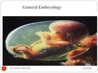
- 1. General Embryology 10/12/2023 By Tesfahun (MSc CM) 1
- 2. Objectives 10/12/2023 By Tesfahun (MSc CM) 2 General objective At the end of this session the students will be able to describe each period of intrauterine life and development of all systems Specific objectives At the end of this course the students will be able to List phases of pre natal period Discuss each period of intrauterine life Explain development of all body systems
- 3. 2 Introduction 10/12/2023 By Tesfahun (MSc CM) 3 Embryology is the study of prenatal development, beginning with gametogenesis up to the formation of full-term fetus. a branch of developmental anatomy that studies the changes that cells, tissues, organs, and the body as a whole undergo from a germ cell of each parent to the resulting adult.
- 4. Significance of embryology 10/12/2023 By Tesfahun (MSc CM) 4 Links the gap between prenatal development and obstetrics, perinatal medicine, pediatrics, and clinical anatomy. Develops knowledge concerning the beginnings of human life and the changes occurring during prenatal development. Is of practical value in helping to understand the causes of variations in human structure.
- 5. 5 Developmental Periods 10/12/2023 By Tesfahun (MSc CM) 5 Human development is a continuous process that begins when an oocyte (ovum) is fertilized by a sperm (spermatozoon).divided into 1. Prenatal/Antenatal development (before birth) a developmental process that represents an amazing integration of increasingly complexity that occurs from fertilization to birth. 2. Postnatal (after birth) periods
- 6. Pre-natal development 10/12/2023 By Tesfahun (MSc CM) 6 Lasts 38 weeks from fertilization to parturition, & divided into three developmental periods 1. Pre-embryonic period (Germinal Period) (0–2 weeks) 2. An Embryonic period (3rd – 8th weeks) 3. Fetal period (9th week - birth)
- 7. 1. Pre-embryonic (Germinal) Period 10/12/2023 By Tesfahun (MSc CM) 7 The first two weeks of development and events of pre-embryonic period include: Fertilization Transportation of zygote down the uterine tube Repeated mitotic divisions/ cleavage or segmentation Implantation The formation of bilaminar embryonic disc Development of the amnion and chorion
- 8. 2. An Embryonic period (3rd – 8th weeks): 10/12/2023 By Tesfahun (MSc CM) 8 Lasts from the beginning of the 3rd Week (day 15) to the end of the 8th week (day 56). During which the primordia of all major organ-systems develop from the 3 germ layers, hence most important period in life. 8
- 9. 3. Fetal period: 10/12/2023 By Tesfahun (MSc CM) 9 Lasts from 9th week to birth (38 weeks, or 40 weeks from LNMP) Period of organ systems growth and differentiation Culminates with parturition and birth of the fetus 9
- 10. Preconditions for the beginning of early development of embryo 10/12/2023 By Tesfahun (MSc CM) 10 Gametogenesis: a process of formation and maturation of the gametes (sperm and ovum) which involves mitosis, meiosis, and cytodifferentiation. Production of gametes Reduction of the number of chromosomes by half. Two types of gametogenesis: Spermatogenesis and Oogenesis
- 11. 10/12/2023 By Tesfahun (MSc CM) 11 Gametes are derived from primordial germ cells (PGCs) that are formed in the epiblast during the second week and that move to the wall of the yolk sac. They are originated from the wall of the yolk sac at the end of 3rd week of embryonic development.
- 12. Cont.. 10/12/2023 By Tesfahun (MSc CM) 12 These cells migrate from the yolk sac toward the developing gonads (primitive sex glands), where they arrive at the end of 4th week and invading the genital ridges in 6th week of development. Hence, the primordial germ cells have an inductive influence on development of the gonadal ridge into ovary or testis.
- 13. Primordial germ cells 10/12/2023 By Tesfahun (MSc CM) 13 Yolk sac The first human germ cells (primordial germ cells) appear in the wall of the yolk sac (3rd week)
- 14. The germ cells, through amoeboid movement, move towards the gonads where they arrive at about 5th week Primordial cells later differentiate into mature gametes i.e. spermatogonia (male) or oogonia (female) Primordialgermcells 10/12/2023 By Tesfahun (MSc CM) 14 Y olksac
- 15. 10/12/2023 By Tesfahun (MSc CM) 15 The sex of the embryo is determined at the time of fertilization. At 5 weeks gonads appear initially as a pair of longitudinal ridges, the genital or gonadal ridges. 6th week: Primordial cell migrate to the ridge, stimulate development and differentiation of gonads from the primitive ridge (into either ovary or testis according to the presence or absence of Y chromosome in the nuclei of those primordial cells)
- 16. During 5th week until 7th week male and female genital systems are similar (development of primitive germ cells) . 7th week: beginning of differentiation of the gonads 10/12/2023 By Tesfahun (MSc CM) 16
- 17. Spermatogenesis (male gametogenesis) 10/12/2023 By Tesfahun (MSc CM) 17 is the sequence of events by which spermatogonia are transformed into mature sperms. Takes place in the male gonads ,approximately 300 million sperm cells are produced daily. At puberty, the testes begin to secrete greatly increased amounts of testosterone, this activates maturation of the seminiferous tubules, and spermatogenesis.
- 18. Oogenesis 10/12/2023 By Tesfahun (MSc CM) 18 is the sequence of events by which oogonia are transformed into mature oocytes (ova). This maturation process begins before birth and is completed after puberty, during child bearing age. Oogenesis continues to menopause, which is permanent cessation of the menses. There are less than two million primary oocytes in the ovaries of a new born female, ranging from 1.2 to 1.6 million, by adolescence no more than 40,000 remain Of these, not more than 500 become secondary oocytes and are expelled at ovulation during the reproductive period
- 19. Abnormal Gametes 10/12/2023 By Tesfahun (MSc CM) 19 The ideal maternal age for reproduction is generally considered to be from 18 to 35 years. The likelihood of chromosomal abnormalities in the embryo increases after the mother is 35. During gametogenesis, homologous chromosomes sometimes fail to separate. As a result of this error of meiotic cell division-nondisjunction some gametes have 24 chromosomes and others only 22
- 20. cont…. 10/12/2023 By Tesfahun (MSc CM) 20 If a gamete with 24 chromosomes unites with a normal one with 23 chromosomes during fertilization, a zygote with 47 chromosomes forms . This condition is called trisomy because of the presence of three representatives of a particular chromosome instead of the usual two.
- 21. 10/12/2023 By Tesfahun (MSc CM) 21 If a gamete with only 22 chromosomes unites with a normal one, a zygote with 45 chromosomes forms. This condition is known as monosomy because only one representative of the particular chromosome pair is present. For a description of the clinical conditions associated with numerical disorders of chromosomes.
- 22. Fertilization 10/12/2023 By Tesfahun (MSc CM) 22 It is a complex process begins with a contact between sperm & ovum, ends up with mixture of the maternal and paternal chromosomes. Time: 12 - 24 hours after ovulation
- 23. Site: ampulla of uterine tube. 10/12/2023 By Tesfahun (MSc CM) 23
- 24. The Laborious Journey of the Sperm 10/12/2023 By Tesfahun (MSc CM) 24 An average ejaculate discharges 40-150 million sperm which eagerly swim upstream toward the fallopian tubes on their mission to fertilize an egg. Only 1% of sperm deposited in the vagina enter the cervix, where they may survive for many hours. Movement of sperm from the cervix to the uterine tube occurs by muscular contractions of the uterus and uterine tube and very little by their own propulsion.
- 25. Cont… 10/12/2023 By Tesfahun (MSc CM) 25 Fast-swimming sperm can reach the egg in a half an hour, while other may take days (30 minutes or 6 day). After reaching the isthmus, sperm become less motile and cease their migration. At ovulation, sperm again become motile, perhaps because of chemo attractants produced by cumulus cells surrounding the egg, and swim to the ampulla, where fertilization usually occurs. The sperm can live up to 48-72 hours.
- 26. Cont… 10/12/2023 By Tesfahun (MSc CM) 26 Only a few hundred will even come close to the egg, due to the many natural barriers and hurdles that exist in the female reproductive tract.
- 27. Cont…. 10/12/2023 By Tesfahun (MSc CM) 27 If a sperm cell meets and penetrates an egg, it will fertilize the egg. The process takes about 24 hours.
- 28. Important events in Fertilization 10/12/2023 By Tesfahun (MSc CM) 28 Before: Capacitation Acrosome reaction: release of enzymes from acrosome induced by, and needed for sperm penetration of, the zona pellucida During: Penetration of corona radiata, zona pellucida and oocyte cell membrane. Recognition: a zona protein ZP3 is responsible for species- specific fertilization
- 29. After entry: Cortical reaction: release of cortical granules by the oocyte. Zona reaction: zona becomes impenetrable to other sperms through enzyme release from cortical granules of oocyte to change structure and composition of the zona pellicuda. Spermatozoa are not able to fertilize the oocyte immediately upon arrival in the female genital tract but must undergo Capacitation and the acrosome reaction to acquire this capability. 10/12/2023 By Tesfahun (MSc CM) 29
- 30. Capacitation 10/12/2023 By Tesfahun (MSc CM) 30 Is a period of conditioning in the female reproductive tract that lasts approximately 7 hours. Much of it occurs in the uterine tube and involves epithelial interactions between the sperm and the mucosal surface of the tube. Destabilize acrosomal sperm head membrane . During this time, a glycoprotein coat and seminal plasma proteins are removed from the plasma membrane that overlies the acrosomal region of the spermatozoa. Only capacitated sperm can pass through the corona cells and undergo the acrosome reaction.
- 31. Acrosome reaction 10/12/2023 By Tesfahun (MSc CM) 31 Occurs after binding to the zona pellucida Is induced by zona proteins. This reaction culminates in the release of enzymes needed to penetrate the zona pellucida, including acrosin- and trypsin-like substances; Acrosin Hyaluronidase Trypsin Colagenase B- galactocidase Esterases
- 32. During fertilization, the spermatozoon must penetrate The corona radiata, The zona pellucida, and The oocyte cell membrane. 10/12/2023 By Tesfahun (MSc CM) 32
- 33. Phases of Fertilization 10/12/2023 By Tesfahun (MSc CM) 33 Fertilization is a sequence of coordinated events Passage of the sperm through the cells of the corona radiata by the effect of: a)Hyaluronidase enzyme secreted from the sperms. b)By movement of its tail. Penetration of the zona pellucida by acrosine (a substance secreted from acrosomal cap). Fusion of the plasma membranes of the oocyte and the sperm.
- 34. 10/12/2023 By Tesfahun (MSc CM) 34 Completion of the second meiotic division of the oocyte & formation of the female pronucleus. Formation of the male pronucleus. As the pronuclei fuse into a single diploid aggregation of chromosomes, becomes a zygote
- 35. 92 10/12/2023 By Tesfahun (MSc CM) 35
- 36. Phases of Fertilization 10/12/2023 By Tesfahun (MSc CM) 36
- 37. The oocyte responds in three ways: 10/12/2023 By Tesfahun (MSc CM) 37 1.Cortical & zona reactions Once a receptor has been activated, a series of reactions to prevent polyspermy (the entrance of more than one sperm) will be initiated. First, the cell surface will be depolarized. Then, cortical granules (lysosomes) released into the perivitelline space will hydrolyze the other receptors. The oocyte membrane becomes impenetrable to other spermatozoa
- 38. Cont… 10/12/2023 By Tesfahun (MSc CM) 38 2. Completion of the second meiotic division of oocyte and formation of female pronucleus. Penetration of the oocyte by a sperm activates the oocyte into completing the second meiotic division and forming a mature oocyte and a second polar body. Following decondensation of the maternal chromosomes, the nucleus of the mature oocyte becomes the female pronucleus.
- 39. 3.Metabolic activation of oocyte 10/12/2023 By Tesfahun (MSc CM) 39 Formation of male pronucleus Tail detaches and degenerates At this stage, the male and female pronuclei are indistinguishable. The two pronuclei fuse eventually, loose nuclear envelops . During growth of male and female pronuclei (both haploid) each replicates its DNA and then undergo first mitotic division.
- 40. Results of fertilization 10/12/2023 By Tesfahun (MSc CM) 40 Restoration of the diploid number of chromosomes, half from the father and half from the mother. Results in variation of human species as maternal and paternal chromosomes intermingle. Determination of the sex of the new individual. Initiation of cleavage. Without fertilization, the oocyte usually degenerates 24 hours after ovulation.
- 41. Cleavage 10/12/2023 By Tesfahun (MSc CM) 41 A series of mitotic cell divisions, increasing the numbers of cells but not size. These cells, which become smaller with each cleavage division, are known as blastomeres. Until the eight-cell stage,they form a loosely arranged clump. After the third cleavage, however, blastomeres maximize their contact with each other, forming a compact ball of cells held together by tight junctions.
- 42. It begins about 30 hours after fertilization. Zygote divides into 2, then 4, then 8, then 16 cells. Zygote lies within the thick zona pellucida during cleavage. It migrates in the uterine tube during cleavage from lateral to medial. Zona pellucida is translucent under the light microscope. 10/12/2023 By Tesfahun (MSc CM) 42
- 43. 10/12/2023 By Tesfahun (MSc CM) 43
- 44. 10/12/2023 By Tesfahun (MSc CM) 44
- 45. Morula 10/12/2023 By Tesfahun (MSc CM) 45 When there are 16-32 blastomeres the developing human is called morula. The Morula reaches the uterine cavity at this stage. Spherical Morula is formed about 3 days after fertilization. It resembles mulberry or blackberry.
- 46. Blastocyst 10/12/2023 By Tesfahun (MSc CM) 46 About the time the morula enters the uterine cavity, fluid begins to penetrate through the zona pellucida into the intercellular spaces of the inner cell mass. A cavity appears within the morula dividing its cells into 2 groups: Outer cell layer called trophoblast. Inner cell layer (mass) attached to one of the poles of the blastocyst. The cavity is called blastocystic cavity or blastocele.
- 47. It is the process by which the Blastocyst penetrates the superficial (Compact) layer of the endometrium of the uterus. The normal site of implantation is the posterior wall of the uterus near the fundus. It begins about the 6th day after fertilization. It is completed by the 11th or 12th day. Implantation 10/12/2023 By Tesfahun (MSc CM) 47
- 48. The Morula reaches the uterine cavity by the 4th day after fertilization, & remains free for one or two days. Fluid passes from uterine cavity to the Morula. Now the Morula is called Blastocyst, its cavity is called blastocystic cavity, its cells divided into Embryoblast & Trophoblast. By the 5th day the Zona pellucida degenerates to allow the blastocyst to increase in size and penetrates the endometrium 10/12/2023 By Tesfahun (MSc CM) 48
- 49. Why implantation is needed ? 10/12/2023 By Tesfahun (MSc CM) 49 The implantation of the blastocyst provides nutrition to the growing embryo from the maternal blood initially by diffusion. Later through the development of the placenta. Then after the development of placenta and umbilical cord the embryo comes out in to the uterine cavity
- 50. Requirements for Implantation 10/12/2023 By Tesfahun (MSc CM) 50 Zona pellucida disappears in time. Normal development and transport of the zygote Endometrium in secretory phase. Normal endocrine regulation
- 51. 10/12/2023 By Tesfahun (MSc CM) 51
- 52. Blastocyst begins implantation by the 6th day. Trophoblast cells penetrate the epithelium of the endometrium. Penetration results from endometrial cells undergo apoptosis (programmed cell death), which facilitates the invasion of the maternal endometrium during implantation. Syncytiotrophoblast engulf these degenerated cells for nutrition of the embryo. 10/12/2023 By Tesfahun (MSc CM) 52
- 53. 10/12/2023 By Tesfahun (MSc CM) 53
- 54. 10/12/2023 By Tesfahun (MSc CM) 54
- 55. Cont… 10/12/2023 By Tesfahun (MSc CM) 55 By 7th day, Trophoblast differentiated into 2 layers: Cytotrophoblast, a mononucleated layer of cells that is mitotically active and forms new cells that migrate into the increasing mass of syncytiotrophoblast, where they fuse and lose their cell membranes. Syncytiotrophoblast, a rapidly expanding, multinucleated mass in which no cell boundaries are discernible.
- 56. It is really amazing ! 10/12/2023 By Tesfahun (MSc CM) 56 By 8th day the blastocyst is superficially embedded in the compact layer of the endometrium.
- 57. Ectopic pregnancy: Implantation outside the uterus: the uterine tube, abdominal cavity, or ovary, cervix. 10/12/2023 By Tesfahun (MSc CM) 57
- 58. 10/12/2023 By Tesfahun (MSc CM) 58
- 59. Second week of development Completion of implantation and continuation of embryonic development. 10/12/2023 By Tesfahun (MSc CM) 59
- 60. Events occur the second week of development 10/12/2023 By Tesfahun (MSc CM) 60 Completion of implantation Formation of the amniotic cavity, embryonic disc, and umbilical vesicle Development of the chorionic sac and primary chronic villi
- 61. Day 8; At this stage the embryo is partly implant in the endometrium. The implantation process initiates the decidual reaction or decidualization in the uterine stroma, the cells of which contribute the maternal component of the placenta. 10/12/2023 By Tesfahun (MSc CM) 61
- 62. The trophoblast begins to differentiate: its inner part becomes a single layer of cells, hence its name the cytotrophoblast The outer layer is more extensive and is the invasive layer is known as the syncytiotrophoblast. It is a syncytium, and at this stage, although it has invaded the endometrium, it has not invaded endometrial blood vessels. 10/12/2023 By Tesfahun (MSc CM) 62
- 63. 10/12/2023 By Tesfahun (MSc CM) 63
- 64. Formation of the amniotic cavity, embryonic disc, and umbilical vesicle. 10/12/2023 By Tesfahun (MSc CM) 64 The inner cell mass of the blastocyst has differentiated into two layers: the upper epiblast and the lower hypoblast. These two layers are in contact and form a bilaminar embryonic disc. Epiblast, the thicker layer, consisting of high columnar cells related to the amniotic cavity Hypoblast, consisting of small cuboidal cells adjacent to the exocoelomic cavity
- 65. Within the epiblast a cavity develops, the amniotic cavity, which fills with amniotic fluid. Some epiblast cells become specialized as amnioblasts, and they secrete the amniotic fluid. 10/12/2023 By Tesfahun (MSc CM) 65
- 66. The exocoelomic membrane is derived from the hypoblast and lines the cavity that appears beneath the hypoblast forming the primary yolk sac. The fluid contained in this sac is the source of nutrition for the embryo before the placenta is fully formed and functional. 10/12/2023 By Tesfahun (MSc CM) 66
- 67. Cont…. 10/12/2023 By Tesfahun (MSc CM) 67 By 12th day of development the blastocyst is completely embedded in the endometrial stroma. The site of the penetration is closed at first by a fibrin plug which later replaced by the epithelial lining of the uterus. The blastocyst lies in the endometrium& bulges gradually in the uterine cavity as the development advances.
- 68. 10/12/2023 By Tesfahun (MSc CM) 68
- 69. By 12 days there has been significant change particularly in the trophoblast. Small clefts appear in the syncytiotrophoblast called lacunae which communicate with the maternal endometrial sinusoids, there by deriving nutritional support for the developing embryo. 10/12/2023 By Tesfahun (MSc CM) 69
- 70. Now blood of maternal capillaries reaches the lacunae so primordial uteroplacental circulation is established by 11th or 12th day. 10/12/2023 By Tesfahun (MSc CM) 70
- 71. Concurrently, extraembryonic mesoderm is formed between the exocoelomic membrane and the cytotrophoblast. 10/12/2023 By Tesfahun (MSc CM) 71
- 72. Small clefts appear within the extraembryonic mesoderm splitting in to two layer These cavity merge to form large extra-embryonic coelom that almost completely surround the embryo and is known as the chorionic cavity 10/12/2023 By Tesfahun (MSc CM) 72
- 73. The extraembryonic coelom splits the extraembryonic mesoderm into two layers; Extraembryonic somatic mesoderm, lining the trophoblast and covering the amnion Extraembryonic splanchnic mesoderm, surrounding the umbilical vesicle The extraembryonic somatic mesoderm and the two layers of trophoblast form the chorion. The chorion forms the wall of the chorionic sac, within which the embryo and its amniotic sac and umbilical vesicle are suspended by the connecting stalk. 10/12/2023 By Tesfahun (MSc CM) 73
- 74. 10/12/2023 By Tesfahun (MSc CM) 74
- 75. The two cavities continue to enlarge, with the amniotic cavity above the epiblast and the yolk sac below the hypoblast, now known as the secondary yolk sac because of the presence of the chorionic cavity 10/12/2023 By Tesfahun (MSc CM) 75
- 76. 10/12/2023 By Tesfahun (MSc CM) 76
- 77. By day 13 the lacunae have enlarged substantially. The cytotrophoblast has begun to form primary chorionic villi, which are finger-like protrusions into the lacunae. The embryo is connected to the cytotropoblast by a connecting stalk of extra-embryonic mesoderm This stalk is the forerunner of the umbilical cord. 10/12/2023 By Tesfahun (MSc CM) 77
- 78. 10/12/2023 By Tesfahun (MSc CM) 78
- 79. Look this guys….. 10/12/2023 By Tesfahun (MSc CM) 79 By the 2nd week the syncytiotrophoblast produces the hormone human chorionic gonadotrophin (HCG) ,which maintains the corpus luteum in the ovary, which in turn sustains the thickness of the endometrium. The hormone is secreted in the urine and thus its presence is an early indicator of pregnancy. This is the basis upon which pregnancy test kits work.
- 80. Formationof germ layers andearly tissue and organ differentiation: thirdweek 10/12/2023 By Tesfahun (MSc CM) 80
- 81. 10/12/2023 By Tesfahun (MSc CM) 81
- 82. Gastrulation 10/12/2023 By Tesfahun (MSc CM) 82 Gastrulation is the formative process by which the three germ layers and axial orientation are established in embryos. It is the beginning of morphogenesis (development of body form) and is the significant event occurring during the third week Gastrulation begins with formation of the primitive streak on the surface of the epiblast
- 83. 10/12/2023 By Tesfahun (MSc CM) 83
- 84. Primitive Streak 10/12/2023 By Tesfahun (MSc CM) 84 The first sign of Gastrulation is the appearance of “primitive streak” By (15-16day). It is groove-like midline depression in the caudal end of the bilaminar embryonic disc. At the cephalic end of the streak the primitive node develops as a small nodular enlargement consists of a slightly elevated area surrounding the small primitive pit.
- 85. 10/12/2023 By Tesfahun (MSc CM) 85
- 86. 10/12/2023 By Tesfahun (MSc CM) 86
- 87. cont… 10/12/2023 By Tesfahun (MSc CM) 87 Cells of the epiblast migrate toward the primitive streak. Upon arrival in the region of the streak, they become flask- shaped, detach from the epiblast, and slip beneath it. This inward movement is known as invagination Once the cells have invaginated, some displace the hypoblast, creating the embryonic Endoderm, and others come to lie between the epiblast and newly created endoderm to form Mesoderm.
- 88. 10/12/2023 By Tesfahun (MSc CM) 88 Cells remaining in the epiblast then form Ectoderm. Thus, the epiblast, through the process of gastrulation, is the source of all of the germ layers, and cells in these layers will give rise to all of the tissues and organs in the embryo.
- 89. 10/12/2023 By Tesfahun (MSc CM) 89
- 90. Cells remaining in the epiblast form ectoderm Some displace the hypoblast to form embryonic endoderm Cells that lie between the epiblast and newly created endoderm form mesoderm. 10/12/2023 By Tesfahun (MSc CM) 90
- 91. The mesoderm germ layer spreads out in all directions to lie between the ectoderm and the endoderm, except in two locations, where the original two germs layers remain in contact: 1. the prechordal plate, at the cephalic end of the disc, a 2. The cloacal plate at caudal end of the disc. 10/12/2023 By Tesfahun (MSc CM) 91
- 92. Prechordal plate 10/12/2023 By Tesfahun (MSc CM) 92 A small circular area of columnar endodermal cells where the ectoderm and endoderm are in contact. It is the primordium of the oropharyngeal membrane, located at the future site of the oral cavity and may also have a role as a signaling center for controlling development of cranial structures
- 93. Cont… 10/12/2023 By Tesfahun (MSc CM) 93 The cloacal plate is replaced by the cloacal membrane. In week 4 this membrane breaks down to establish communication between the gut tube and the amniotic cavity.
- 94. Germ layers 10/12/2023 By Tesfahun (MSc CM) 94 Each of the three germ layers (ectoderm, mesoderm, and endoderm) gives rise to specific tissues and organs. 1. Embryonic ectoderm gives rise to The surface ectoderm. The neuroectodermcentral & peripheral nervous systems. 2. The embryonic mesoderm gives rise to : Axial Skeleton , Straited muscle , dermis, Urogenital system. Connective tissue & smooth muscle of viscera.
- 95. Cont… 10/12/2023 By Tesfahun (MSc CM) 95 3. The embryonic endoderm is the source of the epithelial linings of the respiratory passages & gastrointestinal (GI) tract, including the glands opening into the GI tract & glandular cells of associated organs such as the liver and pancreas.
- 96. Gastrulation 10/12/2023 By Tesfahun (MSc CM) 96
- 97. Notochord 10/12/2023 By Tesfahun (MSc CM) 97 Some mesenchymal cells from the primitive node and pit migrate cranially between the ectoderm and endoderm until it reaches the prechordal plate, forming a median cellular cord, the notochordal process.
- 98. Openings develop in the floor of the notochordal canal and soon coalesce, leaving a notochordal plate. This plate infolds to form the notochord 10/12/2023 By Tesfahun (MSc CM) 98
- 99. Function of notochord 10/12/2023 By Tesfahun (MSc CM) 99 It forms the basis of the axial skeleton (bones of the head and vertebral column). It induces the overlying ectoderm to thicken and form the neural plate; the primordium of the central nervous system. 1/28/2020 170
- 100. Cont… 10/12/2023 By Tesfahun (MSc CM) 100 Some mesenchymal cells from the primitive streak migrate cranially on each side of the notochordal process and around the prechordal plate. Here they meet cranially to form cardiogenic mesoderm in the cardiogenic area where the heart primordium begins to develop at the end of the third week.
- 101. Neurulation 10/12/2023 By Tesfahun (MSc CM) 101 The processes involved in the formation of the neural plate and neural folds and closure of the folds to form the neural tube constitute neurulation. These processes are completed by the end of the fourth week, when closure of the caudal neuropore occurs.
- 102. Neural Plate and Neural Tube 10/12/2023 By Tesfahun (MSc CM) 102 As the notochord develops, the embryonic ectoderm over it thickens to form an elongated, slipperlike plate of thickened epithelial cells, the neural plate. Neural plate formation is induced by the notochord. The ectoderm of the neural plate (neuroectoderm) gives rise to the CNS — the brain and spinal cord.
- 103. On about the 18th day, the neural plate invaginates along its central axis to form a longitudinal median neural groove, which has neural folds on each side. The neural folds become particularly bulging at the cranial end of the embryo and are the first signs of brain development. By the end of the third week, the neural folds have begun to move together and fuse, converting the neural plate into a neural tube, the primordium of the CNS. 10/12/2023 By Tesfahun (MSc CM) 103
- 104. The neural tube soon separates from the surface ectoderm and the free edges of the surface ectoderm fuse so that this layer becomes continuous over the neural tube and the back of the embryo. Subsequently, the surface ectoderm differentiates into the epidermis. Neurulation is completed during the fourth week. 10/12/2023 By Tesfahun (MSc CM) 104
- 105. Neural Crest Formation 10/12/2023 By Tesfahun (MSc CM) 105 Some neuroectodermal cells lying along the crest of each neural fold lose their epithelial affinities and attachments to neighboring cells. As the neural tube separates from the surface ectoderm, neural crest cells migrate dorsolaterally on each side of the neural tube. They soon form a flattened irregular mass, the neural crest,.
- 106. The neural crest soon separates into right and left parts that migrate to the dorsolateral aspects of the neural tube. Neural crest cells migrate in various directions and disperse within the mesenchyme. 10/12/2023 By Tesfahun (MSc CM) 106
- 107. Development of chorionic villi 10/12/2023 By Tesfahun (MSc CM) 107 Shortly after primary chorionic villi appear at the end of the second week, they begin to branch.
- 108. Early in the third week, mesenchyme grows into these primary villi, forming a core of mesenchymal tissue. The villi at this stage-secondary chorionic villi-cover the entire surface of the chorionic sac. 10/12/2023 By Tesfahun (MSc CM) 108
- 109. Some mesenchymal cells in the villi soon differentiate into capillaries and blood cells. They are called tertiary chorionic villi when blood vessels are visible in them. The capillaries in the chorionic villi fuse to form arteriocapillary networks. 10/12/2023 By Tesfahun (MSc CM) 109
- 110. Development of a villus. A. Transverse section of a primary villus showing a core of cytotrophoblastic cells covered by a layer of syncytium. B. Transverse section of a secondary villus with a core of mesoderm covered by a single layer of cytotrophoblastic cells, which in turn is covered by syncytium. C. Mesoderm of the villus showing a number of capillaries and venules. 10/12/2023 By Tesfahun (MSc CM) 110
- 111. By the end of the third week, embryonic blood begins to flow slowly through the capillaries in the chorionic villi Oxygen and nutrients in the maternal blood in the intervillous space diffuse through the walls of the villi and enter the embryo's blood. 10/12/2023 By Tesfahun (MSc CM) 111
- 112. 1/28/2020 220 Concurrently, cytotrophoblastic cells of the chorionic villi proliferate and extend through the syncytiotrophoblast to form a cytotrophoblastic shell, which gradually surrounds the chorionic sac and attaches it to the endometrium. 10/12/2023 By Tesfahun (MSc CM) 112
- 113. 10/12/2023 By Tesfahun (MSc CM) 113
- 114. Function of the villi 10/12/2023 By Tesfahun (MSc CM) 114 Villi that attach to the maternal tissues through the cytotrophoblastic shell (anchoring villi). Main exchange of material between the blood of the mother and the embryo takes place. 222
- 115. Fourth to Eighth Weeks Organogenesis period (4th to 8th weeks) 10/12/2023 By Tesfahun (MSc CM) 115 1/28/2020 232
- 116. All major external and internal structures are established during the fourth to eighth weeks. By the end of this period, the main organ systems have begun to develop; however, the function of most of them is minimal except for the cardiovascular system. As the tissues and organs form, the shape of the embryo changes, and by the eighth week, it has a definitely human appearance. 10/12/2023 By Tesfahun (MSc CM) 116
- 117. 10/12/2023 By Tesfahun (MSc CM) 117 Because the tissues and organs are differentiating rapidly during the fourth to eighth weeks, exposure of embryos to teratogens during this period may cause major congenital anomalies. Teratogens are agents such as drugs and viruses that produce or increase the incidence of congenital anomalies.
- 118. Folding of Embryo 10/12/2023 By Tesfahun (MSc CM) 118 Flat trilaminar disc folds into a somewhat cylindrical embryo. Folding occurs in both median & horizontal planes Results from rapid growth of the embryo Long axis increases rapidly than the sides Occurs simultaneously on both axis. Constriction at the junction of embryo & yolk sac.
- 119. Folding in Median Plane 10/12/2023 By Tesfahun (MSc CM) 119 Occurs in the cranial and caudal ends. Causing head and tail folds. Moving ventrally as the embryo elongates cranially and caudally.
- 120. Head Fold 10/12/2023 By Tesfahun (MSc CM) 120
- 121. 10/12/2023 By Tesfahun (MSc CM) 121 At the beginning of the 4th week neural folds in the cranial region thickened to form primordium of the brain. Initially the developing brain projects dorsally into the amniotic cavity. Later grows cranially beyond the oropharyngeal membrane. Overhangs the developing heart
- 122. Septum transversum, primordial heart, pericardial coelom & oropharyngeal membrane move onto the ventral surface. Endoderm of the yolk sac is incorporated into the embryo as a foregut. The foregut lies between the brain & heart Oropharyngeal membrane separates the foregut from the stomodeum. 10/12/2023 By Tesfahun (MSc CM) 122
- 123. Septum transversum lies caudal to heart after the folding and develops into central tendon of diaphragm. 10/12/2023 By Tesfahun (MSc CM) 123
- 124. Tail Fold 10/12/2023 By Tesfahun (MSc CM) 124 Results primarily from growth of the distal part of the neural tube. This is primordium of the spinal cord. As embryo grows, the caudal eminence projects over the cloacal membrane. During folding, part of endoderm is incorporated into the embryo as a hindgut.
- 125. Terminal part of the hindgut soon dilates to form the cloaca. Cloaca is the primordium of urinary bladder and anorectal canal Before folding primitive streak lies cranial to the cloacal membrane After folding it lies caudal to it 10/12/2023 By Tesfahun (MSc CM) 125
- 126. After Tail Fold 10/12/2023 By Tesfahun (MSc CM) 126 The connecting stalk (primordium of umbilical cord) is attached to the ventral surface of the embryo. Allantois (a diverticulum of yolk sac) is partially incorporated into the embryo.
- 127. 10/12/2023 By Tesfahun (MSc CM) 127
- 128. Allantois 10/12/2023 By Tesfahun (MSc CM) 128 The allantois appears on about day 16 as a small out pouching from the caudal wall of the yolk sac that extends into the connecting stalk.
- 129. Allantois 10/12/2023 By Tesfahun (MSc CM) 129 A sausage-like diverticulum from the caudal part of the yolk sac(endoderm origin) During the second month, the extraembryonic part of the allantois degenerates Although the allantois is not functional in human embryos, it is important for: Blood formation occurs in its wall during the third to fifth weeks. Its blood vessels persist as the umbilical vein and arteries.
- 130. The intraembryonic part of the allantois runs from the umbilicus to the urinary bladder, Form urachus The median umbilical ligament. 10/12/2023 By Tesfahun (MSc CM) 130
- 131. Median umbilical ligament 10/12/2023 By Tesfahun (MSc CM) 131
- 132. Folding in Horizontal Plane 10/12/2023 By Tesfahun (MSc CM) 132 Folding of the sides of the embryo produces right and left lateral folds (the primordia of the ventrolateral wall) Lateral folding is produced by the rapidly growing somites. Lateral folding or rolling the edges of the embryonic disc ventrally toward the median plane and forming a roughly cylindrical embryo
- 133. As the abdominal walls form, part of endoderm is incorporated into the embryo as the midgut Initially there is a wide connection between midgut & yolk sac After folding the connection is reduced to yolk stalk(omphaloenteric duct) (vitelline duct) 10/12/2023 By Tesfahun (MSc CM) 133
- 134. 10/12/2023 By Tesfahun (MSc CM) 134
- 135. By the fifth week, the yolk sac duct, allantois, and umbilical vessels are restricted to the region of the umbilical ring The distal portion of the allantois remains in the connecting stalk. 10/12/2023 By Tesfahun (MSc CM) 135
- 136. 10/12/2023 By Tesfahun (MSc CM) 136 Folding of Embryo
- 137. Control of embryonic development 10/12/2023 By Tesfahun (MSc CM) 137 Several control mechanisms guide differentiation and ensure synchronized development, such as Tissue interactions, Regulated migration of cells and cell colonies Controlled proliferation Programmed cell death. Each system of the body has its own developmental pattern. 1/28/2020 251
- 138. 1/28/2020 25 At the beginning of the fourth week The embryo is almost straight Has 4 to 12 somites. The neural tube is widely open at the rostral and caudal neuropores . Highlights of the fourth to eighth weeks 10/12/2023 By Tesfahun (MSc CM) 138
- 139. 10/12/2023 By Tesfahun (MSc CM) 139
- 140. By 24 days The first two pharyngeal arches are visible. The first (mandibular arch) and the second (hyoid arch) are distinct. The heart produces a large ventral prominence and pumps blood. 1/28/2020 254 10/12/2023 By Tesfahun (MSc CM) 140
- 141. By 26 to 27 days Three pairs of pharyngeal arches are visible The rostral neuropore is closed The forebrain produces a prominent elevation of the head Upper limb buds are recognizable The otic pits (Primordial of internal ear) Ectodermal thickenings (lens placodes) 10/12/2023 By Tesfahun (MSc CM) 141
- 142. Anencephaly 10/12/2023 By Tesfahun (MSc CM) 142 Is a congenital anomaly characterized by the total or partial absence of the cranial vault , the covering skin, and the brain missing or reduced to small mass
- 143. By the end of the fourth week 10/12/2023 By Tesfahun (MSc CM) 143 1/28/2020 257 Embryo has C-shaped curve Rudiments of many of the organ systems( CVS) The fourth pair of pharyngeal arches are visible The lower limb buds are visible Long tail-like caudal eminence ispresent The caudal neuropore is usually closed
- 144. Spina bifida 10/12/2023 By Tesfahun (MSc CM) 144 Spina bifida is a family of congenital anomalies defects in the closure of the spinal column characterized by herniation or exposure of the spinal cord and/or meninges through an incompletely closed spine.
- 145. Fifth Week 10/12/2023 By Tesfahun (MSc CM) 145 Enlargement of the head is caused mainly by the rapid development of the brain and facial prominences. The face soon contacts the heart prominence. The rapidly growing second pharyngeal arch overgrows the third and fourth arches, forming a lateral ectodermal depression on each side-the cervical sinus. Changes in body form are minor during the fifth week compared with those that occurred during the fourth week, but growth of the head exceeds that of other regions. 1/28/2020 259
- 146. Cervical sinus 10/12/2023 By Tesfahun (MSc CM) 146
- 147. 261 Upper limbs begin to show regional differentiation as the elbows and large handplates develop . Digital rays, begin to develop in the handplates Auricular hillocks-develop largely Retinal pigment has formed The intestines enter the extraembryonic coelom in the proximal Sixth Week 10/12/2023 By Tesfahun (MSc CM) 147
- 148. 262 The limbs undergo considerable change Notches appear between the digital rays in the handplates Communication between the primordial gut and umbilical vesicle is now reduced to a relatively slender duct, the omphaloenteric duct. Ossification of the bones of the upper limbs has begun. 1/28/2020 Seventh Week 10/12/2023 By Tesfahun (MSc CM) 148
- 149. The digits of the hand are separated but noticeably webbed. Notches are now clearly visible between the digital rays of the feet. The caudal eminence is still present but thick. The scalp vascular plexus has appeared and forms a characteristic band around the head. Eighth week 10/12/2023 By Tesfahun (MSc CM) 149
- 150. 1/28/2020 By the end of Eighth week 10/12/2023 By Tesfahun (MSc CM) 150 All regions of the limbs are apparent, the digits have lengthened and are completely separated. Purposeful limb movements first occur during this week. Ossification begins in the femur. All evidence of the caudal eminence has disappeared by the end of the eighth week. Both hands and feet approach each other ventrally.
- 151. 1/28/2020 Cont… 10/12/2023 By Tesfahun (MSc CM) 151 The embryo has distinct human characteristics. However, the head is still disproportionately large, constituting almost half of the embryo. The neck region is established, and the eyelids are more obvious. The eyelids are closing, and they begin to unite by epithelial fusion.
- 152. 1/28/2020 Cont… 10/12/2023 By Tesfahun (MSc CM) 152 The intestines are still in the proximal portion of the umbilical cord. The auricles of the external ears begin to assume their final shape. Although there are sex differences in the appearance of the external genitalia, they are not distinctive enough to permit accurate sexual identification.
- 153. Day 52 10/12/2023 By Tesfahun (MSc CM) 153
- 154. Period from the beginning of the ninth week to birth. Characterized by Maturation of tissues and organs Rapid growth of the body. 10/12/2023 By Tesfahun (MSc CM) 154 Fetal period( Ninth week to Birth);
- 155. 9-12 weeks 10/12/2023 By Tesfahun (MSc CM) 155 At the beginning of 9th week Head ½ of crown-ramp length of the fetus Short legs and small thighs The liver is the major site of erythropoiesis. At 9 weeks the face is broad, the eyes are widely separated, the ears are low set, and the eyelids are fused. Urine formation begins between the 9th and 12th weeks.
- 156. ½ of the CRL 1/3 of the CRL ¼ of the CRL 10/12/2023 By Tesfahun (MSc CM) 156
- 157. 10/12/2023 By Tesfahun (MSc CM) 157 The sex can be differentiated by external genitalia by 12 weeks By the 11th week, the intestines have returned to the abdomen By the end of week 12 limbs reach their relative length in comparison with the rest of the body. Primary ossification centers appear in the skeleton (cranium (skull) and long bones) By the end of 12 weeks the spleen will begin erythropoiesis.
- 158. A 9 week fetus the arrow shows that still the intestine is found in the proximal part of the umbilical cord. 10/12/2023 By Tesfahun (MSc CM) 158
- 159. An 11-week fetus :Note its relatively large head and that the intestines are no longer in the umbilical cord 10/12/2023 By Tesfahun (MSc CM) 159
- 160. Omphalocele 10/12/2023 By Tesfahun (MSc CM) 160
- 161. 13 to 16 Weeks 10/12/2023 By Tesfahun (MSc CM) 161 Growth is rapid during this period Ossification of the fetal skeleton is active during this period, and the bones are clearly visible on ultrasound images by the beginning of the 16th week. The lower limbs have lengthened. Limb movements are visible during ultrasound examinations.
- 162. 13 to 16 Weeks 10/12/2023 By Tesfahun (MSc CM) 162
- 163. Scalp hair patterning is also determined during this period. By 16 weeks, the eyes face anteriorly rather than anterolaterally. The external ears are close to their definitive position on the sides of the head. 10/12/2023 By Tesfahun (MSc CM) 163
- 164. 17 to 18 Weeks 10/12/2023 By Tesfahun (MSc CM) 164 Growth slows down during this period, but the fetus still increases its CRL by approximately 50 mm. Fetal movements-quickening-are commonly felt by the mother. The skin is now covered with a greasy, cheese like material- vernix The vernix protects the fetal skin from abrasions and hardening that result from exposure to the amniotic fluid with urine.
- 165. 10/12/2023 By Tesfahun (MSc CM) 165 Eyebrows and head hair are visible at 20 weeks. Brown fat (body fat) forms during this period and is the site of heat production, particularly in the newborn infant.
- 166. The fetuses are usually completely covered with fine downy hair- lanugo-that helps to hold the vernix caseosa on the skin 10/12/2023 By Tesfahun (MSc CM) 166
- 167. Cont… 10/12/2023 By Tesfahun (MSc CM) 167 By 18 weeks, the uterus is formed and canalization of the vagina has begun. By this time, many primordial ovarian follicles containing oogonia are visible. By 20 weeks, the testes have begun to descend.
- 168. 21-25 weeks 10/12/2023 By Tesfahun (MSc CM) 168 There is a substantial weight gain by 24 week, types II pneumocytes in the interalveolar walls of the lung have begun to secrete surfactant. At 21 weeks, rapid eye movements begin and blink- startle responses have been reported at 22 to 23 weeks.
- 169. Fingernails are present by 24 weeks. 10/12/2023 By Tesfahun (MSc CM) 169
- 170. Although a 22- to 25-week fetus born prematurely may survive if given intensive care it may die because its respiratory system is still immature. 10/12/2023 By Tesfahun (MSc CM) 170
- 171. 26-29 weeks 10/12/2023 By Tesfahun (MSc CM) 171 A fetus may survive if born prematurely and given intensive care because lungs are capable of breathing air. Eyes open at the beginning of this period. The central nervous system has matured to the stage where it can direct rhythmic breathing movements and control body temperature. By 28 weeks bone marrow become the major site of erythroposis.
- 172. 30-34 weeks 10/12/2023 By Tesfahun (MSc CM) 172 Pupillary light reflex can be produced Upper and lower limbs have a chubby appearance Fat in the body is about 8% of the body weight
- 173. 35-38 weeks 10/12/2023 By Tesfahun (MSc CM) 173 At 35 weeks fetus will have a firm grasp & show a spontaneous orientation to light. Secondary ossification centers appear in the epiphyses Amount of fat changes to 16% of body weight The nervous system is sufficiently mature to carry out some integrative functions. By 36 weeks, the circumferences of the head and abdomen are approximately equal.
- 174. Cont … 10/12/2023 By Tesfahun (MSc CM) 174 After this, the circumference of the abdomen may be greater than that of the head. There is a slowing of growth as the time of birth approach. At the time of birth Weight of a normal fetus is 3000 to 3400 g CRL is about 36 cm Sexual characteristics are pronounced, and the testes should be in the scrotum.
- 175. Preterm:<37 weeks Term: 37 to 41 weeks Post term:>42 weeks 10/12/2023 By Tesfahun (MSc CM) 175
- 176. The Placenta and Fetal Membranes 10/12/2023 By Tesfahun (MSc CM) 176 The chorion, amnion, umbilical vesicle, and allantois constitute the fetal membranes Fetal membranes separate the fetus from the endometrium and provides protection.
- 177. The Placenta 10/12/2023 By Tesfahun (MSc CM) 177 The placenta is the primary site of nutrient and gas exchange between the mother and fetus. Nutrients and oxygen pass from the maternal blood through the placenta to the fetal blood. Shortly after birth, the placenta and fetal membranes are expelled from the uterus as the afterbirth
- 178. The Decidua 10/12/2023 By Tesfahun (MSc CM) 178 Decidua refers to the gravid endometrium, the functional layer of the endometrium in a pregnant woman that separates from the remainder of the uterus after parturition (childbirth). The three regions of the decidua are named according to their relation to the implantation site:
- 179. 10/12/2023 By Tesfahun (MSc CM) 179 The decidua basalis is the part of the decidua deep to the conceptus that forms the maternal part of the placenta. The decidua capsularis is the superficial part of the decidua overlying the conceptus. The decidua parietalis is all the remaining parts of the decidua
- 180. Decidua 10/12/2023 By Tesfahun (MSc CM) 180 Three regions of the decidua : Decidua basalis Decidua capsularis Decidua parietalis
- 181. Development of the Placenta 10/12/2023 By Tesfahun (MSc CM) 181 Early placental development is characterized by the rapid proliferation of the trophoblast and development of the chorionic sac and chorionic villi. Chorionic villi cover the entire chorionic sac until the beginning of the eighth week. As pregnancy advances, villi on the embryonic pole continue to grow and expand, giving rise to the chorion frondosum (bushy chorion)
- 182. Chorion frondosum 10/12/2023 By Tesfahun (MSc CM) 182
- 183. Villi associated with the decidua capsularis are compressed, reducing the blood supply to them. By the 3 rd month (smooth chorion (chorion laeve) 10/12/2023 By Tesfahun (MSc CM) 183
- 184. By 22 to 24 weeks, the reduced blood supply to the decidua capsularis causes it to degenerate and disappear. The smooth part of the chorionic sac fuses with the decidua parietalis, thereby slowly obliterating the uterine cavity. 10/12/2023 By Tesfahun (MSc CM) 184
- 185. 10/12/2023 By Tesfahun (MSc CM) 185
- 186. The only portion of the chorion participating in the exchange process is the chorion frondosum, which, together with the decidua basalis, makes up the placenta. Fusion of the amnion and chorion to form the amniochorionic membrane obliterates the chorionic cavity. 10/12/2023 By Tesfahun (MSc CM) 186
- 187. It is the amniochorionic membrane that ruptures during labor (the expulsion of the fetus and placenta from the uterus). Preterm rupture of this membrane is the most common event leading to premature labor. 330 10/12/2023 By Tesfahun (MSc CM) 187
- 188. Cont … 10/12/2023 By Tesfahun (MSc CM) 188 Growth in the size and thickness of the placenta continues rapidly until the fetus is approximately 18 weeks old (20 weeks' gestation). The fully developed placenta Covers 15% to 30% of the decidua and Weighs approximately one sixth of the fetus.
- 189. 332 The chorionic villi attach firmly to the decidua basalis through the cytotrophoblastic shell and anchor the chorionic sac to the decidua basalis. Endometrial arteries and veins pass freely through gaps in the cytotrophoblastic shell and open into the intervillous space. 10/12/2023 By Tesfahun (MSc CM) 189
- 190. 334 As the chorionic villi invade the decidua basalis, decidual tissue is eroded to enlarge the intervillous space. This erosion produces several wedge-shaped areas of decidua, placental septa, that project toward the chorionic plate, the part of the chorionic wall related to the placenta 10/12/2023 By Tesfahun (MSc CM) 190
- 191. 335 The placental septa divide the fetal part of the placenta into irregular convex areas cotyledons 10/12/2023 By Tesfahun (MSc CM) 191
- 192. 336 Each cotyledon consists of two or more stem villi and their many branch villi . By the end of the fourth month, the decidua basalis is almost entirely replaced by the cotyledons A number of large arteries and veins, the chorionic vessels, converge toward the umbilical cord 10/12/2023 By Tesfahun (MSc CM) 192
- 193. Full-term placenta 10/12/2023 By Tesfahun (MSc CM) 193 Is discoid with 22 cm in length approximately 3 cm thick weighs about 500 to 600 g Fetal side Smooth, shiny and covered by amnion Umbilical vessels radiate from the umbilical cord They branch on the fetal surface to form chorionic vessels. Maternal side 15 to 20 slightly bulging areas, the cotyledons, covered by a thin layer of decidua basalis, are clearly recognizable.
- 194. 3 Fetal side 10/12/2023 By Tesfahun (MSc CM) 194 39 Maternal side
- 195. 10/12/2023 By Tesfahun (MSc CM) 195 N.B. After birth, the placenta is always inspected for missing cotyledon. Cotyledons remaining attached to the uterine wall after birth may cause severe bleeding.
- 196. Circulation of the placenta 10/12/2023 By Tesfahun (MSc CM) 196 Placental circulation consists of independent circulation of blood in two systems: Utero-placental circulation A mature placenta has a volume of about 500ml of blood being occupied in the villi system and 150 ml lying in the intervillous space. The blood of the intervillous spaces is refilled about 3 or 4 times per minute.
- 197. 343 Feto-placental circulation 10/12/2023 By Tesfahun (MSc CM) 197
- 198. Functions of the placenta 10/12/2023 By Tesfahun (MSc CM) 198 Exchange of Gases Exchange of Nutrients and Electrolytes,amino acids, free fatty acids, carbohydrates, and vitamins Transmission of maternal immunoglobulin G (IgG) Endocrine Synthesis and Secretion HCG The steroid hormones synthesized by the placenta are progesterone and estrogens
- 199. Placental Abnormalities 10/12/2023 By Tesfahun (MSc CM) 199 Abnormal adherence of chorionic villi to the myometrium is called placenta accreta When chorionic villi penetrate the full thickness of the myometrium to or through the perimetrium (peritoneal covering) (placenta percreta). When the blastocyst implants close to or overlying the internal os of the uterus (placenta previa).
- 200. 10/12/2023 By Tesfahun (MSc CM) 200
- 201. Umbilical Cord 10/12/2023 By Tesfahun (MSc CM) 201 It is a soft twisting cord measuring (30- 90) cm in length (average 55) ,(1-2) cm in diameter. It is a pathway between the ventral aspect of the embryo and the placenta (chorion) It has a smooth surface because it is covered by the amnion
- 202. Structure of Umbilical Cord 10/12/2023 By Tesfahun (MSc CM) 202 Connecting stalk: Allantois & Umbilical vessels Wharton’s jelly (extra embryonic mesoderm) Yolk stalk (Vitello-intestinal duct): A narrow, elongated duct which connects gut to yolk sac It contains Vitelline Vessels (Later on , it is obliterated and the vitelline vessels disappear
- 203. 10/12/2023 By Tesfahun (MSc CM) 203
- 204. NormalAttachment of Umbilical Cord 10/12/2023 By Tesfahun (MSc CM) 204 It is attached to a point near the centre of the fetal surface of the placenta.
- 205. Anomalies of Umbilical Cord 10/12/2023 By Tesfahun (MSc CM) 205 Battledore placenta : is attached to the margin of the placenta (it is not dangerous). Velamentous insertion of the cord : is attached to the amnion away from placenta, (It is dangerous to the fetus due to liability of rupture of its blood vessels during labor)
- 206. Abnormalities in Length 10/12/2023 By Tesfahun (MSc CM) 206 Very Long Cord It is dangerous , it may prolapse or coil around the fetus. Prolapsed cord is compressed during labor causing fetal hypoxia or anoxia.
- 207. Very Short Cord 10/12/2023 By Tesfahun (MSc CM) 207 It is dangerous because it may cause premature separation of placenta, or the cord itself may rupture.
- 208. Amnion 10/12/2023 By Tesfahun (MSc CM) 208 It is athin, transparent & tough fluid-filled, membranous sac surrounding the embryo. At First : It is seen as a small cavity lying Dorsal to embryonic plate. At Stage of Chorionic Vesicle: The amnion becomes separated from the chorion by Chorionic Cavity (extra embryonic coelom).
- 209. 10/12/2023 By Tesfahun (MSc CM) 209 After Folding: the amnion expands greatly and is becomes on the ventral surface of the embryo. As a result of expansion of the amnion, the extra embryonic coelom is gradually obliterated and amnion forms the epithelial covering of umbilical cord.
- 210. 10/12/2023 By Tesfahun (MSc CM) 210
- 211. Amniotic Fluid 10/12/2023 By Tesfahun (MSc CM) 211 It is a watery fluid inside the amniotic cavity (sac). It has a major role in fetal growth & development It increases slowly, to become (700-1000) ml by full term (37) weeks. Composition 99% of amniotic fluid is water It contains un-dissolved material of desquamated fetal epithelial cells + organic & inorganic salts As pregnancy advances, composition of amniotic fluid changes as fetal excreta (meconium = fetal feces/& urine) are added
- 212. Sources of amniotic fluid 10/12/2023 By Tesfahun (MSc CM) 212 Maternal source: Diffusion across amnio-chorionic membrane at the decidua parietalis. Diffusion across chorionic plate from the maternal blood in the intervillous spaces of the placenta. Later, it is derived from Fetal source Skin, Fetal Respiratory tract & Mostly by Excreting Urine (at beginning of 11th week)
- 213. Circulation & Fate of amniotic fluid 10/12/2023 By Tesfahun (MSc CM) 213 Amniotic fluid remains constant & in balance Most of fluid is swallowed by fetus, and absorbed into fetal respiratory and digestive tracts, where it is metabolised Part of fluid passes through placental membrane into maternal blood capillaries in intervillus space, Other part of fluid is excreted by fetal kidneys and returned to the amniotic sac through the fetal urinary tract.
- 214. Functions of amniotic fluid 10/12/2023 By Tesfahun (MSc CM) 214 Provides symmetrical external growth of the embryo Acts as a barrier to infection (it is an aseptic medium) Permits normal fetal lung development Prevents adherence of embryo to amnion Protects embryo against external injuries Keeps the fetal body temperature constant Allows the embryo to move freely, aiding muscular development in the limbs Maintain homeostasis of fluids & electrolytes Permits studies on fetal enzymes, hormones and diagnosis of fetal sex and chromosomal abnormalities
- 215. Anomalies of Volume ofAmniotic Fluid 10/12/2023 By Tesfahun (MSc CM) 215 Oligohydramnios A decreased amount of amniotic fluid (less than 400 ml) Causes : Placental insufficiency with low placental blood flow Preterm rupture of amnio-chorionic membrane occurs in 10% of pregnancies Renal Agenesis (failure of kidney development) Obstructive Uropathy (urinary tract obstruction) lead to absence of fetal urine (the main source) Complications : Fetal abnormalities (pulmonary hypoplasia,facial & limb defects)
- 216. Polyhydramnios (Hydramnios) 10/12/2023 By Tesfahun (MSc CM) 216 An excess amount of amniotic fluid (1500–2000 ml). Causes Fetal ( 1-20% ) : Esophageal atresia. Maternal (2-20%) : Defects in maternal circulation. Idiopathic (3-60%) It may be associated with severe anomalies of the CNS
- 217. 371 Amniotic band syndrome 10/12/2023 By Tesfahun (MSc CM) 217 This is a set of congenital malformations attributed to amniotic bands. Occasionally, tears in the amnion result in amniotic bands that may encircle part of the fetus, particularly the limbs and digits. Bands then form from the amnion, like scar tissue, constricting fetal structures Amputations,ring constrictions,and other abnormalities, including craniofacial deformations, may result.
- 218. 372 10/12/2023 By Tesfahun (MSc CM) 218
- 219. 1/28/2020 Yolk sac development 10/12/2023 By Tesfahun (MSc CM) 219 It is large at 32 days. It atrophies as pregnancy advances and detaches itself from the midgut by the end of the 6th week. By 10 weeks age it has shrunk to a pear-shaped remnant about 5 mm in diameter and remains connected with the midgut by a narrow yolk stalk. By 20 weeks age is invisible.
- 220. Dr. L. Tchakarov 37 3 weeks 4 weeks 20 weeks 10 weeks 10/12/2023 By Tesfahun (MSc CM) 220
- 221. Fetal Circulation The main features of the fetal circulation are: Nonfunctioning lungs Course of the blood from the placenta to the heart Three shunts permitting the blood to bypass the liver and lungs: Foramen ovale Ductus venosus Ductus arteriosus 10/12/2023 By Tesfahun (MSc CM) 221
- 222. 10/12/2023 By Tesfahun (MSc CM) 222 The oxygenated blood from the placenta reaches the fetus by umbilical vein Most of the blood bypasses the liver through the ductus venosus, although little blood enters the liver In the inferior vena cava, the oxygenated blood mixes with the deoxygenated blood arriving from the fetus
- 223. 10/12/2023 By Tesfahun (MSc CM) 223 IVC opens into the right atrium. In the right atrium, the caval blood is guided into the left atrium through the foramen ovale. However, little blood remains in the right atrium, which mixes with the blood arriving through the superior vena cava. In the left atrium also, the oxygenated blood mixes with deoxygenated blood arriving from the lungs.
- 224. 10/12/2023 By Tesfahun (MSc CM) 224 Blood enters the left ventricle and then into the ascending aorta. Thus the heart and the brain receive better oxygenated blood. The blood from the right atrium enters into the right ventricle, and from there into the pulmonary artery. Most of the blood from the pulmonary artery enters into the aorta through the ductus arteriosus. From the aorta, the blood is distributed to body tissues and flows through the umbilical arteries into the placenta.
- 225. 10/12/2023 By Tesfahun (MSc CM) 225 The blood circulating in the fetal arterial system is not fully oxygenated There is mixing of oxygenated and deoxygenated blood in the: I. Liver sinusoids II. Inferior vena cava III. Right atrium IV. Left atrium V. Descending aorta. I II V IV III
- 226. 10/12/2023 By Tesfahun (MSc CM) 226 What happens at birth? Oh… let me take a deep breath… and then everything will be OK
- 227. 10/12/2023 By Tesfahun (MSc CM) 227 At birth, dramatic changes occur in the circulatory pattern. The changes are initiated by baby’s first breath. Fetal lungs begin to function Placental circulation ceases The three shunts that short-circuited the blood during the fetal life cease to function
- 228. 1. The infant’s lungs begin to function: The lungs inflate, which tends to draw blood from the right ventricle. Oxygenated blood from the lungs passes through pulmonary veins to left atrium. The increased pressure in the left atrium results in closure of the foramen ovale. effectively separating the two atria. Neonatal Circulation 10/12/2023 By Tesfahun (MSc CM) 228
- 229. 10/12/2023 By Tesfahun (MSc CM) 229 2. The placental circulation ceases. Umbilical vessels are no longer needed they become obliterated Occlusion of the placental circulation causes fall of blood pressure in the inferior vena cava and right atrium 3. The shunts stop to function: Within a day or two of birth, the ductus arteriosus closes off, preventing blood from the aorta from entering the pulmonary artery The ductus venosus closes off so that all blood entering the liver passes through the hepatic sinusoids.
- 230. Adult Derivatives of Fetal Vascular Structures 10/12/2023 By Tesfahun (MSc CM) 230 Ductus venosus becomes the ligamentum venosum, attached to the inferior vena cava. Ductus arteriousus becomes ligamentum arteriousum Foramen ovale closes shortly after birth, fuses completely in first year and becomes fossa ovalis The intra-abdominal portions of the umbilical arteries become the medial umbilical ligaments The intra-abdominal portion of the umbilical vein becomes the ligamentum teres
- 231. Persistence of Fetal Circulation Patent ductus arteriosus and patent foramen ovale each characterize about 8% of congenital heart defects. Both cause a mixing of oxygen-rich and oxygen-poor blood Blood reaching tissues is not fully oxygenated and can cause cyanosis. Many of these defects go undetected until child is at least school age. 10/12/2023 By Tesfahun (MSc CM) 231
- 232. THANK YOU 10/12/2023 By Tesfahun (MSc CM) 232
- 233. 1. Discuss the classification of pre-natal development 2. Write at least two Preconditions for the beginning of early development of embryo 3. List at least 3 events that occur in the first, second and third week of development. 10/12/2023 By Tesfahun (MSc CM) 233 Quiz