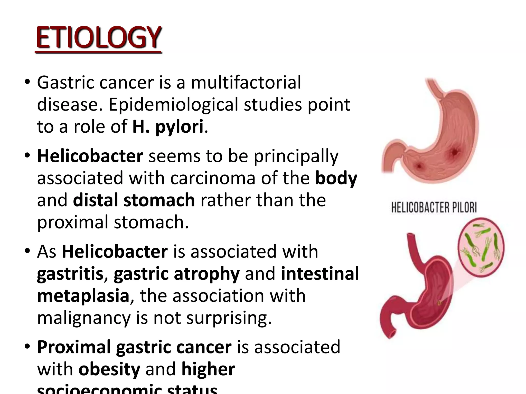
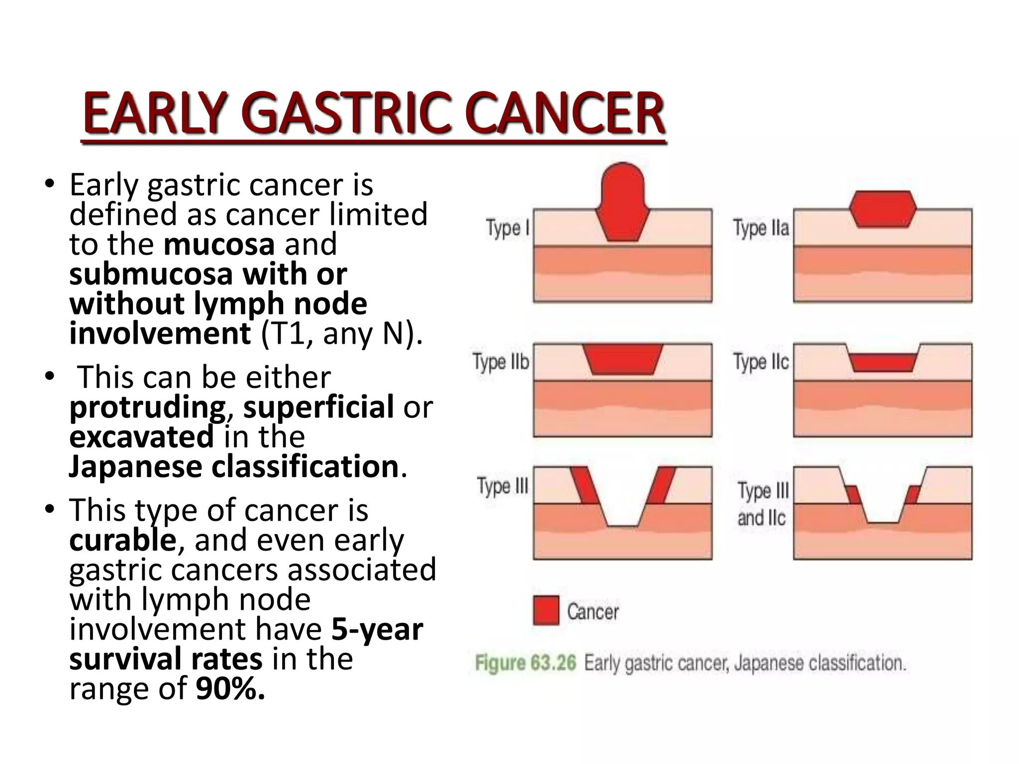
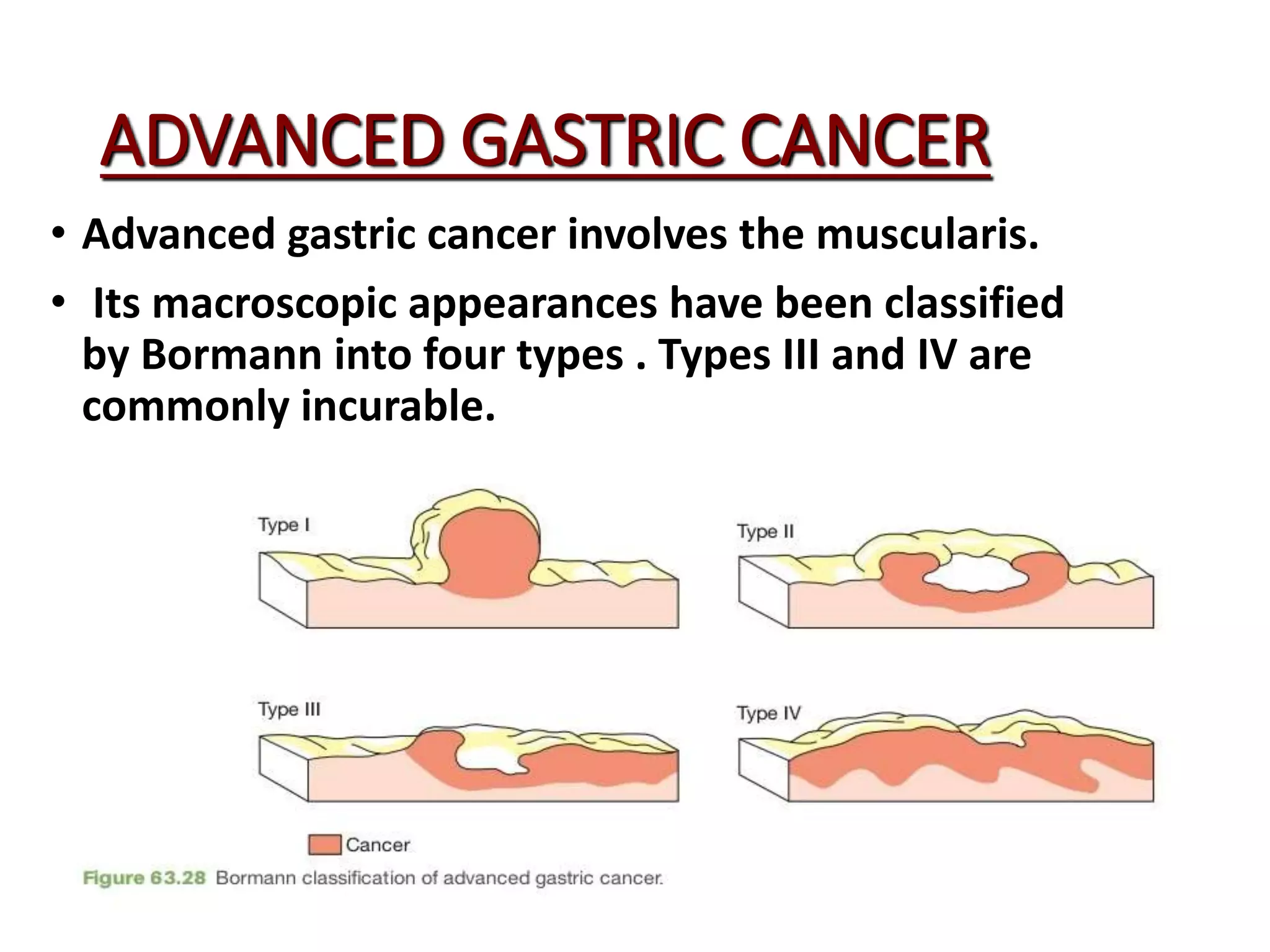
A power point presentation on classification, types and investigations of gastric/stomach cancer presented by students to the faculty of private medical teaching hospital and approved by the surgery department of the college. The resources taken are guyton and hall book of physiology and bailey and Love's short practice if surgery.
2. CA STOMACH
• Carcinoma of the stomach is a major
cause of cancer mortality
worldwide. Its prognosis tends to be
poor although better results are
obtained in Japan, where the
disease is common.
• Gastric cancer is actually an
eminently curable disease provided
that it is detected at an appropriate
stage and treated adequately.
• Early diagnosis is therefore the key
to success with this disease.
• Unfortunately, the late presentation
of many cases is the cause of the
3. ETIOLOGY
• Gastric cancer is a multifactorial
disease. Epidemiological studies point
to a role of H. pylori.
• Helicobacter seems to be principally
associated with carcinoma of the body
and distal stomach rather than the
proximal stomach.
• As Helicobacter is associated with
gastritis, gastric atrophy and intestinal
metaplasia, the association with
malignancy is not surprising.
• Proximal gastric cancer is associated
with obesity and higher
6. LAUREN CLASSIFICATION
INTESTINAL GASTRIC CA
• In intestinal gastric
cancer, the tumour
resembles a carcinoma
elsewhere in the
tubular gastrointestinal
tract and forms
polypoid tumours or
ulcers.
• It probably arises in
areas of intestinal
metaplasia.
DIFFUSE GASTRIC CA
• Diffuse gastric cancer
infiltrates deeply into
the stomach without
forming obvious mass
lesions, but spreads
widely in the gastric
wall.
• Not surprisingly, this
has a much worse
prognosis.
7. EARLY GASTRIC CANCER
• Early gastric cancer is
defined as cancer limited
to the mucosa and
submucosa with or
without lymph node
involvement (T1, any N).
• This can be either
protruding, superficial or
excavated in the
Japanese classification.
• This type of cancer is
curable, and even early
gastric cancers associated
with lymph node
involvement have 5-year
survival rates in the
range of 90%.
8. ADVANCED GASTRIC CANCER
• Advanced gastric cancer involves the muscularis.
• Its macroscopic appearances have been classified
by Bormann into four types . Types III and IV are
commonly incurable.
9. Spread of CA stomach:
• DIRECT SPREAD: The tumour penetrates the muscularis,
serosa and ultimately adjacent organs such as the
pancreas, colon and liver.
• LYMPHATIC SPREAD: This may be extensive, the tumour
even appearing in the supraclavicular nodes (Troisier’s
sign).
• Unlike malignancies such as breast cancer, nodal
involvement does not imply systemic dissemination.
• BLOOD BORNE METASTASIS: These occur first to the liver
and subsequently to other organs, including lung and
bone. This is uncommon in the absence of nodal disease.
10. CONTINUED...
• TRANSPERITONEAL SPREAD: This is a common mode
of spread once the tumour has reached the serosa of
the stomach and indicates incurability.
• Tumours can manifest anywhere in the peritoneal
cavity and commonly give rise to ascites.
• The ovaries may sometimes be the sole site of
transcoelomic spread (Krukenberg’s tumours).
• Tumour may spread via the abdominal cavity to the
umbilicus (Sister Joseph’s nodule).
• Transperitoneal spread of gastric cancer can be
detected most effectively by laparoscopy and cytology.
11. GASTROINTESTINAL STROMAL TUMOR
• Accounts for 1-3% of all gastrointestinal
tumors.
• Arise from the interstitial cells of cajal and
are part of normal autonomic nervous
system of stomach.
• 50-70% occur in stomach, 10-20% in small
intestine and 10% in other parts of GIT
• All GIST are responsive to tyrosin kinase
antagonists like imatinib and they don't
express actin and desmin like other smooth
muscle tumors (differentiating point).
• Difficult to diagnose endoscopically because
mucosa over the tumor appears normal until
it ulcerates.
• The metastasis occurs in liver and
peritoneum but lymph nodes are rarely
involved.
12. GASTRIC LYMPHOMA
PRIMARY GASTRIC LYMPHOMA:
• Arise on the background of chronic gastritis as normal
stomach is devoid of lymphoid tissue.
• In the setting of chronic gastritis stomach acquires MALT
which can undergo lymphoid degeneration. H. pylori is
thought to be the main causative organism.
• They range from low grade to poorly differentiated high
grade tumors.
• CT scan of abdomen, pelvis and thorax should be
performed to rule out systemic lymphoma.
• H.pylori eradication therapy is used for low grade
lymphoma associated with infection (70-95% cases).
14. SYMPTOMS:
• Every patient over the age of 45 presenting
with dyspepsia should undergo endoscopy
before starting treatment.
• Most common cause of late presentation is
liberal use of H2 receptor antagonists and
proton pump inhibitors as these drugs
relieve the symptoms of malignancy as well.
15. SYMPTOMS:
• Palpable epigastric mass which signifies
curable disease.
• Tumor may bleed leading to iron def anemia.
• Perforate leading to pruritus.
• Obstruction leads to dysphagia and
persistent vomiting.
16. PHYSICAL SIGNS
• Signs associated with advanced or metastatic
disease.
• Palpable abdominal mass in epigastrium.
• Palpable periumbilical lymph nodes.
• Palpable ovarian mass (krukenberg tumor)
• Palpable mass on DRE.(blumer shelf)
17. INVESTIGATIONS
• Gastroscopy is the gold
standard for diagnosis and
screening.
• Exact site of tumor can be
located and biopsy can be
obtained to establish tissue
diagnosis.
• In case of high index of
suspicion, if biopsy turned
out to be negative for
malignancy , patient should
be re endoscope and more
aggressive biopsy should be
obtained.
18.
19. INVESTIGATIONS:
Endosopic ultarsonography
helps to define the limit of
spread of gastric cancer into
gastric wall and can also
identify lymph node
involvement.
CT scan of abdomen , pelvis
and chest provides info about
local extrnt of tumor , lymph
node involvement and
systemic metastasis. Play a key
role for treatment planning .