
More Related Content
Similar to final_minerals.pptx
Similar to final_minerals.pptx (20)
Recently uploaded
Recently uploaded (20)
final_minerals.pptx
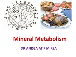
- 1. DR ANISSA ATIF MIRZA Mineral Metabolism
- 2. Introduction
- 4. Minerals Minerals are Inorganic elements Not biosynthesized in human body Widely distributed in nature Present in foods of Plant and Animal origin
- 5. Minerals In Human body
- 6. •Minerals in human body serve for various structural and functional roles •Hence it is essential to ingest Minerals through diet.
- 7. Human Body Ingests Seven Food Nutrients Food Substances Carbohydrates Proteins Water Dietary Fiber Minerals Vitamins Lipids
- 8. Minerals In Human body Minerals are Nutrient Of Human Food Essential Nutrient Micro Nutrient Non Calorific Nutrient
- 10. Minerals • Natural in Occurrence • Solid in nature • Inorganic • Definite chemical composition • Crystal structure due to internal arrangement of atoms
- 11. Minerals ingested are not changed in the body. Minerals are not destroyed by heat, light, acid or mixing
- 13. Body Minerals 30 Chemical elements are identified as Minerals. Important for human growth, development and regulation of vital functions
- 14. •Minerals are classified based on: Functional need to body Its daily requirement
- 15. Two Broad Classes Of Minerals • Macro Minerals – 60-80 % • Micro Minerals- 20%
- 16. •Macro/Principle/Chief Minerals • Body needs Macro Minerals relatively in large quantities • Minerals present in body tissues at concentrations >50 mg/kg • Requirement of these Minerals is >100 mg/day
- 17. 7 Names Of Macro/Chief Minerals 1. Calcium (Ca) 2. Phosphorus (P) 3. Sulfur (S) 4. Magnesium (Mg) 5. Sodium (Na) 6. Potassium (K) 7. Chloride (Cl)
- 18. • Micro Minerals /Trace Elements •Body needs Micro Minerals relatively in less amount •Present in body tissues at concentrations <50 mg/kg •Requirement of these Minerals is ﹤100 mg/day
- 19. Subclasses Of Micro/Trace Minerals •Essential Trace Elements •Possibly Essential Trace Elements •Non Essential Trace Elements
- 20. Name Of 10 Essential Micro/Trace Elements 1. Iron (Fe) 2. Copper (Cu) 3. Cobalt (Co) 4. Chromium (Cr) 5. Fluoride (F) 6. Iodine (I) 7. Manganese (Mn) 8. Molybdenum (Mo) 9. Selenium (Se) 10.Zinc (Zn)
- 21. Possibly Essential Elements for Humans Ni, Si, Sn, V, Ba, Li
- 22. Non Essential Trace Elements Of Humans Pb, Hg, Al, Ag, Bo
- 23. Body Minerals Biological forms of minerals in living systems Na K Ca Mg P Cl C N H O S Fe Zn Cu Mn Se V Si As Mo I Co Br F
- 24. Nutritionally Important Minerals Macro Minerals Trace Elements Element g/kg Element mg/kg Ca P K Na Cl S Mg 15 10 2 1.6 1.1 1.5 0.4 Fe Zn Cu Mo Se I Mn Co 20-50 10-50 1-5 1-4 1-2 0.3-0.6 0.2-0.5 0.02-0.1
- 25. Minerals in the Body
- 26. General Characteristic Features Of Human Body Minerals
- 27. Sources Of Minerals To Human Body
- 28. •A mixed diet of varied foods •Is the best source of Minerals
- 29. Minerals in Foods • Minerals are found in all food groups. • More reliably found in –Fresh Fruits –Vegetables –Animal products
- 30. Factors Affecting Mineral Requirements • Form of Mineral fed - Inorganic vs Organic forms • Interactions with other minerals • Tissue storage • Physiological State
- 31. Site for Mineral Absorption Small intestine Large intestine
- 32. Variable Bioavailability of Minerals
- 33. Bioavailability Of Minerals • Bioavailability (absorption capacity) of Minerals is influenced by : –Genetics – Aging – Nutritional Status –Other food compounds
- 34. Nutrient Interactions Some food components bind with Minerals reducing their bioavailability Mineral interactions can affect another minerals absorption, and excretion
- 35. • Often other substances in foods decrease absorption (bioavailability) of Minerals: –Oxalate, found in spinach, prevents absorption of most Calcium in spinach. –Phytate, in most plants makes minerals poorly available
- 36. Oxalate Phytate
- 37. Factors Affecting Requirements • Interactions with other Minerals
- 38. • Phosphorous binds with Magnesium in the small intestine. • So Magnesium absorption is limited when Phosphorous intakes are high
- 39. Uptake And Transportation Of Minerals
- 40. Some Minerals require no carriers to transport into intestinal wall. Some Minerals require carriers to enter into intestinal wall.
- 41. • Excretion and Regulation Site Of Minerals. –Small intestine –Kidneys
- 42. General Functions of Minerals
- 43. • Minerals with structural functions: Ca, P ,Mg in bones; S in Keratin. • Minerals serve as Inorganic Cofactors: participate with Enzymes in metabolic processes . • Role of Minerals in Acid-Base and Water balance: Na+, K+ and Cl-
- 44. • Minerals have role in Nerve & Muscle Function : Ca, Na, K, Mg • Minerals are components of certain biomolecules: –Fe- Heme, –Co- Vitamin B12 –I2-Thyroid hormones.
- 45. Mineral Deficiencies and Excesses
- 47. • Most Minerals have an optimal range in blood/body. –Minerals below range leads to deficiency symptoms –Minerals above range leads to toxicity symptoms
- 48. •Deficiency and excess of Minerals in human body •Affect the normal health and vitality of human body •Which may lead to suffer from various manifestations.
- 49. Note •Mineral content of soils •Dictates Mineral status of plants.
- 50. • Mineral deficiencies usually are rare • As they are widely distributed and essentially taken through food. • However there are many deficiency cases noted of • Iron , Iodine and Calcium deficiencies.
- 51. •It may take many months to develop Mineral toxicity. –The time taken to develop is impacted by body stores.
- 52. Study Of Specific Minerals
- 55. •Symbol : Ca+2 •Divalent Cation •Atomic Weight : 40 g/mol •Atomic Number: 20 •Nature :Soft Grey Alkaline Earth Metal
- 56. •Calcium is the most essential abundant Macromineral of human body. •Fifth most abundant element in Earth´s crust
- 57. Calcium Occurrence In Nature • Naturally Calcium does not exist freely • Calcium occurs in form of Salts –Limestone (CaCO3) –Gypsum (CaSO4*2H2O) –Fluorite (CaF2)
- 58. Calcium In the Human Body Calcium is the most abundant Macro Mineral Average adult body contains approx. 1.5 kg of Calcium.
- 59. • 99% of the body Calcium is associated to skeleton (Bones and Teeth). • 1% Calcium is present in other tissues and body fluids.
- 60. Calcium in bones is in dynamic state Calcium of bones may serve as large reservoirs storing excess Calcium Bones releases Calcium when extracellular fluid Calcium concentration decreases.
- 62. Calcium Dietary Requirements –Adult : 800 mg/day –Pregnancy, lactation and post-menopause: 1500mg/day/1.5 g/day –Growing Children: (1-18 yrs): 1200 mg/day –Infants: (< 1 year): 300-500 mg /day
- 63. Ca – Daily Requirements Age/ sex Ca (mg) 1-3 350 4-6 450 7-10 550 11-18 M 1000 11-18 F 800 19 + 700
- 64. Dietary Sources Of Calcium
- 65. Dietary Calcium sources • Rich Calcium Sources - Milk and Milk Products - Millet (Ragi) - Wheat-Soy flour - Black strap molasses
- 67. • Calcium Good sources - Yoghurt, sour cream, ice cream - Tofu - Gauva ,Figs - Cereals - Egg yolk - Legumes
- 68. - Green leafy vegetables as collard, kale , Broccolli, Cabbage and raw turnip - Small Fish as trout, salmon and sardines with bones - Meat - Almonds, brazil nuts, dried figs, hazel nuts - Also soybean flour and cottonseed flour
- 69. Ca – Dietary Sources • Milk – 100 ml =120mg • Cheese – 15gm = 110mg • Yoghurt pot – 80gm = 160mg
- 71. •Absorption of Calcium occurs in the small intestine •In Duodenum and first half Jejunum
- 72. •Calcium must be in a soluble and ionized form for its absorption. •Calcium salts are unabsorbable forms.
- 73. Calcium Absorption •Absorption depends on need of Calcium to body: •Particularly high during growth, pregnancy and lactation
- 74. Calcium Transport Mechanism Across Intestinal Mucosal Membrane
- 75. Ca Absorption Mechanism Simple diffusion/Passive An active transport involving Ca pump
- 76. Calcium Passive Transport • Is a non saturable, paracellular • It is less efficient process • Is not affected by calcium status or parathyroid hormone
- 77. •Active Absorption of Calcium: • Against electrical and concentration gradient, by an energy dependent active process.
- 78. Calcium Active Transportation Regulated by the active form of Vitamin D/Calcitriol. Which involves Calbindin (Calcium-Binding Protein) –
- 80. Parathyroid Hormone (PTH) indirectly enhances Ca absorption through the increased activation of Calcitriol.
- 81. Calcitriol Calcitriol /activated Vitamin D , induces the synthesis of Ca binding protein Calbindin Calbindin in the intestinal epithelial cells then promotes Ca absorption.
- 82. Acidity (low pH) Acidity is more favorable for Ca absorption. Calcium salts are soluble in acid solutions So acidity increases the absorption of Calcium.
- 83. Lactose , Citric acid promotes Calcium uptake by intestinal cell.
- 84. • Amount of Proteins in Diet: • Amino acids Lysine and Arginine form soluble complexes with Calcium • Hence high protein diet favors the absorption of Calcium.
- 85. • Concentration of Calcium in diet: • Higher the concentration of Calcium • More is the absorption of Calcium.
- 87. • Phytates and Oxalates present in plant origin diet form insoluble salts and interfere with Ca absorption.
- 88. • The high content of dietary Phosphates results in the formation of insoluble Ca phosphate and prevent Ca uptake. • Dietary ratio of Ca : P ---1:1 / 2:1 • is ideal for Ca absorption.
- 89. •The Free Fatty acids react with Ca to form insoluble Ca soaps.
- 90. •The Alkaline condition (high pH) is unfavorable for Ca absorption.
- 91. •Low Estrogen levels in postmenopausal women lowers Calcium absorption. •Since Estrogen increases Calcitriol levels
- 92. •High content of Dietary fiber,Caffeine,Sodium interferes with Ca absorption.
- 93. • Amount of Magnesium in diet: Excess Magnesium in diet inhibits Calcium absorption. • As Magnesium competes with Calcium for absorption.
- 94. Calcium Absorption and Excretion at GIT
- 95. •Usual Ca intake is1000 mg/day. •About 35 % is absorbed (350 mg/day) by the intestine.
- 96. •Remaining Calcium in the intestine is excreted in the feces •250 mg/day enters intestine via secreted gastrointestinal juices and sloughed mucosal cells
- 97. 90 % (900 mg/day) of the daily intake is excreted in the feces 10 % (100 mg/day) of the ingested calcium is excreted in the urine.
- 99. •Total content of Calcium in an Adult body is 1-1.5 Kg. •Calcium constitutes 2% of total body weight.
- 100. BODY CALCIUM •99% of Calcium is in Bones •0.8% of Calcium is in soft tissues (ICF) •0.1% in Blood ( ECF)
- 101. PLASMA CALCIUM
- 102. Three Forms of Circulating Ca2+
- 103. Diffusible Calcium • 50% Ca2+ Ionized/Physiologically active form. • 10% combined with anions (Citrate, Phosphate) –Non-dissociated/Non ionizable form.
- 104. Non diffusible Calcium • 40% combined with plasma proteins • Combination with proteins depends on pH 0.2 mmol/l ,Ca2+ on each pH unit
- 105. Blood Calcium Levels •The normal serum total calcium is: – 9-11 mg/dL – 2 -3 mmol/L
- 106. • Normal levels of the ionized/free/diffusible/physiological form of Calcium is –4.5-5.6 mg/dL –1.1-1.4 mmol/L
- 107. •Protein bound Calcium (Mostly bound to Albumin)/Non diffusible/Bound form of Calcium: 4 mg%.
- 108. •Calcium Salts /Bound form/Inorganic Salts/Diffusible: • Calcium Phosphate and Calcium Citrate=1mg%
- 110. Calcium In Alkalosis •Alkalosis favors binding of more Calcium with Proteins. •This consequently lowers ionized Calcium.
- 111. Acidosis Favors Ionization of Calcium
- 112. Multiple Biological Functions of Calcium
- 113. •Calcium is widely distributed in the body • Involved with many functions to keep the body vital and active.
- 114. 1. Structural Role Of Calcium •Calcium is a major structural element in the vertebrate skeleton forms bones and teeth.
- 115. • Calcium along with Phosphorous, Magnesium forms the inorganic matrix of the bone as Hydroxyapatite crystals • Which gives the tensile strength to the bones and teeth.
- 116. •In the form of Calcium Phosphate(Ca10(PO4)6(OH)2 known as Hydroxyapatite
- 117. •Osteoblasts are responsible for bone formation •While Osteoclasts are for bone resorption.
- 118. •Bones undergo mineralization during osteoblastic activity • Demineralization during osteoclastic activity.
- 119. Bone Act As Major Reservoir Of Calcium
- 120. • Osteoclasts secrete acid, causing the release of calcium and phosphate into the bloodstream. • There is constant exchange of calcium between bone and blood.
- 121. Leg bone of a horse showing the trebecular (spongy) bone and the cortical (solid) bone. This bone is able to withstand forces generated by this 1,500 lb animal Trebecular bone of the lower spine. Changes with aging. Cortical bone with Halversion system (a series of channels supplying nutrients). Black dots are osteocytes Cross section through trebecular and cortical bone revealing the internal architecture surrounded by marrow tissue.
- 122. Demineralized bone: Shown is the organic matrix consisting mostly of collagen upon which the bone crystals are laid.
- 123. Hydroxyapatite (crystal structure) Ca10(PO4)6 OH2 Ca P O H
- 124. Remember/Note • During growth , pregnancy and lactation phase • To give strength for building bones and teeth. • One should take adequate amounts of dietary Calcium and Phosphorous
- 125. 2. Calcium Role in Muscle Contraction •The ionized free form of Calcium interacts with •Muscle Protein Troponin C to trigger muscle contraction.
- 126. • Calcium also activates Ca-ATP ase and increases the interaction between Actin and Myosin during muscle contraction. • Thus Calcium has role in excitation and contraction of muscle fibers.
- 127. 3. Role Of Calcium In Nerve Impulse Conduction •Ionized Calcium transmits nerve impulses •From pre-synaptic to post- synaptic region.
- 128. 4. Role of Calcium in Hormonal Actions •Calcium serves as second and third messenger for certain hormonal activities. •Calcium –Calmodulin complex mediates the hormonal action.
- 129. • Calmodulin is a Calcium binding regulatory Protein which binds with 4 Calcium ions. • Calmodulin serve as messenger during hormonal action by stimulating Protein Kinases.
- 130. •Epinephrine require Calcium as second messenger at the time of its action. •ADH require Calcium as third messenger during its action.
- 131. 5. List Of Enzymes Activated By Calcium and Mediated By Calmodulin • Adenyl Cyclase • Glycerol-3-PO4 Dehydrogenase • Glycogen Synthase • Pyruvate Carboxylase • Pyruvate Dehydrogenase • Pyruvate Kinase
- 132. 6. Calcium as Chelating Agent In Blood Clotting Mechanism. • Calcium as Clotting factor IV serves as a cofactor for several reactions in the Cascade of blood clotting process. • Calcium serves as chelating agent during Thrombin formation.
- 133. 7.Calcium act as a Cofactor of Enzymes • Calcium serve as an inorganic cofactor of: (Direct action) –Pancreatic Lipase –ATPase –Succinate Dehydrogenase
- 134. 8.Calcium Role in Secretion of Hormones • Calcium stimulates to release of following Hormones: –PTH –Insulin –Calcitonin –Vasopressin/ADH
- 135. 9.Calcium Transport Across The Biomembranes • The cell membrane is generally impermeable to Calcium ions. • Calcium influx into cells is via Calcium channels by Na /Ca exchange mechanism.
- 136. •There are different Calcium Channels located in the membranes of various cell organelles.
- 137. 10. Calcium Prolongs Systole •Calcium acts on Heart and prolongs Systole. •Hypercalcemia may lead to Cardiac arrest in Systole.
- 138. Remember •When Calcium is administered intra venously •It should be infused very slowly to avoid the cardiac arrest.
- 139. 11.Calpains – Calcium Dependent Cysteine Proteases •Calpains are involved in: •Cell mobility •Cell cycle progression •Cell membrane fusion events
- 140. •Cell fusion in Myoblasts •Neural vesicle Exocytosis •Platelet aggregation
- 141. •Increased concentration of Calcium in cells. •Increases Calpain activation.
- 142. •Increased Calpains causes unregulated proteolysis . • Hyperactivity of Calpains consequent leads to irreversible tissue damage.
- 143. • Calcium is a key component in the maintenance of the cell structure • Membrane rigidity, permeability and viscosity are partly dependent on local calcium concentrations
- 144. •11. Calcium promotes • Transportation of water and ions across the membranes. • Excitability of cell membranes.
- 145. •Calcium regulates cellular secretory process such as : •Endocytosis •Exocytosis •Cell motility
- 146. • Calcium has role in: –Cell to Cell contact –Cell to cell communication –Cell adhesion in tissues
- 147. •Calcium is added to mothers milk during lactation phase of women.
- 148. Calcium Active Role: - In the relaxation and constriction of muscles - In nerve impulse transmission - As an intracellular signal - In cell aggregation and movement - In secretion of hormones - In cell division
- 149. Calcium Passive Role: - As a cofactor for many enzymes (e.g. Lipase) and proteins - As component in the blood clotting cascade
- 151. Homeostasis Of Blood Calcium OR Regulation of Blood Calcium
- 152. • The normal levels of total serum Calcium is 9-11 mg%. • It is very essential to maintain the constant range of Calcium. • For normal health and survivallence of human body.
- 153. • Most important is the ionized or physiological form of Calcium present in blood • This plays an important roles in various physiological and metabolic functions of human body.
- 154. • Maintenance of calcium homeostasis. –Regulation in dietary absorption –Storage –Excretion of Ca
- 155. Factors Regulating Blood Calcium Levels •Parathyroid Hormone (PTH) •Vitamin D- Calcitriol •Calcitonin
- 156. •The PTH , Calcitriol and Calcitonin cooperatively works •To regulate the transiently increased and decreased levels of serum Calcium .
- 157. Parathyroid hormone (PTH) PTH is secreted by two pairs of parathyroid glands. PTH is initially synthesized as a precursor, preProPTH. Two proteolytic cleavages produce the ProPTH and the secreted form of PTH (84 aa).
- 159. •The secretion of PTH are promoted •By low Ca2+ concentration in blood.
- 160. Regulation of PTH Secretion and Biosynthesis • Extracellular Ca 2+ regulates secretion of PTH –Low Ca 2+ increases PTH levels –High Ca 2+ decreases PTH levels
- 161. Mechanism of action of PTH •PTH is the most important endocrine regulator of Ca and Phosphorous concentration. •Function: –Elevate serum Ca level.
- 162. •PTH has 3 independent tissues to exert its action. •Intestine (Indirectly) •Bone (Directly) •Kidney (Directly)
- 163. • PTH Regulates through 3 Main Effects: -Stimulating activation of vitamin D → ↑ intestinal Ca absorption -Stimulating bone resorption -Increasing renal tubular calcium reabsorption
- 164. Actions of Parathyroid Hormone On Bone
- 165. •Parathyroid hormone acts directly on bone to stimulate resorption •This releases Ca2+ into the extracellular space and fluids (slowly)
- 166. PTH Action on the Bone • Decalcification or Demineralization of bone, carried out by osteoclasts. • →blood Ca level ↑ • Note: this is being done at the expense of loss of Ca from bone, particularly in dietary Ca deficiency.
- 167. • Gs protein-coupled receptors in osteoblasts increase cAMP and activate Protein Kinase Activity (PKA) • This Inhibits osteoblast function • This occurs when PTH is secreted continuously.
- 169. Circulating Forms of PTH
- 170. Action Of PTH on the Kidney and Intestine •Action on the Kidney: increase the Ca reabsorption. •Action on the Intestine: indirect, increase the intestine absorption of Ca by promoting the synthesis of Calcitriol.
- 171. • PTH Effects in Kidney –Parathyroid hormone acts directly on kidney –To increase calcium reabsorption and phosphate excretion (rapid)
- 172. • Gs protein-coupled receptors • Parathyroid hormone acts on distal tubule • Increases renal reabsorption of Calcium. • Adds Calcium to blood regulating its levels.
- 174. Role Of Calcitriol/ Activated Vitamin D - Calcitriol several effects on the intestine and kidneys that increase absorption of calcium and phosphate into the extracellular fluid - Important effects on bone deposition and bone absorption
- 175. PTH and Calcitriol By their Activity Increases Blood Calcium Levels
- 176. •Calcium levels below subnormal levels •Stimulates the secretion of PTH •PTH then stimulates the Vitamin D activation to Calcitriol.
- 179. Activation of Vitamin D3 - Cholecalciferol formed in the skin by sun - Converted in liver and Kidney to - 1,25 DHCC Controlled by PTH - Plasma calcium concentration inversely regulates 1,25 DHCC
- 180. •PTH and Calcitriol then acts on three target organs •They try to increase the blood Calcium levels by their Hypercalcemic action.
- 182. Action On Intestinal Mucosal cells • Calcitriol enters intestinal mucosal cells. • Acts like Steroidal hormone • Stimulate the biosynthesis of Calbindin a Calcium binding Protein by gene expression. • Calbindin binds with dietary Calcium in GIT, promotes it absorption and diffuse in blood.
- 183. Calcitriol Action On Renal Tubules • Calcitriol acts on renal tubules and increases tubular renal absorption of Calcium from plasma ultra filtrate there by decreasing excretion of Calcium. • The reabsorbed Calcium by renal tubules add Calcium to blood these by increasing blood Calcium levels.
- 184. Action On Bones • PTH hormone directly acts on bones causing decalcification of bones • To release bound form of Calcium into free form, catalyzed by increased activity of ALP • Which increases the levels blood Calcium to blood there by increasing blood Calcium levels to attain a normal level of 9-11 mg%
- 185. Remember • The low intake of dietary Calcium may increase the bone resorption by PTH to regulate blood Calcium levels. • This may decrease the blood Calcium content of bones • Leading to weakness in bones manifesting bone pain and recurrent bone fractures.
- 186. Calcitonin • Calcitonin a peptide hormone (32 aa) secreted by the parafollicular cells of Thyroid gland - Calcitonin tends to decrease plasma Calcium concentration
- 187. Role Of Calcitonin In Decreasing The Blood Calcium Views • When ever the blood Calcium goes above 11 mg% • The Calcitonin by its Hypocalcemic action • Tries to lower the increased the blood Calcium levels.
- 188. •Calcitonin promotes the bone mineralization or Calcification of bones. •The blood Calcium is taken up by bones and reserved.
- 189. • Thus Calcitonin increases Osteoblasts activity • Enhances bone mineralization. • Promotes bone growth • Reduces increased blood Calcium levels to attain 9-11 mg%.
- 190. -Calcitonin adds Calcium to bones and increases bone mineralization.
- 191. •CT has the ability to decrease blood Ca and P levels and its major target cells also in bone, kidney and intestine. 1. Bone: Stimulate Osteogenesis. 2. Intestine: Inhibit absorption of Ca. 3. Kidney: enhance of Ca excretion from urine. Role Of Calcitonin (CT)
- 192. •PTH and Calcitonin are antagonistic in actions. •OR •Action of Calcitonin is opposite to that of PTH.
- 193. Hormonal Regulators •Calcitonin (CT) –Lowers Ca++ in the blood –Stimulates Osteoblasts –Inhibits Osteoclasts
- 194. •Parathormone (PTH) –Increases Ca++ in the blood –Stimulates Osteoclasts
- 195. • Calcitriol –Increases Ca++ in the blood –Increase Ca++ uptake from the gut –Stimulates osteoclasts
- 196. (+)
- 197. Regulation of Calcium Homeostasis
- 198. Calcium Turnover
- 199. Calcium Balance • Calcium Intake = Calcium output • Negative calcium balance: Output > intake –Negative Ca2+ balance leads to osteoporosis • Positive calcium balance: Intake > output –Positive Ca balance occurs during
- 200. Calcium Balance
- 201. Exercise and Calcium •Normal bone function requires weight-bearing exercise •Total bed-rest causes bone loss and negative calcium balance.
- 202. Calcium Homeostasis
- 203. Calcium and the Cell • Translocation across the plasma membrane • Translocation across the ER and mitochondrion; Ca2+ ATPase in ER and plasma membrane
- 206. bone 1000 g Ca++ stored in bone Calcium Homeostasis Blood Ca++ small intestine kidney Ca++ lost in urine Calcium in the diet calcium lost in feces Ca++ absorbed into blood calcium resorption calcium deposition
- 207. intake excretion 1000 g Ca++ stored in bone Calcium Homeostasis Blood Ca++ small intestine kidney Ca++ lost in urine Calcium in the diet calcium lost in feces Ca++ absorbed into blood calcium resorption calcium deposition storage bone
- 208. Calcium Homeostasis Blood Ca++ small intestine kidney Ca++ lost in urine Calcium in the diet calcium lost in feces bone Ca++ absorbed into blood calcium resorption calcium deposition
- 209. Calcium Homeostasis Blood Ca++ small intestine kidney Ca++ Ca++ Ca++ bone 1,25 Vit. D3 (+) 1,25 Vit D3 deposition Calcitonin (-) Ca++ PTH Parathormone (+) resorption
- 210. 1,25 Vitamin D3 Cholesterol precursor 7-dehydrocholesterol UV Vitamin D3 25 Vitamin D3 1,25 Vitamin D3 Low plasma Ca++ increase kidney enzymes
- 211. Excretion Of Calcium
- 212. Excretion of Ca •Mostly through the intestine. •Partly through the kidney.
- 213. Calcium Excretion In feces: 80% In urine: 20%
- 214. •Unabsorbed dietary Calcium is mostly excreted out through feces.
- 215. •Excretion of Ca into the feces is a continuous process •This is increased in vitamin D deficiency
- 216. When Will Calcium Excreted In Urine?
- 217. • The renal threshold for Calcium is 10 mg%. • When blood Calcium crosses more than 10 mg% it is excreted in Urine.
- 218. Excretion of Calcium under influence of PTH.
- 219. •The excretion of Calcium and Phosphorous is reciprocally regulated. •If Phosphorous excretion is increased Calcium excretion is decreased.
- 220. • Conditions Increasing Excretion Calcium - Low Parathyroid hormone (PTH) - High extracellular fluid volume - High blood pressure - Low plasma Phosphate - Metabolic Alkalosis
- 221. • Excretion Of Calcium Is decreased by: - High Parathyroid hormone - Low extracellular fluid volume - Low blood pressure - High plasma phosphate - Metabolic acidosis - Low Vitamin D3
- 222. Clinical Significance Of Calcium
- 224. Defect In Following Factors Leads to Calcium Related Disorders • Dietary Intake Of Calcium • Role of PTH , Calcitriol and Calcitonin • Status of Parathyroid , Thyroid , Liver and Kidney
- 225. Investigations To Diagnose Calcium Related Disorders • Serum Ca and Pi levels • PTH • Vit D ( 1,25 Dihydroxy levels) • Mg • Urinary Ca/ Cr ratio
- 227. Hypercalcemia •Hypercalcemia is the condition where there is •Persistent high levels of blood Calcium above 11 mg%.
- 228. Conditions Leading To Hypercalcemia •Excessive intake of Calcium •Hyperparathyroidism- Increased PTH •Parathyroid Adenoma •Hypervitaminosis D
- 229. • Pagets Disease (Increased Release From Bones) • Addisons Disease (Decreased Excretion Of Calcium) • Bone Tumors (Leak of Calcium From Bones) • Multiple Myeloma-Leukemia , Polycythemia • Milk Alkali Syndrome( Calcium+Alkali)
- 230. –Excessive use of antacids with phosphate-binding –Prolonged immobility –Thiazide diuretics –Thyrotoxicosis
- 231. Hypercalcemia Signs and Symptoms •Muscle weakness •Personality changes •Nausea and Vomiting •Polyuria •Extreme thirst
- 232. • Anorexia • Constipation • Pathological fractures • Calcifications in the skin and Cornea • Cardiac arrest(prolonged Systole)
- 233. Clinical manifestations of Hypercalciemia Osteodystrophy (Recklinhauzen disease) Cystosis swelling in the distal ends of both fibula bones Mechanism Hyperparatireosis – increasing of Са in blood – waste of Са from bones by resorbtion – osteoporosis – overgrowth of connective tissue (but Са isn’t deposited) - osteofibrosis
- 234. Hypocalcemia • Hypocalcemia is the condition where there is persistent low levels of blood Calcium below 9 mg %. • Hypocalcemia is more dangerous and life threatening if not corrected and managed timely.
- 235. Conditions Causing Hypocalcemia •Malnutrition and Malabsorption •Diarrhea •Acute Pancreatitis •Hypoparathyroidism •Hypovitaminosis D
- 236. • Rickets • Osteomalacia • Renal Rickets (Deficiency of 1α Hydroxylase) • Fanconis Syndrome • Hypoalbuminemia( Decreases Protein bound Calcium)
- 237. • Chronic kidney failure • Low blood magnesium level (in cases with severe alcoholism) • Diet high in Phytate
- 238. Hypocalcaemia – Clinical Features • Neuromuscular excitability • Paraesthesia (tingling sensation) around mouth, fingers and toes • Tetany • Muscle cramps, Carpopedal spasms
- 239. • Seizures – focal or generalised • Laryngospasm, Stridor and apneas (neonates) • Cardiac Rhythm disturbances (prolonged QT interval) • Chvostek’s and Trousseau’s signs – latent hypocalcemia
- 241. Calcium Deficiencies • Tetany • Rickets –In growing children's • Osteomalacia (Osteoporosis) –In adult animals
- 242. Tetany •Tetany is the manifestations caused due to hypocalcemia. •Serum Calcium below 7 mg % causes Tetany.
- 243. • Tetany is a life threatening condition. • Tetany may be suffered in persons whom –Parathyroid gland is surgically removed –Parathyroid dysfunction due to auto immune disorder.
- 244. •Low Calcium levels directly affects neuromuscular activity. •Leads to increased neuromuscular irritability of muscles.
- 245. •Twitching and spasm of muscles of face,hand,feet neck •Carpopedal and Laryngeal and Stridor Spasm.
- 246. Clinical Sign Of Tetany
- 247. •Chvostek’s Sign (Tapping over facial nerve causes facial contraction) •Trousseaus Sign (Inflation of BP Cuff for 3 minutes causes Carpopedal spasm).
- 248. ECG Changes In Tetany •Increased Q-T interval in ECG.
- 249. •Low blood Calcium •Increased Phosphate in blood •Low urine Calcium and Phosphorous
- 250. Treatment Of Tetany •Intravenous infusions of Calcium salts.
- 251. Cramps Clinical manifestations of Hypocalcaemia Tetany The process of tetany potentiation at the motor neurons and interneuron of spinal cord violate Conduction of impulses at reflex arch become easier Activate a reflex muscles contraction on mechanical and other stimuli Spasm of larynx, bronchus asphyxia death Coronarospasm (cardiotetanus) angina Stop of heart
- 252. Calcium Deficiencies -Rickets weakness and deformity of the bones that occurs from vitamin D deficiency or dietary deficiency of Ca and P in a growing person or animal.
- 253. Гіпокальциемія Ricket Clinical manifestations of Hypocalcaemia
- 254. Calcium and Osteoporosis • Around age 40, bone breakdown exceeds formation. • By age 65, some women have lost 50% of bone mass.
- 256. Calcium Deficiencies -Osteoporosis progressive loss of bone density, thinning of bone tissue and increased vulnerability to fractures in the elderly people of both sexes.
- 257. Calcium and Osteoporosis • Bone growth is greatest during “linear growth” – Peaks out at around age 30
- 258. How does Osteoporosis Look?
- 259. •Binding of Calcium to Albumin is pH dependent •Acute alkalosis increases calcium binding to protein and decreases ionized calcium Effect Of pH On Extracellular Calcium
- 260. • Patients who develop Acute Respiratory Alkalosis • Have increased neural excitability and are prone to seizures • This is due to: – Low ionized calcium in the extracellular fluid –Increased permeability to Sodium ions
- 261. Prevention is the Key •Maintain adequate Calcium and Vitamin D intake— •Perform weight-bearing exercise •Take Estrogen supplements?
- 262. Severe Symptomatic: •IV 10% Calcium Gluconate @ 0.11 mmol/kg (0.5 mls/kg – max 20 mls) over 10 minutes •Continuous IV infusion of Calcium Gluconate @ 0.1 mmol/kg (Max 8.8 mmols) over 24 hours Severe Asymptomatic: Oral Calcium Supplements @ 0.2 mmol/kg (Max 10 mmols or 400 mg Ca) 4 x a day Treatment of Hypocalcaemia
- 263. Calcium Toxicity • Calcium deposition in soft tissue • Impaired kidney function • Interference of other nutrient absorption –Iron & zinc
- 264. •Toxicity – Hypercalcemia (normally does not to occur) •Hyperparathyroidism, vitamin D intoxication, cancer are few causes.
- 265. Toxicity Of Calcium • MAS (Milk Alkali Syndrome) - Rare and potentially life threatening condition in individuals consuming large quantities of calcium and alkali - Characterized by renal impairment, alkalosis and Hypercalcemia: cause progressive depression of the nervous system
- 266. Pathogenesis unknown Metabolic calcinosis (інтерстиціальне звапнення) Limestone deposits in skin, tendons, fascias, muscles, along nerves and vessels
- 267. It arises in necrotic and dystrophic tissues - tuberculosis center , infarctions, dead fetus, chronic focus of inflamations (lungs and heart like an armor ), focuses of atherosclerosis, scar tissue Dystrophic static calcinosis (petrification) Mechanism: alkalinity conditions – increased absorption Са from blood – The increased activity of phosphatases, which prodused from necrotic cells – formation of insoluble salts of Са
- 268. Metastasic calcinosis Calcinosis of aortic valve
- 270. Phosphorous • Phosphorous is a Macromineral /Chief/Principle Element of human body. • It is a second most abundant Mineral of human body.
- 271. Daily Requirement/RDA of Phosphorous • The dietary Ca:P ratio ideally should be 1:1 for optimal absorption and functions. • Thus the requirement of dietary Phosphorous is more or less same as that of Calcium.
- 272. •Adults= 800 mg/day •Growing Children=1000 mg/day
- 273. Dietary Sources Of Phosphorous • Milk and Milk Products • Cereals • Egg • Meat • Green Leafy Vegetables , Cabbage , Cauliflower
- 274. Absorption Of Dietary Phosphorous •Absorption of Phosphorous is along with Calcium. •Hence factors promoting and inhibiting Calcium absorption are likely with Phosphorous.
- 275. • The Calcium and Phosphorous ratio in diet affects absorption and excretion of Phosphorous. • If any one of this is excess in diet the excretion of the other is increased.
- 276. Body Distribution Of Phosphorous •Total content of Phosphorous in an adult body is 1 Kg. •Phosphorous is present in each and every cell of body cell.
- 277. • 80 % of Phosphorous is present in bones and teeth along with Calcium as Hydroxyapatite crystals. • 10% of Phosphorous is present in Muscles and blood associated to Proteins, Lipids and Carbohydrate moieties.
- 278. •10 % of Phosphorous is component of various Phosphorylated biomolecules.
- 279. Phosphorous In Blood • In blood Phosphorous is present in following forms: • Free/Ionized Phosphorous: 40% –H2PO4- –HPO4- -
- 280. • Bound/Complex forms of Phosphorous: • Phosphorous bound and present as organic forms- Non diffusible form. • Phosphorous bound to other Cations /Inorganic salt : Calcium Phosphate
- 281. •Total Phosphorous levels in Whole blood= 40 mg% •Serum Inorganic Phosphorous •Adults= 2-4 mg% •Children's= 4- 6 mg%
- 282. • Fasting levels of serum inorganic Phosphorous are higher than post prandial values. • Since after meals the inorganic Phosphorous from blood are drawn into cells • Where it is utilized for phosphorylation of Glucose , Fructose and Galactose during metabolism.
- 283. • After a rich Carbohydrate diet there decreases serum inorganic Phosphorous levels. • In Diabetes mellitus the levels of serum inorganic Phosphorous get increased due to low utilization within cells.
- 284. Functions Of Phosphorous • Phosphorous along with Calcium has important role in bone mineralization and bone development. • Phosphorous and Calcium are components of Hydroxyapatite crystals of bone inorganic matrix.
- 285. • Phosphorous is important component during biosynthesis of certain Phosphorylated biomolecules viz: –Phospholipids –Nucleotides- Components of DNA and RNA –Phosphoproteins
- 286. • Phosphorylation reactions of metabolism and forming Esters ex Glucose-6-PO4,Fru-6-PO4 etc • High energy phosphorylated compounds. –Creatine Phosphate ATP,GTP ,CTP ,UTP. • Nucleotide Coenzymes :NAD+,NADP,FAD,PLP
- 287. • Phosphorous is a component of Phosphate Buffer system which participate in Acid Base Balance. –KH2PO4/K2HPO4 • Phosphorous is involved in phosphorylation of certain enzymes and bringing covalent modification.
- 288. Excretion Of Phosphorous •About 500 mg of Phosphorous is excreted through urine per day. •Renal threshold for Phosphorous is 2 mg%.
- 289. •PTH hormone stimulates the excretion of Phosphorous •By inhibiting tubular renal reabsorption of Phosphorous.
- 290. • Thus there is inverse relationship of PTH activity and serum Phosphorous levels. • Hyperparathyroidism decreases serum inorganic Phosphorous levels.
- 291. •Excretion of Phosphorous and Calcium is reciprocally regulated. •When Phosphorous is excreted Calcium is retained and vice a versa.
- 292. Disorders Associated To Phosphorous
- 293. Hyperphosphatemia •Hyperphosphatemia is abnormally persistent high levels •Of serum Inorganic Phosphorous above the normal range.
- 294. Conditions Causing Hyperphosphatemia • Increased dietary intake of Phosphorous • Hypoparathyroidism (Decreased PTH decreased excretion) • Hypervitaminosis D (Increased Calcitriol increased absorption)
- 295. • Bone Tumors (More turn over of Phosphorous) • Diabetes mellitus (Decreased utilization) • Renal Failure • Chronic Nephritis • Intake of Steroids (Decreased excretion)
- 296. Hypophosphatemia •Hypophosphatemia is abnormally persistent low levels •Of serum Inorganic Phosphorous below the normal range.
- 297. Conditions Of Hypophosphatemia • Starvation and Malabsorption Syndrome • Hyperparathyroidism • Hypovitaminosis D • Rickets • Osteomalacia • Rich Carbohydrate diet • Intake of antacids, contraceptives and Diuretics
- 298. Sulfur Metabolism
- 299. Sulfur •Sulfur is an essential Macromineral. •Third most abundant Mineral of human body.
- 300. RDA of Dietary Sulfur •No specific dietary requirement for Sulfur. •Sulfur as free element cannot be utilized.
- 301. • Sulfur is mainly associated to Sulfur containing compounds viz: –Sulfated Amino acids and Proteins –Sulfolipids –Mucopolysaccharides (Sulfated) –Sulfated Vitamin B complex members: Thiamine, Pantothenic acid, Biotin and Lipoic acids
- 302. • Proteins contains about 1% Sulfur by weight. • The ingestion of dietary Proteins rich in Sulfur containing amino acids is sufficient source of Sulfur.
- 303. Dietary Sources Of Sulfur • Dietary sources of Sulfated Proteins: • Egg • Fish • Meat • Liver • Legumes • Cereals
- 304. Dietary Absorption •The sulfated Amino acids are absorbed from intestine •Through active transport mechanism.
- 305. Body Distribution Of Sulfur • The total content of Sulfur in an adult body is 150-200 gm. • Very small amount of inorganic Sulphate occurs in tissues and body fluids. • Sulfur levels in blood=0.1-1 mg%
- 306. Functions Of Sulfur
- 307. • Sulfur in the body is present in organic form as various biomolecules carrying following functions: • Sulfated Proteins ,Enzymes containing Sulfur containing amino acids possess – SH groups serves as functional parts. • The SH groups are responsible for forming S-S bonds in the structures.
- 308. Sulfated Compounds Of Human Body • Immunoglobulins • Keratin of Nail and Hair • Glutathione Peroxidase • FAS Complex • Coenzymes-TPP , Biotin ,CoA
- 309. PAPS •Phospho Adenosine Phospho Sulfate(PAPS) is an active Sulfate
- 310. •PAPS is a conjugating agent involved in: – Detoxification process – In Conjugation reaction – By Sulfuration
- 311. • Substances like Phenol , Indole , Skatole and Steroids are detoxified by Sulfuration Conjugation reaction • To form Organic Sulfates like Etheral Sulfates: Indoxyl Sulfate, Skatoxyl Sulfate to get excreted in urine.
- 312. • PAPS is also used during biosynthesis of Glycosaminoglycans/MPS: –Heparin –Chondritin Sulfate –Dermatan Sulfate – Keratan Sulfate
- 313. • SAM activated Methionine a Sulfated Amino acid • Is an active donor of Methyl groups • SAM is actively involved in Transmethylation reactions.
- 314. •Iron Sulfur Proteins are components of ETC (Respiratory Chain).
- 315. Excretion Of Sulfur •The Sulfur from different sulfated compounds is oxidized in Liver and excreted through Urine.
- 316. •Urine excretes both inorganic and organic forms of Sulfur. •In the form of Thiocynates and Sulfur containing amino acid.
- 317. Forms Of Sulfur Excreted •Inorganic Sulfate = 80%. •Organic Sulfate/Etheral Sulfate-10 % •Unoxidized Sulfur =10%
- 318. Magnesium Metabolism
- 319. Magnesium • Magnesium (Mg) is a Macromineral of human body • It is the fourth most abundant mineral and important Cation of human body.
- 320. Daily Requirement/RDA of Magnesium •Adults = 300-400 mg/day •High doses of Mg above 600mg/day orally causes diarrhea.
- 321. Dietary Sources Of Magnesium • Cereals • Nuts, Beans , Almonds • Meat, Milk • Green Leafy Vegetables (Chlorophyll- Mg) • Cabbage, Cauliflower
- 322. Absorption of Mg •About 50-80 % of dietary Magnesium is absorbed by intestinal mucosal cells. •Through a specific carrier system.
- 323. Factors affecting Mg Absorption
- 324. Factors promoting Mg absorption: •PTH •Calcitriol
- 325. Factors inhibiting Mg absorption: High Calcium and Phosphorous in diet Phytates Fatty acids Alcohol consumption
- 326. Body Distribution Of Magnesium • The content of Mg in an adult body is 20 gm. • 70 % of Mg is in bones along with Ca and P • 30 % of Mg is in soft tissues and body fluids.
- 327. Blood Magnesium Levels •Free/Ionized form of Mg -60% •Mg bound to Proteins- 30% •Salts of Mg-10%
- 328. Normal range of Serum Magnesium- 2-3 mg%
- 329. Functions Of Magnesium •Magnesium along with Calcium and Phosphorous. •Is a component of inorganic matrix of Bones and Enamel of teeth
- 330. •Ionized form of Magnesium has role in neuro muscular function.
- 331. • Mg++ is inorganic cofactor of Enzyme Kinases: •Hexokinase •PFK •PK •Glucokinase
- 332. •Mg has role in sensitizing Insulin •Which Promotes Glucose uptake by cells.
- 333. • Mg is a component of Chlorophyll pigments of plants. • Hence green leafy vegetables are good sources of Mg.
- 334. Disorders Associated To Magnesium
- 335. Hypomagnesemia • Hypomagnesemia low levels of Mg(< 2mg%) leads to : • Neuromuscular irritability • The manifestations are managed by oral dosage of Mg +2
- 336. Hypomagnesemia Conditions • Starvation and Malnutrition • Malabsorption • Chronic Alcoholism • Liver Cirrhosis • Uncontrolled Diabetes mellitus(Osmotic diuresis) • Hyperthyroididsm • Rickets
- 337. Hypermagnesemia • Hypermagnesemia is increased levels of serum Mg. • Hypermagnesemia depresses nerve conduction.
- 338. Hypermagnesemia Conditions •Hypothyroidism •Advanced Renal Failure (Less excretion)
- 339. Sodium Metabolism
- 340. Sodium •Sodium is an essential Macromineral •Sodium serves as a body Electrolyte. •Sodium (Na +) is the chief Cation of ECF.
- 341. RDA Of Sodium • Sodium is taken through diet in the form of NaCl. • 5-10 gm of NaCl per day provide the required amount of Na. • 10 gm of NaCl contains 4 gm of Na.
- 342. Remember •Hypertensive patients /Patients having history of Hypertension should limit their intake of NaCl. •For them RDA is 1 gm NaCl/day.
- 343. Dietary Sources of Na • Common Salt (NaCl) • Bread • Whole grains • Nuts • Eggs • Milk • Green Leafy Vegetables
- 344. Absorption Of Sodium • Sodium is readily absorbed from GIT. • Less than 2 % is normally excreted through feces. • However in diarrhea large quantities of Na is lost through feces.
- 345. Body Distribution Of Sodium •50% of Sodium is in bones •40 % of Sodium is in ECF •10% of Na is in Soft tissues.
- 346. •Na + is estimated by Electrolyte Analyzers •Normally in Serum Na + - 136-146 mEq/L. •Na + inICF is 35 mEq/L
- 347. Biomedical Functions Of Na •Na+ along with other electrolytes in ECF exerts osmotic pressure and maintains fluid balance.
- 348. •Na+ has role in neuromuscular function.
- 349. •Na is a component of ECF buffer system plays role in acid base balance.
- 350. • Na+ is involved in Sodium dependent active transport mechanism • For Glucose , Galactose and Amino acids absorption from GIT lumen into the intestinal mucosal cells.
- 351. • Sodium has role in maintenance of cell permeability. • Sodium initiates and maintains heart beat . • Hence high Sodium content in hypertensives aggravate the condition of BP.
- 352. Excretion Of Sodium • Sodium absorbed from GIT after its functional role it is excreted out through Urine. • Sodium metabolism is influenced by Aldosterone a Mineralocorticoids.
- 353. •Aldosterone act to: –Increase renal reabsorption of Na from ultrafiltrate. –Retain blood Sodium. –Decrease Na excretion.
- 354. •In Adrenocortical insufficiency there is decreased Aldosterone •Which decreases renal reabsorption of Na leading to Hyponatremia.
- 355. •Na is alternatively excreted out through Skin sweating.
- 356. Disorders Of Sodium Metabolism
- 357. Hypernatremia •Sodium levels above 150 mEq/L in ECF is termed as Hypernatremia.
- 358. Conditions Causing Hypernatremia • Parenteral Therapy (IV infusion) with Saline Solution. • High intake of Salt without corresponding in take of Water • Hyperaldosteronism (Increased renal reabsorption of Na)
- 359. •Cushing's Syndrome (Hyper Adrenal Cortex Activity) •Osmotic diuresis •Decreased ADH secretion (Causes Hemoconcentration)
- 360. Hyponatremia • Hyponatremia is decreased levels of blood Na . • Low Sodium levels is an emergency critical condition which has to be managed at earliest.
- 361. Conditions Causing Hyponatremia • Diarrhea • Excessive Sweating • Nephrotic Syndrome • Addison's Disease (Decreased Na+ renal reabsorption) • Malnutrition
- 362. Potassium Metabolism
- 363. Potassium •Potassium (K) is a Macromineral and a body Electrolyte. •K+ is a chief cation of ICF.
- 364. RDA and Dietary Sources Of Potassium • 3-4 gm/day is the RDA for Potassium.
- 365. Dietary Rich Sources Of K+ • Fruits: Banana ,Oranges ,Pineapple • Tender Coconut water • Potatoes • Beans • Chicken,Liver
- 366. Absorption Of Potassium • 90% of K is efficiently absorbed from GIT and very little is lost through feces. • During diarrhea there is significant loss of K+ ions out from the body.
- 367. Blood Levels Of Potassium •Whole blood contains K+ level upto = 50 mEq/L •K+ is the chief Cation of ICF •The serum /plasma K+ is 3.5- 5.0mEq/L
- 368. Biochemical Functions Of Potassium • Potassium along with other blood Electrolytes • Exerts Osmotic pressure and maintains fluid balance in E.C.F and I.C.F.
- 369. •K+ has role in neuromuscular function. •K+ of E.C.F influences Cardiac muscle activity.
- 370. •K+ is component of I.C.F buffer system •Plays important role in acid base balance.
- 371. •K+ is cofactor for Enzyme Pyruvate Kinase of Glycolysis.
- 372. •K+ of I.C.F is necessary for proper Protein biosynthesis by Ribosomes.
- 373. Excretion Of Potassium Excretion of Na+ and K+ are reciprocally regulated. •If Na+ is excreted K+ is retained vice a versa.
- 374. •Aldosterone increases K+ excretion . •Aldosterone inhibits tubular renal reabsorption of K+ and promotes its excretion.
- 375. •Thus in Adrenal Cortex insufficiency decreased Aldosterone levels . •Decreased K+ excretion and leads to Hyperkalemia.
- 376. Disorders Of K + Metabolism
- 377. Hyperkalemia •Hyperkalemia is increased K+ levels more than 5 mEq/L .
- 378. Hyperkalemia Conditions • Dehydration Conditions • Violent Muscular Activity • Intravascular Hemolysis • Addisons Disease (Adrenal Cortex Insufficiency) • Acidosis • Renal Failure (Decreased Excretion)
- 379. Hypokalemia •Hypokalemia is decreased K+ levels more than 3 mEq/L .
- 380. Hypokalemia Conditions •Starvation •Insulin Therapy •Cushing’s Syndrome (Increased Adrenal Cortex Activity) •Alkalosis
- 381. Chloride Metabolism
- 382. • Chloride is a Macromineral and an Electrolyte of human body. • Chloride is negatively charged anion liberated from NaCl.
- 383. The metabolism of Na+ and Cl- goes parallel
- 384. RDA OF Chloride •The daily requirement of Chloride is in the form of NaCl is 5-10 gm/day.
- 385. Dietary Sources • Common Salt (NaCl) • Whole grains • Green Leafy Vegetables. • Eggs • Milk • Chlorinated Water
- 386. Absorption Of Chloride •Dietary Chloride is almost totally absorbed from the GIT.
- 387. Blood And CSF Chloride • Serum Chloride Levels= 95-105 mEq/L • CSF Chloride Levels= 125- 130 mEq/L
- 388. • C.S.F Chloride is higher than serum Chloride • Since in CSF the concentration of Proteins is very low as compared to Serum Protein Levels. • The higher CSF Chloride maintains the osmotic pressure and Donan Membrane Equilibrium.
- 389. Functions Of Chlorides • Chloride is an anion, serves as an electrolyte of body • It maintains osmotic pressure along with other Electrolyte and regulate water balance.
- 390. •Chloride has role in Acid Base Balance by Chloride Shift related to RBC’s.
- 391. •Cl- is essential for production of gastric HCl for digestion process.
- 392. •Enzyme Amylase requires Chloride as cofactor.
- 393. Excretion Of Chloride •The excretion of Cl- and Na+ is parallel. •The renal threshold for Cl- is about 110 mEq/L
- 394. •The retention of Na+ will retain Cl- in the body. •Aldosterone has influence on Na + retention which retains Cl-
- 395. Disorders Of Chloride Metabolism • The Chloride and Sodium ions goes simultaneously. • Conditions increasing Sodium also increases Chloride and vice versa. • Chloride and Sodium has direct relationship.
- 396. • The Chloride(Cl-) and Bicarbonate (HCO3-) ions have inverse relationship. • In Acidosis there is decreased HCO3- and increased Cl- • In Alkalosis there is increased HCO3- and decreased Cl-
- 397. Hyperchloremia • Hyperchloremia is increased Chlorides in serum. • Excess intake of salt with insufficient of water. • Parenteral infusion of Saline (I.V infusion)
- 398. • Dehydration without loss of Salts. • Cushings Syndrome( Retention of Na+ and Cl-) • Acidosis increases Cl- • Nephritis (Decreased excretion of Cl- by kidneys
- 399. Hypochloremia •Hypochloremia is decreased Chlorides in serum •Less Intake of Salt •Severe vomiting and Diarrhoea (Loss of Salt)
- 400. •Congestive Cardiac Failure (Sweating looses Salt) •Addisons Disease (Decreased Renal Reabsorption)
- 401. •Alkalosis (Increases HCO3- Decreases Cl-) •Kidney Dysfunction where there is no renal reabsorption of Cl-
- 403. Iron Metabolism
- 404. Iron •Iron is an essential trace element of human body. •It is an important component of many essential vital biomolecules vital for human body.
- 405. RDA of Dietary Iron •Adult Man =10 mg/day •Menstruating Women = 18mg/day •Pregnant and Lactating Women= 40 mg/day
- 406. Dietary Type Of Iron
- 407. Two Types of Iron in Food
- 408. Heme Iron Derived from the Hemoproteins viz Hemoglobin and Myoglobin Present in Animal Foods Meat ,Liver tissue Plant foods do not contain any Heme Iron .
- 409. Non-Heme Iron Derived mainly from Plant foods Cereals ,Legumes, Nuts, Dates Fruits and vegetables.
- 410. The Iron in Meat is approximately 40% Heme Iron 60% Non-Heme Iron
- 411. Dietary Sources Of Iron • Rich Sources Of Iron- • Organ Meat Like Liver ,Heart Kidney and Jaggery. • Good Sources of Iron- • Dates, Nuts, Green Leafy Vegetables, Pulses, Cereals, Apples and Spinach. • Poor Sources • -Milk , Wheat and Polished Rice.
- 412. IRON IN VEGETABLES VEGETABLES IRON IN /mg Mushroom, pleurote 1.74 Potatoes 0.76 Cabbage, Collards 0.19 Cabbage, Green 0.59 Roasted Pumpkin and Squash Seeds 15 Spinach 2.71 Sesame Butter(Tahim) and Seeds 14.8 Sundried Tomatoes 9.1 Dried Apricot 2.2 Lentils 6.20
- 413. IRON IN FRUITS FRUITS IRON IN/mg Apples, without skin 0.07 Blackberries 0.57 Dates 1.15 Pears, without skin 0.25 Pineapple 0.37 Raspberries 0.57
- 414. IRON IN GRAINS GRAINS SERVING IRON IN /mg Wheat Flour, White Cake, Enriched 1 cup 10.03 Wheat, Soft White 1 cup 9.02 Wheat, Hard White 1 cup 8.76 Sorghum 1 cup 8.45 Corn flour, Masa, Enriched White 1 cup 8.22 Corn flour, Masa, Enriched Yellow 1 cup 8.22 Millet 1 cup 6.02 Oats 1 cup 7.36 Quinoa 1 cup 2.36 Rice Bran, crude 1 cup 21.88
- 415. HEME IRON RICH FOODS Meat IRON IN/mg Beef Lean Chuck 2.9mg Turkey Meat(Dark) 2.3mg Chicken Leg(Roasted) 1.3mg Tuna(Bluefin) 1.3mg Halibut 1.3mg Pork Chops(Loin) 1mg White Tuna 0.9mg Shrimp(Prawns/Camarones) 1mg Liver 30.5mg Clams, Oysters and Mussels 28mg
- 416. Absorption Of Dietary Iron •Only 10% of Dietary Iron is absorbed (1-2 mg/day).
- 417. •The site of absorption is: • Duodenum and Jejunum of GIT by active transport process.
- 418. The Absorption of Iron is Regulated at GIT level
- 419. • The absorption of Iron is proportionately increased where Iron stores are depleted. –In growing Children's –In Iron Deficiency Anemia
- 420. HEME IRON Absorption Heme Iron is well absorbed and relatively unaffected by other factors . It is influenced to some extent by the body’s Iron stores. The average absorption of Heme Iron in meat is about 25%.
- 421. NON-HEME IRON Absorption Non Heme Iron is not so well absorbed as Heme Iron It is affected by both the Iron status of an individual Components in foods eaten at the same time.
- 422. Absorption of non-Heme Iron can vary : 1% in an individual with replete stores 20% in an individual with depleted Iron stores . Generally Non-Heme Iron absorption is less than 5%.
- 423. • Dietary Iron is mostly found in Ferric form associated with food Proteins and organic acids. • Gastric HCl releases Ferric form of Iron in the GIT lumen.
- 424. • Ferric form of Iron (Fe+3) is unabsorbable form of Iron. • Ferric form is transformed to Ferrous form of Iron at GIT in presence of Vitamin C (Ascorbic acid). Fe+3 Fe+2 Vitamin C Glutathione-SH
- 425. •Thus Ascorbic acid transform non absorbable Ferric form of Iron to absorbable Ferrous form •Vitamin C is the most potent enhancer Iron absorption.
- 426. Factors Affecting Iron Absorption
- 427. Iron Absorption Promoting Factor • Gastric acidity- HCl facilitates in releasing the dietary bound form of Iron to free form. • Vitamin C, Glutathione –Cys –SH help in reduction of Ferric to Ferrous in GIT and make it absorbable.
- 428. •Gastroferrin a Glycoprotein of gastric juice facilitates the uptake of Fe+2 Iron from Duodenum and Jejunum.
- 429. •Dietary items promotes and facilitates Iron absorption. –Small peptides –Amino acids and –Low phosphate
- 430. Factors Inhibiting Iron Absorption • Alkalinity • Phytates and Oxalates • Long free Fatty acids (In Steatorrhoea) • Dietary fibers
- 431. • High concentration of dietary Calcium and Phosphorous inhibits Iron absorption. • Low Copper and high lead in body affects Iron metabolism.
- 433. • Tea and Eggs decrease Iron absorption to some extent.
- 434. • Iron absorption is severely impaired in patients who has undergone partial or total surgical removal of Stomach /Intestine.
- 435. • Absorption of Non-Heme iron (plant sources) increased by: – Vitamin C – Meat in diet (MFP factor) – Citric acid and lactic acid from foods – HCl in the stomach – Sugars • Absorption is decreased by: – Phytates and fibers (grain products) – Polyphenols (tea, coffee) – Oxalates – Calcium and phosphorus in milk – Tannic acid – Other minerals (calcium, zinc)
- 436. Uniqueness Of Iron • Iron is one way element • Iron once absorbed and enter in body not excreted out through Urine. • Iron is not excessively absorbed and then get excreted in urine • Hence Iron is little absorbed and little/no loss.
- 437. Regulation Of Iron Metabolism In GIT
- 438. Remembering •Iron is not excessively absorbed from GIT •Iron is not excreted out through Urine.
- 439. •Regulation of Iron metabolism takes place at GIT level •By Intestinal Mucosal block /Mucosal block theory of Iron absorption.
- 440. Mucosal Block Theory Of Iron Absorption
- 441. • For Iron absorption at GIT level Garnick Proposed Mucosal block theory
- 442. •Mucosal block theory explains the regulation of the bodies Iron content within normal state
- 443. •Ferrous (Fe+2) form of Iron from the intestinal lumen is absorbed •Made its entry into intestinal mucosal cells through receptor mediated uptake.
- 444. •Inside the cytosol of Intestinal mucosal cells, Intestinal Iron Carrier (I.I.C) bind with absorbed Fe+2 form of Iron.
- 445. • Fe+2 form of Iron in intestinal mucosal cells is then oxidized to Fe+3 by Ferroxidase I activity . • This Ferric form of Iron is temporarily stored as Ferritin form, in intestinal mucosal cells.
- 446. •The Iron absorption from GIT lumen is regulated by: –The saturation of IIC (Carrier Iron Pool) and –Adequate mucosal Ferritin content.
- 447. • As per the bodies requirement the temporarily stored Iron as Ferritin is released in Ferrous form by Ferroreductase activity. • The Ferrous form of Iron from intestinal mucosal cells is then diffused in blood.
- 448. • Ferrous form Fe+2 form of Iron diffused in blood circulation is transformed to Ferric Fe+3 form in blood circulation • By Ferroxidase II activity of Ceruloplasmin (A Copper containing Protein) .
- 451. Effect of Iron Status on Iron Absorption
- 452. HEME IRON UPTAKE HEME IRON HEME IRON TRANSPORT ENDOCYTOSIS FERROUS IRON LIBERATED WITH IN ENDOSOME
- 453. NON-HEME IRON UPTAKE FERRIC IRON REDUCED BY ASCORBIC ACID FERROUS IRON INCLUDE DUODENAL CYTOCHROME B TRANSFERRIN FERROPORTIN HEPHAESTIN
- 454. VITAMIN C IMPROVE NON-HAEM IRON ABSORPTION
- 455. IRON ABSORPTION IN HUMAN BODY
- 457. Iron Absorption
- 458. Transport Of Iron In Blood
- 459. Transport Of Iron •Transport of Iron through blood is accomplished •With the help of a specific Iron Transport Protein Transferrin.
- 460. Transferrin is chemically a Glycoprotein with mol.weight 90,000 daltons. Transferrin is a beta Globulin plasma Protein.
- 461. Iron Transported By Transferrin • Apo transferrin is a Protein, not bound with Iron. • Apo transferrin binds with two atoms of Fe+3 form of Iron and get transformed to Transferrin/Siderophillin .
- 462. • Transports Iron in the blood • Contains only 2 atoms of Iron in Ferric state • Transferrin is the only source of Iron for Hemoglobin • Transferrin saturation is clinically useful for Iron metabolism studies Transferrin
- 464. •The plasma Transferrin concentration is 250 mg% •Transferrin can bind 400 μg of Iron/dl of plasma. •This is known as Total Iron Binding Capacity Of Iron (TIBC).
- 465. • Transferrin Saturation: • Normal about 30-50 % • Transferrin saturation under 15 %= Iron deficiency
- 467. •TIBC is reduced in patients suffering from Iron Deficiency Anemia and Liver Diseases.
- 468. •High concentration of TIBC is noted in Iron toxicity.
- 469. Iron Uptake By Cells • The Cells of various tissues have specific receptors for Transferrin. • An Iron Transferrin Receptor Complex is formed and Iron is internalized within the cells.
- 470. •Transferrin receptors are richly present on: •Liver •Spleen •Bone Marrow • Pancreas
- 471. Iron Circulation, Uptake Into Cells, & Storage • Transferrin –Delivers Iron to body cells through –Transferrin Receptors
- 473. Storage Of Iron As Ferritin • Iron is normally stored in Liver, Spleen and Bone marrow • Iron is temporarily stored in the form of Ferritin till it get utilized.
- 474. • Apoferritin is a Glycoprotein of 500,000 daltons mol.wt • Apoferritin is not bound to Iron.
- 475. •An Apoferritin can bind with 4,000 atoms of Ferric form of Iron and form Ferritin.
- 476. •Ferritin protein consists of 24 subunits •Ferritin stores are approx. 25% of Iron on weight basis.
- 477. •Inside the Ferritin shell, Iron ions form crystallites together with phosphate and hydroxide ions.
- 478. Ferritin: Iron Storage protein In men, Ferritin contains up to 1 gram of Iron
- 479. • Ferritin levels reflects the amount of BODY IRON STORES • Men: 20-275 μg/litre • Women: 15-200 μg/litre • 15 μg/ litre and less: insufficient Iron stores
- 480. Hemosiderin • Hemosiderin is Iron complex Proteins • Found in tissues in Iron toxic conditions. • Hemosiderin is Ferritin with partially stripped shell.
- 481. • Hemosiderin contains Ferric form of Iron stored around 35% on weight basis. • Hemosiderin is rather insoluble form and mobilization of Iron is much slower from Hemosiderin than Ferritin.
- 482. Body Distribution Of Iron
- 483. Body Distribution Of Iron • Total Iron content of an adult body varies and ranges from 2-5 grams.
- 484. • About 70% of Iron is present in RBC’s associated to Hemoglobin • 5% of Iron is present in Muscles associated to Myoglobin. • Remaining 25 % in other cells associated to Heme and non Heme compounds.
- 485. Role Of Iron In Human Body
- 486. Functions Of Iron •Iron is an essential trace element •Iron is utilized for the biosynthesis of various Iron containing functional biomolecules.
- 487. • Iron is a component of Prosthetic group Heme which in turn forms various Hemoproteins: •Hemoglobin •Myoglobin •Cytochromes •Glutathione Peroxidase •Catalase •Xanthine Oxidase
- 488. •Iron Involved In Oxygen Transport & Storage: –Hemoglobin –Myoglobin
- 489. •Iron Involved In Electron Transport & Energy Metabolism(ATP) –Cytochromes –Fe-S proteins
- 490. •Iron In Drug Detoxification: –Cytochrome P450
- 491. • Substrate Oxidation & Reduction – Iron dependent Enzyme- –Ribonucleotide reductase –Amino acid Oxidases –Fatty acid Desaturases –Nitric oxide Synthetase –Peroxidases
- 492. • Iron serve as an inorganic cofactor for following Enzymes: –NADH Dehydrogenase-FeS Proteins –Succinate Dehydrogenase-Iron Sulfur Proteins –Aconitase –Cytochrome Oxidase –Acyl-CoA Dehydrogenase –NADH Reductase
- 493. Iron Content in Hemoproteins • Hemoglobin: more than one half of total body iron (2.5 grams) Myoglobin: about 0.3 grams Fe, muscle oxygen storage protein • Cytochromes of the mitochondrial respiratory chain (100 mg of iron) • Cytochrome P450: most abundant Hemoprotein of the liver (about 1 mg) detoxifies foreign compounds
- 494. Flavoproteins Hemeproteins Fe-sulfur Nzms Other Fe Nzms Other Fe Proteins Fe-Containing Proteins Transferrin & Others Ferritin & Hemosiderin Other Nzms Heme Flavoproteins Hemoglobin Other Nzms Iron Activated Nzms 2Fe-2S 4Fe-4S Nzms Myoglobin Cytochromes Other Nzms Function
- 495. Non - Heme Iron Proteins Of Body • Ferritin - Iron storage protein • Transferrin: Iron transport protein
- 496. Excretion Of Iron
- 497. Conservation Of Iron In Human Body OR Iron Is One Way Element
- 498. • Iron is a rare element • It is produced and present in deep core of Earth surface . • Since it contains comparatively little Iron, hence Iron is considered as very precious element for biological system.
- 499. • The dietary Iron has to face many interferences in GIT with many factors. • Only 1-2 % of dietary Iron succeed to get absorbed inside the intestinal mucosal cells.
- 500. Note •Iron absorption and release in blood stream is regulated by Hepcidin to maintain the body Iron stores.
- 501. •Iron is conserved recycled and reutilized within the body cells.
- 502. • Iron is said to be one way element since –The dietary absorbed Iron at GIT level(approx 10%) once entered in the body –Iron is stored and functionally reutilized. –Almost no Iron is excreted out through urine.
- 503. • The Hemoglobin (Hb) and Heme (Iron containing compounds)released from lysed RBC’s get bound to –Haptoglobin (Hp) –Hemopexin respectively. • Which prevent Iron excretion through Urine.
- 504. • Hb-Hp complex and Heme- Hemopexin complex prevents the excretion of Iron through Urine and conserve the Iron within body. • Iron is restored as Ferritin and reutilized.
- 505. •To prevent Iron overload and toxicity in the body. •Only 10 % absorbed Iron at GIT is recycled, reutilized and conserved
- 506. Remember • Iron absorption is regulated at GIT level depending upon: • Bodies demand and requirement of body cells • Since Iron is not excreted through Urine • There is no excess absorption of Iron at GIT level
- 507. •Generally Body Iron stores are greater in men than in women
- 508. Routes Of Iron Loss • Physiologically during Menstruation Iron is lost (1mg/day). During Parturition Iron loss is 1gm/pregnancy Loss of Iron in males is less than 0.5 mg/day.
- 509. Only 1 mg of Iron is lost daily from the body (about 0.025% of total body iron) Nonspecific pathways (sloughing of dead cells, iron excretion in bile) In women, additional 30 mg of iron is lost monthly by menstruation (about 1% of total body iron) Loss of Iron is more from a Women's body than Men’s body.
- 510. • Feces contains unabsorbed Iron and Iron lost due to desquamation of intestinal cells (about 30%). • The upper layers of skin cells contain Iron which are being lost and becomes another source of Iron loss.
- 511. Pathological Loss Of Iron
- 512. •Excess of blood loss in cases of Hemorrhage due to accidents •Hemorrhoids is another major source of Iron loss.
- 513. Iron Recycling Copyright 2005 Wadsworth Group, a division of Thomson Learning
- 514. Disorders Of Iron Metabolism
- 515. Disorders Due To Iron Deficiency
- 516. Iron Deficiency Anemia (IDA) •Iron Deficiency Anemia is most common •Nutritional deficiency disease of world population.
- 517. Prevalence Of IDA • 30 % of World population is anemic due to IDA • 70% of Indian population is suffering from IDA. • 85% of pregnant women suffer from IDA. • 15% of maternal deaths are attributed due to IDA.
- 518. Six Causes Of IDA 1. Malabsorption Syndrome: – Gastrectomy – Achlorhydria – Vitamin C deficiency 2. Nutritional deficiency of Iron: –Poverty –Ignorance –Faulty food habits
- 519. 3. Chronic loss of blood –Hook worm Infections (0.3 ml/day/hookworm) –Bleeding Hemorrhoids (Piles) –Peptic Ulcers –Uterine Hemorrhage 4. Repeated Pregnancies ( 1gm/delivery)
- 520. 5. Nephrosis -Kidney dysfunction leads to loss of Haptoglobin, Hemopexin ,Transferrin loss through Urine. 6. Copper and Ceruloplasmin deficiency: Affects Iron transport and Heme biosynthesis .
- 521. Consequences and Manifestations Of IDA
- 522. •Iron deficiency Anemia is characterized by: –Microcytic Hypochromic Anemia –Hb less than 10 gm%
- 523. • Low Iron content in human body Lowers: –Hb levels which in turn • Decreases low Oxygen supply to tissues and cells. –Cytochrome function in ETC –ATP production –Cell activity
- 524. Manifestations Of IDA • Apathy (Uninterested in Surroundings) • Sluggishness ,Fatigue • Impaired attention • Irritability • Poor Memory • Palpitation
- 525. Diagnosis Of IDA •Hb Concentration •Peripheral Smear (PS) of blood •Serum Iron Levels •TIBC Levels
- 526. Other Parameters: Measuring Iron Status •Serum Ferritin •Hematocrit •Ceruloplasmin levels •Vitamin C
- 527. Treatment Of IDA • Dietary Sources Containing Rich Concentration Of Iron • Vitamin C Supplementation • Oral Iron Supplementation
- 529. Hemosiderosis Iron Toxic Condition
- 530. Hemosiderosis • Hemosiderosis is Iron overload condition • Where there is increased Iron Stores as Hemosiderin • In Liver, Spleen, Bone marrow etc without associated tissue injury and cellular dysfunction.
- 531. •Hemosiderosis is an initial Stage of Iron overload. •Hemosiderin are golden brown granules
- 532. Causes of Hemosiderosis •Prolonged Parenteral Iron supplements •Repeated Blood Transfusions
- 533. • Hemosiderosis occurs during treatment of Hemophilia and Beta Thalassemia • Since these patients receive repeated blood transfusions. • GIT level of regulation is bypassed in the parenteral infusion of blood.
- 535. Primary Hemosiderosis • Genetic cause due to presence of abnormal gene on short arm of 6th Chromosome. • In these cases Iron absorption is increased at GIT level and • Transferrin levels in serum are elevated.
- 536. Acquired Hemosiderosis/ Nutritional Siderosis / Bantus Siderosis
- 537. • Bantus are tribal people of Africa. • Who cooked their food in Iron pots. • Staple food of them contained low Phosphate and High Iron content.
- 538. •The Iron absorption is high in the Bantus •Gradually leading to Hemosiderosis termed as Nutritional/Bantus Siderosis
- 539. Hemochromatosis
- 540. What Is Hemochromatosis? •Hemochromatosis is much more severe condition of Iron overload.
- 541. • Deposition of large concentrations of Hemosiderin • In functional stores of organs causing dysfunction and injury to these organs.
- 542. • In Hemochromatosis Hemosiderin is spilled out of tissues and found in blood circulation. • Thus there is Hemosiderin, also deposited under skin.
- 543. Consequences And Clinical Manifestations Of Hemochromatosis
- 544. Iron Poisoning • Acute or over dosage of Iron may lead to Iron poisoning this manifests with: • Vomiting • Nausea • Diarrhea • Hematemesis (Blood Vomits) • Liver Damage • Organ Dysfunctions • Coma
- 545. • Liver Cirrhosis • Pancreatic Damage-Diabetes mellitus • Skin Pigmentation-Bronze Diabetes • Hypothyroidism • Arthritis • Arrhythmia • Heart failure
- 546. • Severe Hemochromatosis leading to organ dysfunctions lead to death. • 90% of affected individuals are Males.
- 547. Clinical sequelae of Iron overload Pituitary → Impaired growth, infertility Thyroid → Hypothyroidism Heart → Cardiomyopathy, cardiac Liver → Hepatic cirrhosis Pancreas → Diabetes mellitus Gonads → Hypogonadism Organ systems susceptible to Iron overload
- 548. •Liver is the principal site for iron storage and has the largest capacity for excess Iron storage. •When the Liver capacity is exceeded, Iron is deposited in other organs.
- 549. •In patients with β-Thalassemia, Iron loading of the anterior pituitary •Is primarily responsible for disrupted sexual maturation.
- 550. • Hemochromatosis also leads to Growth failure due to : –Growth hormone deficiency –Defective synthesis of Insulin-like growth factor
- 551. Hepcidin And Its Role
- 552. Discovery of HEPCIDIN (2000) Hepcidin: “Iron Regulatory Hormone"
- 553. What Is Hepcidin? •Hepcidin is a natural protein hormone of human body •Encoded by the HAMP gene.
- 554. Hepcidin is 25 amino-acid peptide hormone. Hepcidin is synthesized by Hepatocytes. It is then transported in the blood stream for its function. Hepcidin regulates Iron absorption in blood.
- 555. Hepcidin blocks Iron Export from: MACROPHAGES And ENTEROCYTES IN THE SMALL INTESTINE
- 556. • Hepcidin is the principal regulator of systemic/blood Iron homeostasis
- 557. •Hepcidin blocks Iron export from Macrophages and Enterocytes into blood circulation.
- 558. • Hepcidin Reduces • Dietary Iron absorption by reducing Iron transport across the gut mucosa (enterocytes) • Iron exit from Macrophages the main site of Iron storage • Iron exit from the Liver
- 559. Specific Action Of Hepcidin • Hepcidin inhibits iron transport by binding to the Iron export channel Ferroportin • Which is located on the basolateral surface of –Gut Enterocytes –Plasma membrane of Reticuloendothelial cells Macrophages
- 560. • Hepcidin controls Blood Iron concentration • Tissue distribution of Iron by: –Inhibiting intestinal Iron absorption –Iron recycling by macrophages –Iron mobilization from hepatic stores.
- 561. HEPCIDIN In Inflammation • In states of inflammation the Hepcidin level is abnormally high. • In inflammation serum Iron falls down • Due to Iron trapping within Macrophages and Liver cells • Decreased Gut iron absorption.
- 562. • Hepcidin elevated during infections and inflammation, • Causing a decrease in serum Iron levels • Contribute to the development of anemia of inflammation • Probably as a host defense mechanism to limit the availability of Iron to invading microorganisms.
- 563. Regulation Of Hepcidin Synthesis Hepcidin is released from the Liver according to body Iron status: • Iron overload increases Hepcidin expression • Iron deficiency decreases Hepcidin expression
- 564. • Due to mutations in the Hepcidin gene itself or due to mutations in the regulators of Hepcidin synthesis. • Hepcidin defects appears to be the ultimate cause of most forms of Hemochromatosis.
- 571. Iodine Metabolism
- 572. Iodine •Iodine is an essential trace element •Iodine is very vital for normal health , growth and reproduction of human body.
- 573. RDA For Iodine • For Adults – 100- 150 μg/day • Pregnant Women-200 μg/day
- 574. Dietary Sources • Iodized Salt • Sea Foods • Fruits Vegetables grown on sea beds • Onions • Drinking Water
- 575. Absorption Of Iodine •Absorption of Iodine is mainly from small intestine. •Small amounts of Iodine are absorbed through Skin and Lungs.
- 576. Body Distribution Of Iodine • Total body content of Iodine= 25-30 mg. • 80 % of Iodine is taken up by Thyroid gland. • Skin and Skeleton contains small amount of Iodine. • Blood levels of Iodine- 5-10μg%
- 577. Functions Of Iodine •Iodine is mainly taken up by Thyroid gland. •Iodine is utilized for the biosynthesis of Thyroid Hormones .
- 578. • The Iodine is activated and added to Tyrosine residues of Thyroglobulin Protein • To form MIT and DIT which in turn forms T3 and T4.
- 579. • Iodine metabolism requires Selenium. • T4 is transformed to T3 in presence of Se containing Enzyme DeIodinase.
- 580. Functions Of Thyroid Hormones • Thyroid Hormones Regulate Basal Metabolism • Thus Iodine regulate Carbohydrates, Lipids and Protein Metabolism
- 581. •Iodine develops Brain •Regulate Body Temperature
- 582. Excretion Of Iodine • Nearly 70-80% of Iodine is excreted through Urine. • Small amount of Iodine may get excreted through Bile ,Skin and Saliva. • Milk of lactating women contains some Iodine.
- 583. Disorders Of Iodine Deficiency
- 584. •Iodine is generally scarce in soil of Mountanious regions. •Upper regions of mountains contain less Iodine such areas are called as Goiterous belt.
- 585. • Deficiency of Iodine to an adult human body causes Goiter. • Deficiency of Iodine in Children leads to Cretinism,
- 586. • Severe Iodine deficiency in pregnant mothers leads to –Intrauterine hypothyroidism resulting in Cretinism. • The condition is characterized by mental retardation ,slow body development-Dwarfism, Characteristic facial Structure.
- 587. Endemic Goitre • Severe Iodine deficiency in adults leads to Endemic Goitre. • Goitre is a condition of enlarged Thyroid gland • With decreased Thyroid hormone production due to Iodine deficiency.
- 589. • In Goitre enlargement of Thyroid gland • Due to proliferation of Thyroid epithelial cells. • Enlarged Thyroid gland in Iodine deficient state is significant • To extract Iodine from blood more efficiently.
- 590. Types of Goitre •Simple Goitre •Toxic Goitre
- 591. • A simple goiter can occur without a known reason. • In this person thyroid gland is not able to make enough thyroid hormone to meet the body's needs. • This can be due to a lack of iodine in a person's diet. • To make up for the shortage of thyroid hormone, the thyroid gland grows larger.
- 592. •Simple goiters may occur in people •Who live in areas where the soil and water do not have enough Iodine.
- 593. • Toxic Nodular Goiter is an enlarged thyroid gland that has a small, rounded growth or many growths called nodules. • One or more of these nodules produce too much thyroid hormone.
- 594. Goitrogens • These are compounds present in food stuffs • Which prevent utilization of Iodine • Goitrogens leads to Iodine deficient disorder Goiter.
- 595. •Cabbage and Tapioca contain Thiocyanate •This inhibits uptake of Iodine by Thyroid gland.
- 596. •Mustard seed contain Thiourea •Which inhibit Iodination of Thyroglobulin during T3 and T4 hormone synthesis.
- 597. Copper Metabolism
- 598. Copper •Copper is an essential trace element •Required for varied functions of human body keeping it vital and active.
- 599. RDA Of Copper •Adults 2-3 mg/day •Infants and Childrens- 0.5-2 mg/day
- 600. Dietary Sources Of Copper • Organ Meat • Liver • Kidney • Eggs • Cereals • Nuts • Green Leafy Vegetables.
- 601. Absorption Of Copper • About 10% of dietary Copper is absorbed mainly by Duodenum. • Metallothionein facilitates Copper absorption by mucosal cells.
- 602. •Phytate ,Zinc, Molybdenum (Mo+2) decreases Copper uptake into intestinal mucosal cells.
- 603. Body Distribution Of Copper • Total body content of Copper is 100 mg distributed in different organs. • The Copper concentration of Plasma=100-200μg%.
- 604. •95% of Copper in blood is tightly bound to a Copper containing Protein Ceruloplasmin. •1 molecule of Ceruloplasmin contains 8 atoms of Copper.
- 605. Functional Role Of Copper
- 606. Enzymes and Proteins Containing Cu •Copper is an essential constituent of several Enzyme and Proteins.
- 607. Cu- Containing Enzymes •Cytochrome Oxidase (In E.T.C) •Catalase (H2O2 Detoxification)
- 608. • Tyrosinase (Melanin Biosynthesis) • Super oxide Dismutase ( SOD) an Antioxidant. • ALA Synthase (Heme Biosynthesis) • Ascorbic acid Oxidase • Monoamine Oxidase • Phenol Oxidase • Lysyl Oxidase( Collagen Synthesis)
- 609. • Since Cu containing Lysyl Oxidase Enzyme is involved cross linking of Collagen Fibers of bone • Copper has indirect role bone development.
- 610. Copper Containing Proteins • Ceruloplasmin (Ferroxidase II Activity) • Storage form of Copper (Liver RBCs and Brain Cells) –Hepatocuperin –Hemocuperin –Cerebrocuperin
- 611. Role Of Copper In Iron Metabolism
- 612. Ceruloplasmin • Ceruloplasmin is a Copper containing Glycoprotein. • Ceruloplasmin is blue colored. • Ceruloplasmin contains both Cuprous and Cupric forms of Copper in its structure.
- 613. Remember •Ceruloplasmin is not Copper transport Protein.
- 614. • Normal Concentration of Ceruloplasmin -25-50mg%. • 5% of Copper in blood is loosely bound to Protein Albumin.
- 615. • Function of Ceruloplasmin in blood is Ferroxidase II activity. • Ceruloplasmin converts Ferrous to Ferric in blood and added to Apotransferin for its transport.
- 616. • Thus Copper has role in Iron metabolism. • Copper deficiency affects the function of Ceruloplasmin
- 617. • Low Ceruloplasmin levels affects Iron Metabolism: –Transport of Iron –Storage of Iron –Utilization of Iron –Heme Biosynthesis
- 618. • Copper helps in maintaining Myelin Sheaths of Nerve Fibers • Role in development of Nervous system
- 619. •Recently found out Copper helps to protect the Heart •By increasing HDL activity (Scavenging Action-Reverse Transport Of Cholesterol) •Reduces risk of Atherosclerosis.
- 620. •Copper is necessary for the biosynthesis of: –Phospholipids –Melanin –Skin and hair pigment
- 621. Excretion Of Copper •Normally 85-99 % of ingested Copper is excreted through feces via bile •Remaining 1-15% may get excreted through Urine.
- 622. Disorders Associated To Copper Metabolism
- 623. Deficiency Of Copper
- 624. •Deficiency of Copper consequently lowers: –Saturation of Transferrin –Ferritin Levels –Hb concentration –Oxygen supply to tissues –ATP production in body
- 625. Deficiency Of Copper • Deficiency of Copper in body directly and indirectly affects: –Iron Metabolism –Heme Biosynthesis –Leads to Iron deficiency Anemia –Melanin Biosynthesis –Collagen Biosynthesis
- 626. Copper Deficiency Manifestations •Bone disorders –Thin Cortices –Deficient Trabeculae –Wide Epiphyses
- 627. •Weakness ,Weight Loss •Atropy of Myocardium •Demyelination •Non Coordinated movements
- 628. Menkes Disease Kinky /Steel Hair Syndrome A Copper Deficiency Disorder
- 629. •Menkes Disease is associated to Copper Metabolism. •It is inherited X linked disorder affects male only.
- 630. Biochemical Defects •Defects in intestinal Copper absorption. •Leads to Copper deficiency in human body.
- 631. • Due to absence of Copper binding ATPase • Defective Transport of Copper across the Serosa of mucosal cell membrane.
- 632. •In Menkes Disease the serum and Urine Copper levels are markedly decreased.
- 633. • Copper deficiency affects the Melanin biosynthesis. • Causes hypopigmentation of Skin and hair • Leads to greying of hair • Flag type of hair growth (alternate grey and white patches on hair)
- 634. Clinical Manifestations Of Cu Deficiency •Iron Deficiency Anemia •Depigmentation of hair •Mental Retardation •Abnormal Bone formation •Susceptible to Infections.
- 635. Copper Toxicity Disorder Wilsons Disease Hepatolenticular Degeneration
- 636. Wilsons Disease •Wilsons Disease is an inherited disorder associated to Copper metabolism.
- 637. Inheritance •Wilsons Disease is inherited as Autosomal Recessive.
- 638. Incidence •Incidence of Wilsons Disease is 1 in 50,000 of live births.
- 639. Biochemical Defect • Gene present on Chromosome 13 • Encoding for Copper binding ATPase /ATP 7B Gene in cells is defective • Which affects the normal excretion of Copper, through bile out from Liver cells. • Copper is not excreted through bile.
- 640. • In Wilson disease, the Copper builds up in toxic levels in Liver, • Liver releases the copper directly into blood stream.
- 641. •In Wilsons disease due to high toxic levels of Copper in Liver •There occurs defect in incorporation of Copper into Apoceruloplasmin to form Ceruloplasmin.
- 642. • Thus in Wilsons disease the Copper atoms are underutilized • Not incorporated into Apoceruloplasmin to form Ceruloplasmin. • Wilsons disease has low Ceruloplasmin levels in blood which affects its Ferroxidase activity.
- 643. • In Wisons disease due to low Ferroxidase activity of Ceruloplasmin • The Iron transport and Storage is indirectly affected.
- 644. • In Wilsons disease the unutilized Copper liberated out from damaged hepatocytes • Copper is markedly excreted out through Urine.
- 645. Clinical Manifestations • Due to retention of Copper in functional organs like Liver ,Brain ,Kidneys and Eyes. • In Wilsons disease following manifestations are noted.