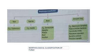
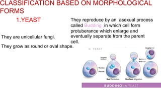
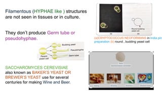
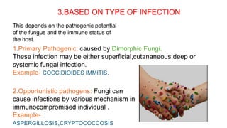
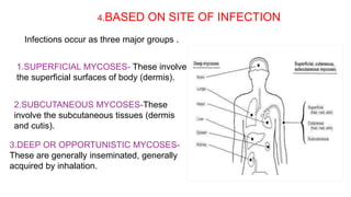
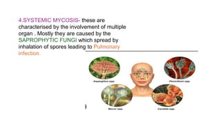
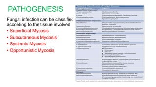
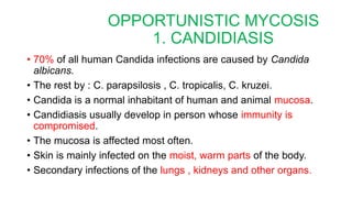
mycology 12345.pptx development of mmmmmbbbbbsssssssssAnuragKumarKumar4
This document provides an overview of mycology, which is the study of fungi. It discusses the classification of fungi based on morphological forms, sporulation, type of infection, and site of infection. The key classifications described are yeasts, dimorphic fungi, and filamentous fungi. It also covers various fungal infections including superficial mycoses of the skin, subcutaneous mycoses, and systemic mycoses. Laboratory methods for fungal identification and diagnosis are mentioned such as microscopy, culture, and staining techniques.
This document provides an overview of mycology, which is the study of fungi. It discusses the classification of fungi based on morphological forms, sporulation, type of infection, and site of infection. The key classifications described are yeasts, dimorphic fungi, and filamentous fungi. It also covers various fungal infections including superficial mycoses like tinea versicolor and tinea nigra, subcutaneous mycoses like mycetoma and sporotrichosis, and systemic mycoses. Laboratory methods for fungal identification and diagnosis are also mentioned such as microscopy, culture, and staining techniques.
Fungi are eukaryotic organisms that can be single-celled or multicellular. Their cell walls contain chitin and glucans. Fungi can cause superficial infections of the skin and nails, subcutaneous infections beneath the skin, and systemic infections of internal organs. Common fungal diseases include ringworm, athlete's foot, histoplasmosis, and coccidiomycosis. Fungi are classified based on their modes of reproduction and microscopic structures. Identification and diagnosis involves microscopic examination, culturing, and serological tests.
Fungi are eukaryotic organisms that include molds, yeasts and dermatophytes. They obtain nutrients through absorption and can reproduce both sexually through spores and asexually through budding or fragmentation. Fungal infections, or mycoses, are a major cause of disease in humans and include cutaneous, subcutaneous, systemic and opportunistic infections. Laboratory diagnosis of fungal infections involves microscopic examination, culture and other tests to identify the causative fungus.
This document provides an introduction to mycology, the study of fungi. It discusses the history of fungi being recognized as pathogens and outlines the key characteristics of fungi such as cell walls containing chitin. Fungi can be classified based on cell morphology into yeasts, molds, and dimorphic fungi. They can also be classified based on sexual reproduction into four classes. Common fungal infections like dermatophytosis and opportunistic infections are described. The document concludes by noting some useful properties of fungi such as food production and antibiotic production.
This document provides an introduction to medical mycology for nurses. It discusses the basic characteristics of fungi, including their cell structure, modes of growth, and reproduction. It then covers various types of fungal infections like cutaneous, subcutaneous, opportunistic, and systemic mycoses. Specific opportunistic infections like candidiasis, cryptococcosis, aspergillosis, and mucormycosis are explained in terms of their causative agents and clinical presentations. The document emphasizes that immunosuppressed patients are more susceptible to deep and opportunistic fungal infections.
Introduction to Mycology 2020 (2).pptxDawn Junkere
shows the different fungal infections human can be exposed to. how medical treatment can be carried out to treat
use of antofungals. the different parts of the fungus
1. Fungi are eukaryotic organisms that do not contain chlorophyll and have cell walls. They can grow as filaments called hyphae and reproduce through spores.
2. Around 300 fungal species are known to be pathogenic to humans, causing infections of the skin, nails, mucous membranes, and various internal organs depending on factors like host immunity.
3. Common pathogenic fungi include Candida, Aspergillus, Cryptococcus, and dermatophytes that cause superficial infections. Opportunistic fungi can cause serious disease in immunocompromised individuals.
This document provides information on deep cutaneous mycosis, specifically focusing on subcutaneous mycoses. It discusses several types of subcutaneous mycoses including sporotrichosis, mycetoma, and chromoblastomycosis. For each condition, it describes the causative organisms, epidemiology, clinical presentation, diagnosis, and treatment. Sporotrichosis is caused by Sporothrix schenckii and can manifest as lymphatic or fixed cutaneous lesions. Mycetoma is characterized by grain formation and can be caused by fungi or actinomycetes. Chromoblastomycosis features slow growing exophytic lesions caused by pigmented fungi that form sclerotic bodies in tissue.
Classification of Fungi. Superficial Mycoses & Systemic Mycoses. Dermatophyte...Eneutron
This document discusses medical fungi that can cause superficial and systemic mycoses. It covers the general characteristics of fungi including their morphology, classification, and modes of infection. Specific types of mycoses are described such as dermatophytoses caused by fungi like Trichophyton that infect the skin, hair, and nails. Deeper mycoses like sporotrichosis that infect subcutaneous tissue via minor trauma are also outlined. The document provides details on laboratory diagnostics and culturing of different fungi as well as antifungal therapies.
This document provides an overview of fungi. It discusses that fungi are eukaryotic organisms classified in the kingdom Fungi. They can exist as molds, yeasts, or dimorphic fungi. Fungi have cell walls containing chitin and reproduce asexually through spores or budding or sexually through the fusion of hyphae. The document outlines the morphological characteristics, differences from bacteria, classification systems based on cell structure and reproduction, and types of fungal infections like superficial, cutaneous, subcutaneous, and systemic mycoses.
This document provides an overview of mycology, which is the study of fungi. It discusses the classification, morphology, and types of fungal infections that affect the skin, nails, hair (dermatophytes), subcutaneous tissues, and internal organs/systems (systemic and opportunistic fungi). Common fungal infections discussed include dermatophytosis, tinea versicolor, tinea nigra, sporotrichosis, chromomycosis, mycetoma, blastomycosis, histoplasmosis, and coccidioidomycosis. The document also covers the laboratory diagnosis and treatment of various mycoses. The lecture objectives are to describe the classification of fungal infections,
This document discusses several types of deep fungal infections:
1. Subcutaneous mycoses like sporotrichosis, chromoblastomycosis, and mycetoma which are caused by fungi entering through the skin.
2. Systemic mycoses like histoplasmosis which are acquired by inhalation and can disseminate through the bloodstream. Histoplasmosis is caused by Histoplasma capsulatum and presents as pulmonary infection, disseminated infection affecting organs, or cutaneous lesions. Treatment involves antifungals like amphotericin B or itraconazole.
3. Rarer infections like lobomycosis caused by Lacazia loboi which
This document summarizes various fungal diseases that affect humans and animals. It describes five main groups of fungal diseases: superficial mycoses, cutaneous mycoses, subcutaneous mycoses, systemic mycoses, and opportunistic mycoses. For each group, it provides examples of pathogenic fungi, the locations they infect, and the resulting diseases. It also discusses the transmission routes and typical symptoms for some of the major fungal diseases like blastomycosis, coccidioidomycosis, cryptococcosis, and histoplasmosis.
This document provides information on fungal diseases of medical importance. It defines fungi and their characteristics, including that they are eukaryotic organisms that absorb nutrients and reproduce both sexually and asexually. The document classifies fungi into phyla and discusses some common fungal infections affecting humans, including superficial infections like tinea versicolor and piedra, as well as deeper infections and opportunistic infections in immunocompromised individuals. It provides details on the causative agents, symptoms, and treatment of various medically important fungal diseases.
This document discusses mycology, which is the study of fungi. It describes the key characteristics of fungi and how they differ from bacteria. Some of the major types of fungi are yeasts, molds, and dimorphic fungi, which can exist in both yeast and mold forms. Fungal diseases are classified as superficial mycoses, mucocutaneous mycoses, subcutaneous mycoses, and deep mycoses. Common fungal infections in humans include candidiasis, dermatophytosis, and various respiratory fungal infections.
This document discusses Coccidioidomycosis, an endemic mycosis caused by the dimorphic fungus Coccidioides immitis. It is found in arid regions of the Americas. In soil, it forms hyphae and arthrospores that can be inhaled and cause infection in the lungs, forming spherules filled with endospores. Most infections are asymptomatic or cause flu-like symptoms, but some can disseminate to bones, meninges or skin. Diagnosis involves identifying spherules microscopically or culturing the fungus from specimens. Serologic tests can also detect antibodies. There is no treatment for mild cases but disseminated disease requires antifungal therapy
This document discusses various mycoses (fungal infections) that affect humans. It describes 10 specific mycoses - mycetoma, chromomycosis, sporotrichosis, rhinosporidiosis, subcutaneous phycomycosis, cryptococcosis, blastomycosis, paracoccidioidomycosis, coccidioidomycosis, and histoplasmosis. For each, it provides details on the causative agents, clinical manifestations, pathogenesis, diagnosis, and laboratory identification. Key diagnostic methods mentioned include microscopy, histopathology, culture, and serology. The document is intended as an educational reference for medical professionals regarding these important fungal diseases.
This document provides an introduction to mycology and virology for public health second-year students. It defines key terms related to fungi and outlines the objectives, characteristics of fungi, medical importance of fungi, and types of fungal infections including superficial, cutaneous, subcutaneous, and systemic mycoses. Specific fungi that cause different types of infections are described along with their symptoms, transmission, and clinical presentation.
Fungus is a group of eukaryotic organisms that exist as saprophytes. There are four main types of fungi: yeasts, yeast-like fungi, molds, and dimorphic fungi. Yeasts are unicellular and reproduce by budding, yeast-like fungi grow partly as yeasts and chains of elongated budding cells, molds form true mycelium and branching filaments, and dimorphic fungi can exist as molds or yeasts. Fungal diseases in humans are known as mycoses and can be superficial, subcutaneous, or systemic infections. Common fungal genera that cause disease include Candida, dermatophytes, and dimorphic fungi such as Histoplasma
Fungal infections can be caused by yeasts, molds, or dimorphic fungi. Superficial fungal infections include dermatophytosis (ringworm) and pityriasis versicolor. Subcutaneous fungal infections like sporotrichosis present as nodules that can drain or spread. Systemic fungal infections are caused by true pathogens that infect healthy hosts or opportunistic pathogens that infect immunocompromised individuals. Common opportunistic fungal infections are caused by Candida, Aspergillus, Cryptococcus, and Mucor molds. Diagnosis involves microscopy, culture, or antigen detection of fungi in clinical samples from infected sites.
This document discusses fungal infections. It begins by listing the characteristics of fungi and describing the differences between molds and yeasts. The major classifications of human mycoses are described including superficial infections like tinea capitis and tinea versicolor. Deeper infections like sporotrichosis, mycetoma, and systemic mycoses caused by dimorphic fungi such as coccidiomycosis and histoplasmosis are also outlined. Opportunistic fungal infections in immunocompromised individuals that are described include Pneumocystis jiroveci pneumonia, oral and vaginal candidiasis, and cryptococcosis. Common treatment regimens for several of the infections are also provided.
This document provides an overview of mycology (the study of fungi). It discusses that fungi are eukaryotic organisms that lack chlorophyll and can exist as unicellular or multicellular forms. It describes the cell structure of fungi including their cell walls made of chitin and cell membranes containing ergosterol. It covers the taxonomic classification of fungi into phyla and discusses the structures and reproduction of different types of fungi including yeasts, molds, and thermally dimorphic fungi. It also addresses the laboratory diagnosis of fungal infections through microscopy, staining, culture and the clinical manifestations of different fungal infections.
The document discusses medical mycology, which is the study of pathogenic fungi that cause disease. It covers topics such as the classification of fungi, their modes of reproduction, pathogenesis of fungal infections, diagnosis of fungal diseases, and antifungal therapies. The major classes of antifungal drugs are discussed including polyenes, azoles, and echinocandins which act on the fungal cell membrane or cell wall.
This document discusses mycology and dermatophytosis. It defines mycology as the study of fungi and fungal diseases. Dermatophytosis is a superficial fungal infection of the skin, hair, and nails, caused by dermatophyte fungi. Common symptoms include round, scaly lesions known as ringworm. The document outlines the structure and types of fungi that cause dermatophytosis, epidemiology, pathogenesis, clinical features, diagnosis including microscopic examination of skin scrapings and cultures, and treatment with topical and oral antifungal medications.
Fungal Infections/ Mycoses ppt by Dr.C.P.PRINCEDR.PRINCE C P
PPT prepared by :
DR.PRINCE C P
Associate Professor &HOD
Department of Microbiology,
Mother Theresa Post Graduate & Research Institute of Health Sciences (Government of Puducherry Institution)
According to tissue involved, MYCOSES are classified into:
Superficial (Surface )
Cutaneous
Subcutaneous
Deep Cutaneous
Systemic (Primary )
Systemic ( Opportunistic)
Mycotic Poisoning
most of the fungal infections are opportunistic in nature.
candida albicans is the common Fungal pathogen.
This document provides information about fungi and mycosis. It discusses that fungi are eukaryotic, aerobic organisms that can be saprophytic, symbiotic, or parasitic. Fungi can be beneficial in producing foods or antibiotics. The document classifies fungi taxonomically into phyla, classes including yeasts, molds and dimorphic fungi. It also describes laboratory diagnosis of fungi through microscopy and culture. Finally, it discusses different types of fungal infections that can affect the skin, subcutaneous tissues, internal organs, or be opportunistic.
How to Manage Your Lost Opportunities in Odoo 17 CRMCeline George
Odoo 17 CRM allows us to track why we lose sales opportunities with "Lost Reasons." This helps analyze our sales process and identify areas for improvement. Here's how to configure lost reasons in Odoo 17 CRM
1. Fungi are eukaryotic organisms that do not contain chlorophyll and have cell walls. They can grow as filaments called hyphae and reproduce through spores.
2. Around 300 fungal species are known to be pathogenic to humans, causing infections of the skin, nails, mucous membranes, and various internal organs depending on factors like host immunity.
3. Common pathogenic fungi include Candida, Aspergillus, Cryptococcus, and dermatophytes that cause superficial infections. Opportunistic fungi can cause serious disease in immunocompromised individuals.
This document provides information on deep cutaneous mycosis, specifically focusing on subcutaneous mycoses. It discusses several types of subcutaneous mycoses including sporotrichosis, mycetoma, and chromoblastomycosis. For each condition, it describes the causative organisms, epidemiology, clinical presentation, diagnosis, and treatment. Sporotrichosis is caused by Sporothrix schenckii and can manifest as lymphatic or fixed cutaneous lesions. Mycetoma is characterized by grain formation and can be caused by fungi or actinomycetes. Chromoblastomycosis features slow growing exophytic lesions caused by pigmented fungi that form sclerotic bodies in tissue.
Classification of Fungi. Superficial Mycoses & Systemic Mycoses. Dermatophyte...Eneutron
This document discusses medical fungi that can cause superficial and systemic mycoses. It covers the general characteristics of fungi including their morphology, classification, and modes of infection. Specific types of mycoses are described such as dermatophytoses caused by fungi like Trichophyton that infect the skin, hair, and nails. Deeper mycoses like sporotrichosis that infect subcutaneous tissue via minor trauma are also outlined. The document provides details on laboratory diagnostics and culturing of different fungi as well as antifungal therapies.
This document provides an overview of fungi. It discusses that fungi are eukaryotic organisms classified in the kingdom Fungi. They can exist as molds, yeasts, or dimorphic fungi. Fungi have cell walls containing chitin and reproduce asexually through spores or budding or sexually through the fusion of hyphae. The document outlines the morphological characteristics, differences from bacteria, classification systems based on cell structure and reproduction, and types of fungal infections like superficial, cutaneous, subcutaneous, and systemic mycoses.
This document provides an overview of mycology, which is the study of fungi. It discusses the classification, morphology, and types of fungal infections that affect the skin, nails, hair (dermatophytes), subcutaneous tissues, and internal organs/systems (systemic and opportunistic fungi). Common fungal infections discussed include dermatophytosis, tinea versicolor, tinea nigra, sporotrichosis, chromomycosis, mycetoma, blastomycosis, histoplasmosis, and coccidioidomycosis. The document also covers the laboratory diagnosis and treatment of various mycoses. The lecture objectives are to describe the classification of fungal infections,
This document discusses several types of deep fungal infections:
1. Subcutaneous mycoses like sporotrichosis, chromoblastomycosis, and mycetoma which are caused by fungi entering through the skin.
2. Systemic mycoses like histoplasmosis which are acquired by inhalation and can disseminate through the bloodstream. Histoplasmosis is caused by Histoplasma capsulatum and presents as pulmonary infection, disseminated infection affecting organs, or cutaneous lesions. Treatment involves antifungals like amphotericin B or itraconazole.
3. Rarer infections like lobomycosis caused by Lacazia loboi which
This document summarizes various fungal diseases that affect humans and animals. It describes five main groups of fungal diseases: superficial mycoses, cutaneous mycoses, subcutaneous mycoses, systemic mycoses, and opportunistic mycoses. For each group, it provides examples of pathogenic fungi, the locations they infect, and the resulting diseases. It also discusses the transmission routes and typical symptoms for some of the major fungal diseases like blastomycosis, coccidioidomycosis, cryptococcosis, and histoplasmosis.
This document provides information on fungal diseases of medical importance. It defines fungi and their characteristics, including that they are eukaryotic organisms that absorb nutrients and reproduce both sexually and asexually. The document classifies fungi into phyla and discusses some common fungal infections affecting humans, including superficial infections like tinea versicolor and piedra, as well as deeper infections and opportunistic infections in immunocompromised individuals. It provides details on the causative agents, symptoms, and treatment of various medically important fungal diseases.
This document discusses mycology, which is the study of fungi. It describes the key characteristics of fungi and how they differ from bacteria. Some of the major types of fungi are yeasts, molds, and dimorphic fungi, which can exist in both yeast and mold forms. Fungal diseases are classified as superficial mycoses, mucocutaneous mycoses, subcutaneous mycoses, and deep mycoses. Common fungal infections in humans include candidiasis, dermatophytosis, and various respiratory fungal infections.
This document discusses Coccidioidomycosis, an endemic mycosis caused by the dimorphic fungus Coccidioides immitis. It is found in arid regions of the Americas. In soil, it forms hyphae and arthrospores that can be inhaled and cause infection in the lungs, forming spherules filled with endospores. Most infections are asymptomatic or cause flu-like symptoms, but some can disseminate to bones, meninges or skin. Diagnosis involves identifying spherules microscopically or culturing the fungus from specimens. Serologic tests can also detect antibodies. There is no treatment for mild cases but disseminated disease requires antifungal therapy
This document discusses various mycoses (fungal infections) that affect humans. It describes 10 specific mycoses - mycetoma, chromomycosis, sporotrichosis, rhinosporidiosis, subcutaneous phycomycosis, cryptococcosis, blastomycosis, paracoccidioidomycosis, coccidioidomycosis, and histoplasmosis. For each, it provides details on the causative agents, clinical manifestations, pathogenesis, diagnosis, and laboratory identification. Key diagnostic methods mentioned include microscopy, histopathology, culture, and serology. The document is intended as an educational reference for medical professionals regarding these important fungal diseases.
This document provides an introduction to mycology and virology for public health second-year students. It defines key terms related to fungi and outlines the objectives, characteristics of fungi, medical importance of fungi, and types of fungal infections including superficial, cutaneous, subcutaneous, and systemic mycoses. Specific fungi that cause different types of infections are described along with their symptoms, transmission, and clinical presentation.
Fungus is a group of eukaryotic organisms that exist as saprophytes. There are four main types of fungi: yeasts, yeast-like fungi, molds, and dimorphic fungi. Yeasts are unicellular and reproduce by budding, yeast-like fungi grow partly as yeasts and chains of elongated budding cells, molds form true mycelium and branching filaments, and dimorphic fungi can exist as molds or yeasts. Fungal diseases in humans are known as mycoses and can be superficial, subcutaneous, or systemic infections. Common fungal genera that cause disease include Candida, dermatophytes, and dimorphic fungi such as Histoplasma
Fungal infections can be caused by yeasts, molds, or dimorphic fungi. Superficial fungal infections include dermatophytosis (ringworm) and pityriasis versicolor. Subcutaneous fungal infections like sporotrichosis present as nodules that can drain or spread. Systemic fungal infections are caused by true pathogens that infect healthy hosts or opportunistic pathogens that infect immunocompromised individuals. Common opportunistic fungal infections are caused by Candida, Aspergillus, Cryptococcus, and Mucor molds. Diagnosis involves microscopy, culture, or antigen detection of fungi in clinical samples from infected sites.
This document discusses fungal infections. It begins by listing the characteristics of fungi and describing the differences between molds and yeasts. The major classifications of human mycoses are described including superficial infections like tinea capitis and tinea versicolor. Deeper infections like sporotrichosis, mycetoma, and systemic mycoses caused by dimorphic fungi such as coccidiomycosis and histoplasmosis are also outlined. Opportunistic fungal infections in immunocompromised individuals that are described include Pneumocystis jiroveci pneumonia, oral and vaginal candidiasis, and cryptococcosis. Common treatment regimens for several of the infections are also provided.
This document provides an overview of mycology (the study of fungi). It discusses that fungi are eukaryotic organisms that lack chlorophyll and can exist as unicellular or multicellular forms. It describes the cell structure of fungi including their cell walls made of chitin and cell membranes containing ergosterol. It covers the taxonomic classification of fungi into phyla and discusses the structures and reproduction of different types of fungi including yeasts, molds, and thermally dimorphic fungi. It also addresses the laboratory diagnosis of fungal infections through microscopy, staining, culture and the clinical manifestations of different fungal infections.
The document discusses medical mycology, which is the study of pathogenic fungi that cause disease. It covers topics such as the classification of fungi, their modes of reproduction, pathogenesis of fungal infections, diagnosis of fungal diseases, and antifungal therapies. The major classes of antifungal drugs are discussed including polyenes, azoles, and echinocandins which act on the fungal cell membrane or cell wall.
This document discusses mycology and dermatophytosis. It defines mycology as the study of fungi and fungal diseases. Dermatophytosis is a superficial fungal infection of the skin, hair, and nails, caused by dermatophyte fungi. Common symptoms include round, scaly lesions known as ringworm. The document outlines the structure and types of fungi that cause dermatophytosis, epidemiology, pathogenesis, clinical features, diagnosis including microscopic examination of skin scrapings and cultures, and treatment with topical and oral antifungal medications.
Fungal Infections/ Mycoses ppt by Dr.C.P.PRINCEDR.PRINCE C P
PPT prepared by :
DR.PRINCE C P
Associate Professor &HOD
Department of Microbiology,
Mother Theresa Post Graduate & Research Institute of Health Sciences (Government of Puducherry Institution)
According to tissue involved, MYCOSES are classified into:
Superficial (Surface )
Cutaneous
Subcutaneous
Deep Cutaneous
Systemic (Primary )
Systemic ( Opportunistic)
Mycotic Poisoning
most of the fungal infections are opportunistic in nature.
candida albicans is the common Fungal pathogen.
This document provides information about fungi and mycosis. It discusses that fungi are eukaryotic, aerobic organisms that can be saprophytic, symbiotic, or parasitic. Fungi can be beneficial in producing foods or antibiotics. The document classifies fungi taxonomically into phyla, classes including yeasts, molds and dimorphic fungi. It also describes laboratory diagnosis of fungi through microscopy and culture. Finally, it discusses different types of fungal infections that can affect the skin, subcutaneous tissues, internal organs, or be opportunistic.
Similar to final mycology.pptxbsbsjsbbshsjsbsvsjsbh (20)
How to Manage Your Lost Opportunities in Odoo 17 CRMCeline George
Odoo 17 CRM allows us to track why we lose sales opportunities with "Lost Reasons." This helps analyze our sales process and identify areas for improvement. Here's how to configure lost reasons in Odoo 17 CRM
This presentation includes basic of PCOS their pathology and treatment and also Ayurveda correlation of PCOS and Ayurvedic line of treatment mentioned in classics.
How to Add Chatter in the odoo 17 ERP ModuleCeline George
In Odoo, the chatter is like a chat tool that helps you work together on records. You can leave notes and track things, making it easier to talk with your team and partners. Inside chatter, all communication history, activity, and changes will be displayed.
A review of the growth of the Israel Genealogy Research Association Database Collection for the last 12 months. Our collection is now passed the 3 million mark and still growing. See which archives have contributed the most. See the different types of records we have, and which years have had records added. You can also see what we have for the future.
Main Java[All of the Base Concepts}.docxadhitya5119
This is part 1 of my Java Learning Journey. This Contains Custom methods, classes, constructors, packages, multithreading , try- catch block, finally block and more.
Strategies for Effective Upskilling is a presentation by Chinwendu Peace in a Your Skill Boost Masterclass organisation by the Excellence Foundation for South Sudan on 08th and 09th June 2024 from 1 PM to 3 PM on each day.
Exploiting Artificial Intelligence for Empowering Researchers and Faculty, In...Dr. Vinod Kumar Kanvaria
Exploiting Artificial Intelligence for Empowering Researchers and Faculty,
International FDP on Fundamentals of Research in Social Sciences
at Integral University, Lucknow, 06.06.2024
By Dr. Vinod Kumar Kanvaria
3. MYCOLOGY??
It is a branch of Microbiology that deals with study
of FUNGI.
Mycology derived form the Greek word “Mycos” meaning
“FUNGUS”
Medical Mycology deals with the Etiology, Pathogenesis,
Detection, Prevention and Treatment of FUNGAL
INFECTION
4. FUNGI
1. Fungi are Eukaryotic Protista that differ from bacteria and other
Prokaryotes.
CHARACTERISTICS
*They posses rigid cell wall containing CHITIN,
MANNAN , other POLYSACCHARIDES.
*Their Cytoplasmic Membrane Contains STEROL.
*They posses true nuclei with a nuclear membrane
and paired chromosomes.
*They divide Asexually, Sexually or by both
processes.
*They may be Unicellular or
Multicellular.
5. CLASSIFICATION OF FUNGI
• Based on Morphological Forms
• Based on Sporulation
• Based on type of infection
• Based on site of Infection
prodced by fungus.
7. CLASSIFICATION BASED ON MORPHOLOGICAL
FORMS
1.YEAST
They are unicellular fungi.
They grow as round or oval shape.
They reproduce by an asexual process
called Budding ,in which cell form
protuberance which enlarge and
eventually separate from the parent
cell.
8. Filamentous (HYPHAE like ) structures
are not seen in tissues or in culture.
They don’t produce Germ tube or
pseudohyphae.
SACCHAROMYCES CEREVISIAE
also known as BAKER’S YEAST OR
BREWER’S YEAST use for several
centuries for making Wine and Beer.
(a)CRYPTOCOCCUS NEOFORMANS in India pink
preparation (b) round , budding yeast cell
9. IN CULTURE ,Macroscopic appearance of
colony is Pasty,resembling bacterial
colonies.
Example -
SACCHAROMYCES CEREVISIAE CRYPTOCOCCUS NEOFORMANS
10. 2.YEAST LIKE FUNGI
• Unicellular Fungi
• Reproduce by Budding and Fission.
• They may be oval ,spherical forms
or filamentous forms.
Example-
• Colonies in culture Pasty Appearance.
In CANDIDA ,bud remains attached to
mother cell, elongates and undergo
repeated budding to form chains of
repeated cells called
PSEUDOHYPAHE
a) Oval budding cell of candida albicans in Gram Smear
b) Pseudohyphae
c) Oval, budding yeast cell
11. 3.FILAMENTOUS FUNGI OR MOULDS
SEPTATE
They grow as long branching
filament of 2-10 micron meter
width called “HYPHAE”
HYPHAE are of two types-
• HYPHAE having cross wall or
septa ,they are multicellular.
ASEPTATE
They devoid of septa called
Coenocytic(mutlinucleated).
12. HYPHAE grow continuously and
form a branching tangled mass of
growth called MYCELIUM.
• Reproduce by Asexual means (
Spore formation),some exhibit
Sexual reproduction.
Based on growth pattern in culture
medium ,the mycelia may be
categorised into two categories.
• AERIAL MYCELIUM-part of mycelium
which project above the surface of
culture medium
• VEGETATIVE MYCELIUM –part of mycelium
that grows on the surface of culture media.
13. HYPHAE may be Racquet shaped ,
Nodular, Pectinate,Spiral ,Root like or
Chandelier.
Example-
• DERMATOPHYTE
• ASPERGILLUS
• RHIZOPUS
• MUCOR
• PENICILLIUM
ASPERGILLUS MUCOR
RHIZOPUS
14. 4.DIMORPHIC FUNGI
They exist as Hyphal form Molds in the environment at ambient temperature
25.C and as a Yeast in human tissues at body temperature 37.C
Example-
• BLASTOMYCES DERMATITIDIS
• PARACOCCIDIOIDES
BRASILIENSIS
• COCCIDIOIDES IMMITIS
• HISTOPLASMA CAPSULATUM
• SPOROTHRIX SCHENCKII
• PENICILLIUM MARNEFFEI
HISTOPLASMA CAPSULATUM
MYCELIAL
(filamentous) form
YEAST form in tissue
section
15. 2.BASED ON SPORULATION
Also called Taxonomical basis of classification.
Classify on basis either they exhibit, whether Asexual or Sexual Spores.
They divide into 4 phyla on the basis of spores.
1.PHYLUM ZYGOMYCOTA they are
lower fungi produce Sexual Spores
Called ZYGOSPORE,and posses
Aseptate hyphae.
2.PHYLUM ASCOMYCOTA –they produce
Sexual Spores called ASCOSPORES ,and
posses Septate hyphae.
MUCOR
17. 3.BASED ON TYPE OF INFECTION
This depends on the pathogenic potential
of the fungus and the immune status of
the host.
1.Primary Pathogenic: caused by Dimorphic Fungi.
These infection may be either superficial,cutananeous,deep or
systemic fungal infection.
Example- COCCIDIOIDES IMMITIS.
2.Opportunistic pathogens: Fungi can
cause infections by various mechanism in
immunocompromised individual .
Example-
ASPERGILLOSIS,CRYPTOCOCCOSIS
18. 4.BASED ON SITE OF INFECTION
Infections occur as three major groups .
1.SUPERFICIAL MYCOSES- These involve
the superficial surfaces of body (dermis).
2.SUBCUTANEOUS MYCOSES-These
involve the subcutaneous tissues (dermis
and cutis).
3.DEEP OR OPPORTUNISTIC MYCOSES-
These are generally inseminated, generally
acquired by inhalation.
19. 4.SYSTEMIC MYCOSIS- these are
characterised by the involvement of multiple
organ . Mostly they are caused by the
SAPROPHYTIC FUNGI which spread by
inhalation of spores leading to Pulmonary
infection.
36. PATHOGENESIS
Fungal infection can be classified
according to the tissue involved
• Superficial Mycosis
• Subcutaneous Mycosis
• Systemic Mycosis
• Opportunistic Mycosis
37. SUPERFICIAL MYCOSES
Superficial mycoses are the fungal infections involving the
skin, hair, nail and mucosa.
examples include:
• Tinea versicolor
• Tinea nigra
• Piedra
• Dermatophytoses
38. Tinea versicolor / Pityriasis versicolor
• It is caused by lipophilic fungus
Malassezia furfur , which is
characterised by flat- round
scaly hypopigmented to hyperpigmented
patches of skin.
• Patches mainly seen on trunk region –
Chest, abdomen, and back.
• Lesions are confined to superficial
layers of skin
(stratum corneum)
39. Tinea nigra
• It is characterised by painless
, black, nonscaly patches
present on palm and sole .
• It is caused by a black
colored yeast like fungus
Hortaea werneckii.
• Females are more commonly
affected than males.
40. Piedra
• It is characterised by nodule
formation on the hair shaft, which
may either be black or white in
color.
• It is of 2 types;
Black piedra – characterised by
black colored , gritty,firmly
attached nodules on the hair
shaft.
It is caused by Piedraia hortae.
41. • White piedra –
It is characterised by soft, grey- white nodules
which are less firmly attached to the hair shaft.
It is caused by Trichosporon beigelii.
Trichosporon beigelii produces creamy white
colonies.
42. Dermatophytoses (or tinea or ringworm)
• It is the most common superficial
mycoses affecting skin, hair,and
nail.
• These include:
Trichophyton species: infect
skin, hair and nails
Microsporum species: infect
skin and hair
Epidermophyton species:
infect skin and nail.
43. Classification:
1. Anthropophilic dermatophytes
-cause mild lesions
-infect human only
- respond poorly to treatment
2. Zoophilic dermatophytes
- infect animals
3. Geophilic dermatophytes: soil
-humans get infected when contact with soil
44. . Lesions on nails: fungi invade the nails through
lateral/ superficial nail plates then spread
throughout the nail.
Types of clinical diseases:
• Tinea capitis
• Tinea corporis
• Tinea cruris
• Tinea pedis
• Tinea unguum
• Tinea barbae
• Tenia faciei etc.
45. SUBCUTANEOUS MYCOSES
Subcutaneous mycoses are the mycotic infections of the skin,
subcutaneous tissue and sometimes bone, resulting from
inoculation of saprophytic fungi of soil or decaying matter.
They are mainly confined to the tropics and subtropics.
46. Mycetoma
• Mycetoma, also known as maduramycosis
or madura foot is a chronic , slowly
progressive granulomatous infection of the
skin and subcutaneous tissues.
• Clinically manifested as a triad of swelling,
discharging sinuses and presence of
granules in the discharge.
47. Clinical features:
- more common in males than females
- lower limbs are commonly affected than
upper limbs.
- sometimes, underlying bones may be
involved.
Mycetoma is of two types:
1. Eumycetoma – caused by fungal agents
such as Madurella mycetomatis
2. Actinomycetoma – caused by bacterial
agents such as Nocardia.
48. Sporotrichosis
• It is also known as Rose picker’s
disease /Gardner’s disease/
carpenter’s diseae
• Caused by a thermally dimorphic
fungus, sporothrix schenckii.
• It is pesent on woods and plants
(rose) and mode of infection is
traumatic inoculation.
• It presents as subcutaneous
noduloulcerative lesions.
• It is common in tropical countries
with high humidity.
• It is also present in the soil and
hence walking barefoot also
predisposes to infection.
49. Chromoblastomycosis
• It refers to slow growing chronic
subcutaneous lesions caused by goup of
phaeoid fungi (i.e. darkly pigmented fungi)
that produce a characteristic morphology
called sclerotic body.
• Agents include : Fonsecaea, Phialophora
cladosporium and Rhinocladiella.
• It affects more males than females.
• Treatment: cryotherapy or laser therapy
followed by antifungals like itraconazole.
50. Rhinosporidiosis
• It is caused by Rhinosporidium
seeberi.
• It produces large friable polyps in
the nose (most common site),
conjuctiva, bronchus, larynx, eyes
genitalia.
• Source of infection : stagnant water
bodies.
- infection occurs via inhalation of
spores while bathing in
contaminated rivers and ponds.
51. • Fungi that are able to cause systemic illness in healthy people
are rare and confined to specific geographic location across the
world.
• Fungal infection of internal organs.
• Primarily involve the Respiratory System.
• Infection occurs by inhalation of air-borne conidia.
• Change in temperature determines the form. The fungus is a
mold when grown at 25 C but grows as yeast at body
temperature (Thermal Dimorphism).
SYSTEMIC MYCOSIS
52. PRIMARY SYSTEMIC
MYCOSIS
• Infections of internal organs of the body.
• Caused by dimorphic fungi
• The following are the types of Systemic mycosis :
1. Blastomycosis
2. Coccidiomycosis
3. Histoplasmosis
4. Paracoccidioidomycosis
53. 1. BLASTOMYCOSIS
• Caused by : Blastomyces dermatitidis.
• Inhalation of conidial spores.
• Primary infection : Pulmonary blastomycosis.
• Secondary infection : May spread to other organs including skin
(Cutaneous mycosis).
• Osteoarticular blastomycosis : Occurs in about 30% of patients
with the spine, pelvis, cranial bones, ribs and long bones most
commonly involved.
54.
55. Diagnosis
• Specimens : Bronchial secretion, urine, Scrapings from infection,
Sputum, Skin Scrapings, Bone marrow, Pleural fluid , Blood and
cerebrospinal fluid.
• Microscopic Examination :
- 10% KOH and Parker ink or calcofluor white mounts.(Skin, Body
fluids).
- Positive direct microscopy demonstrating characteristics yeast- like
cells.
• Culture : On Blood or Sabouraud agar must be incubated for several
weeks.
• Treatment :
- Amphotericin B
56. 2. Coccidiomycosis
• Caused by : Cocxidiodes immitis (Dimorphic)
• Inhalation of Arthrospores.
• Primary infection : Lungs.
• Secondary infection :
- May spread to other organs including skin.
- Other silent infections – severe pneumonia, lesions in
skin,
bones , joints, and meninges.
57.
58. DIAGNOSIS
• Specimens : Bronchial secretion, urine, Scrapings from infection, Sputum,
Skin Scrapings, Bone marrow, Pleural fluid , Blood and cerebrospinal fluid.
• Microscopic Examination :
- 10% KOH and Parker ink or calcofluor white mounts.(Skin, Body
fluids).
- Positive direct microscopy demonstrating spherules with endospores.
• Culture : On Blood or Sabouraud agar must be incubated for several
weeks.
• Treatment :
- Amphotericin B
- Anoralazole derivative may be used
59.
60. 3. HISTOPLASMOSIS
• Caused by : Histoplasma capsulatum (Dimorphic fungus).
• Natural habitat : Soil .
• Grow as a filamentous form in culture.
• In human tissues it forms : Yeast cells.
• Inhalation of Spores ( Conidial ) into the respiratory tract.
• It affects the reticulo-endothelial system(RES).
• Observed commonly in AIDS patients.
61. DIAGNOSIS
• Specimens : Bronchial secretion, urine, Scrapings from infection, Sputum,
Skin Scrapings, Bone marrow, Pleural fluid , Blood and cerebrospinal fluid.
• Microscopic Examination :
- 10% KOH and Parker ink or calcofluor white mounts.(Skin, Bodyfluids).
- Positive direct microscopy demonstrating characteristics yeast-like
cells.
• Culture : On Blood or Sabouraud agar must be incubated for several
weeks.
• Treatment :
- Amphotericin B
62.
63. 4.
PARACOCCIDIOIDOMYCOSIS
• Caused by Paracoccidioidies brasiliensis (Dimorphic fungus).
• Inhalation of Spores.
• Natural habitat : soil.
• Primary : Chronic granulomatous infection are found in the lung
• Occasionally infect Gastro intestinal mucosa.
• The fungus can disseminate hematogenously or
lymphogenously into the skin, mucosa or lymphoid organs.
64.
65. DIAGNOSIS
• Specimens : Bronchial secretion, urine, Scrapings from infection, Sputum,
Skin Scrapings, Bone marrow, Pleural fluid , Blood and cerebrospinal fluid.
• Microscopic Examination :
- 10% KOH and Parker ink or calcofluor white mounts.(Skin, Body
fluids).
- Positive direct microscopy demonstrating 20-60 um , round, narrow
base
budding yeast cells with multiple budding.
• Culture : On Blood or Sabouraud agar must be incubated for several
weeks.
• Treatment :
- Amphotericin-B
- Itraconazole
66.
67. OPPORTUNISTIC MYCOSIS
1. CANDIDIASIS
• 70% of all human Candida infections are caused by Candida
albicans.
• The rest by : C. parapsilosis , C. tropicalis, C. kruzei.
• Candida is a normal inhabitant of human and animal mucosa.
• Candidiasis usually develop in person whose immunity is
compromised.
• The mucosa is affected most often.
• Skin is mainly infected on the moist, warm parts of the body.
• Secondary infections of the lungs , kidneys and other organs.
68.
69. DIAGNOSIS
• Specimens : Bronchial secretion, urine, Scrapings from infection, Sputum,
Skin Scrapings, Bone marrow, Pleural fluid , Blood and cerebrospinal fluid.
• Microscopic Examination :
- 10% KOH and Parker ink or calcofluor white mounts.(Skin, Body
fluids).
- Grocott’s methenamine silver(GMS) or Gram stain (Tissue sections).
• Culture : On Blood or Sabouraud agar must be incubated for several
weeks.
- After 48 hours of incubation on agar mediums, round, whitish,
rough
surfaced colonies form.
• Treatmet : Topical – Nystatin and Azoles
- Amphotericin B with 5- fluorocytosine
70.
71. 2. ASPPERGILLOSIS
• Aspergilloses are most frequently Caused by Aspergillus
fumigatus, A. flavus , A. niger.
• By inhalation of Spores or Ingestion of products contaminated
with Aspergillus.
• Pathway of entry : Bronchial system , but the organism can also
invade the body through injuries in the skin or mucosa.
• Aspergillosis of respiratory tract is common.
• Cerebral Aspergillosis
72. DIAGNOSIS
• Samples Collection : Sputum, Bronchial washings and Tracheal aspirates
• Microscopical Examination :
- Sputum, washings and aspirates make wet mounts in 10%
KOH and Parker ink or Gram stained smears.
- Tissue sections should be stained with H&E ,GMS.
• Culture : Sabouraud Dextrose agar
• Treatment :
- High dose of Amphotericin B
- Azoles
73. 3.CRYPTOCOCCOSIS
• Caused by Cryptococcus neoformans (Encapsulated Yeast).
• The individual cell is surrounded by a polysaccharide capsule
several micrometers wide.
• Normal habitat : Soil . Frequently found in bird droppings.
• The organisms are inhaled and enter the lungs , resulting in a
pulmonary cryptococcosis
• Pathogen can reach to Brain(CNS) and can cause Cryptococcal
Meningitis.
• Commonly found in immuno compromised patients. Ex – AIDS.
77. Questions
Ques 1- Which of following organisum is not been isolated artificial culture media?
a) Crytococcus neoformans
b) Rhinospordium seeberi
c) Histoplasma capsulatum
d) Penicillium marneffi
Ques 2 – Which of the following added in SDA to suppress the growth of saprophytic fungi?
a) Cycloheximide b) Chloromphenicol c) Gentamicin
d ) Amhotericin B
Ques 3 - Which of the following statements is not true for DERMATOPHYTE?
a) The fungi affect the keratinised layer of skin
b) They affect the hair and nail
c) They may be geophilic
d) They do not grow at room temperature
78. Ques 4- The following are saprophytic fungi associated with infection in
immunocompromised individual except
a) Candida albicans
b) Aspergillus fumigatus
c)Cocciodes immitis
d) Rhizopu Arrhizus
Ques 5- Fungal agent that does not infect nail-
a) Trichophyton b) Epidermophyton
c) Microsporum d) Candida albicans
Ques 6- Fungi which do not have Sexual stage-
a) Zygomycota b) Ascomycota
. c) Basidiomycota d) Fungi imperfecti
79. Short Answer Questions-
Ques 7- Write a short note on Dermatophytoses.
Ques 8- What are Dimorphic fungi? Explain with an Example.
Long Answer Questions-
Ques 9- Discuss the Clinical Manifestations and Laboratory diagnosis of
Sporotrichosis.