What do youthink
humans have in
common with the
pig?
http://www.clipartpanda.com/categories/pig-in-mud-cartoon
https://ferrebeekeeper.files.wordpress.com/2014/03/farmer-clip-art-4.gif
3.
Humans and Pigsmay be
closer than you think!
Both are mammals
We share common
body systems
The anatomy of
the pig is close to
that of humans
The fetal pigs will
tell us more about
our own bodies
and give us a way
to explore!
http://www.fanpop.com/clubs/human-anatomy/images/10358267/title/human-anatomy-photo
http://www.biologycorner.com/pig/fetal_pig02.jpg
4.
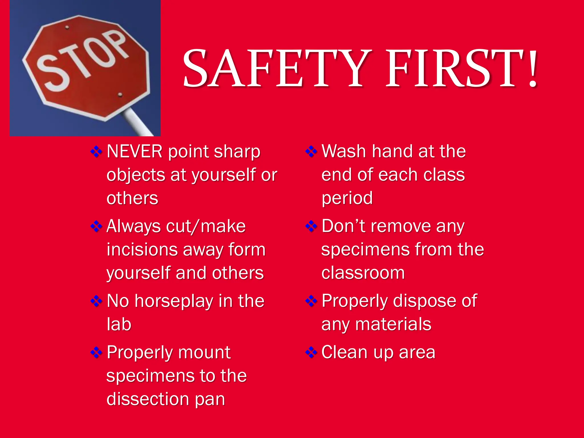
SAFETY FIRST!
❖ NEVERpoint sharp
objects at yourself or
others
❖ Always cut/make
incisions away form
yourself and others
❖ No horseplay in the
lab
❖ Properly mount
specimens to the
dissection pan
❖ Wash hand at the
end of each class
period
❖ Don’t remove any
specimens from the
classroom
❖ Properly dispose of
any materials
❖ Clean up area
5.
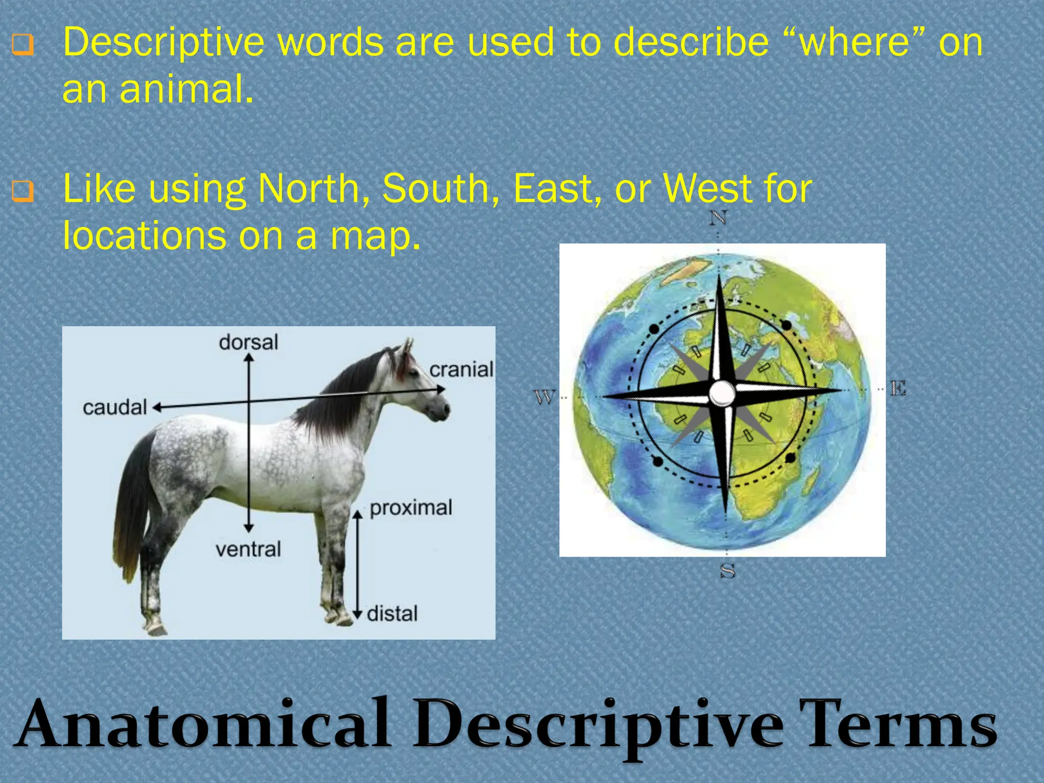
❑ Descriptive wordsare used to describe “where” on
an animal.
❑ Like using North, South, East, or West for
locations on a map.
6.
Dorsal --toward the back
Ventral -- toward the front/belly
Separated by the frontal plane
V
7.
Cranial --toward the head
Rostral -- toward the nose/beak
Caudal -- toward the tail
Separated by the transverse plane
8.
Medial –directed toward the
midline (sagittal plane)
Lateral -- directed away from the
midline (sagittal plane)
Sagittal Plane
9.
Proximal --located close to the
sagittal line of the body.
Distal -- located away from the
sagittal line of the body
10.
External Anatomy
❖ Skin
❖Nose
❖ Tongue
❖ Eyelids
❖ External Ear
❖ Digits
❖ Umbilical Cord
❖ 2 umbilical arteries
❖ Umbilical vein
❖ Teats
❖ Anus
❖ Identify the Sex
❖ Male – Scrotal Sac (ventral to anus)
and Urogenital Opening
❖ Female – Urogenital opening (ventral
to anus) and genital papilla
11.
DEMO SLIDE BOX23 Commercial slide. Eye, monkey
Alternative human eye
anterior
chamber
posterior
chamber
vitreous
space
lens
iris
Sclera
Cornea
Sclera
12.
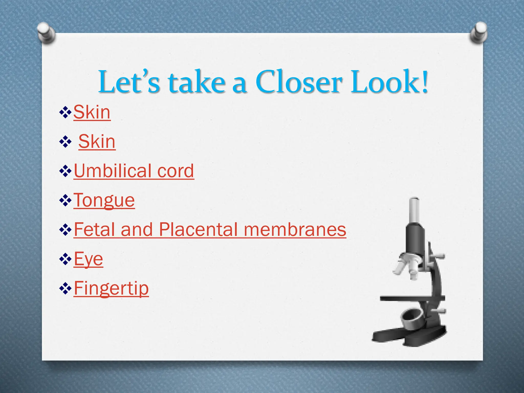
Let’s take aCloser Look!
❖Skin
❖ Skin
❖Umbilical cord
❖Tongue
❖Fetal and Placental membranes
❖Eye
❖Fingertip
13.
Digestive Tract
(Gastrointestinal Tract)
❖Mouth
❖ Teeth
❖ Tongue
❖ Parotid Gland
❖ Sublingual Gland
❖ Mandibular Gland
❖ Epiglottis
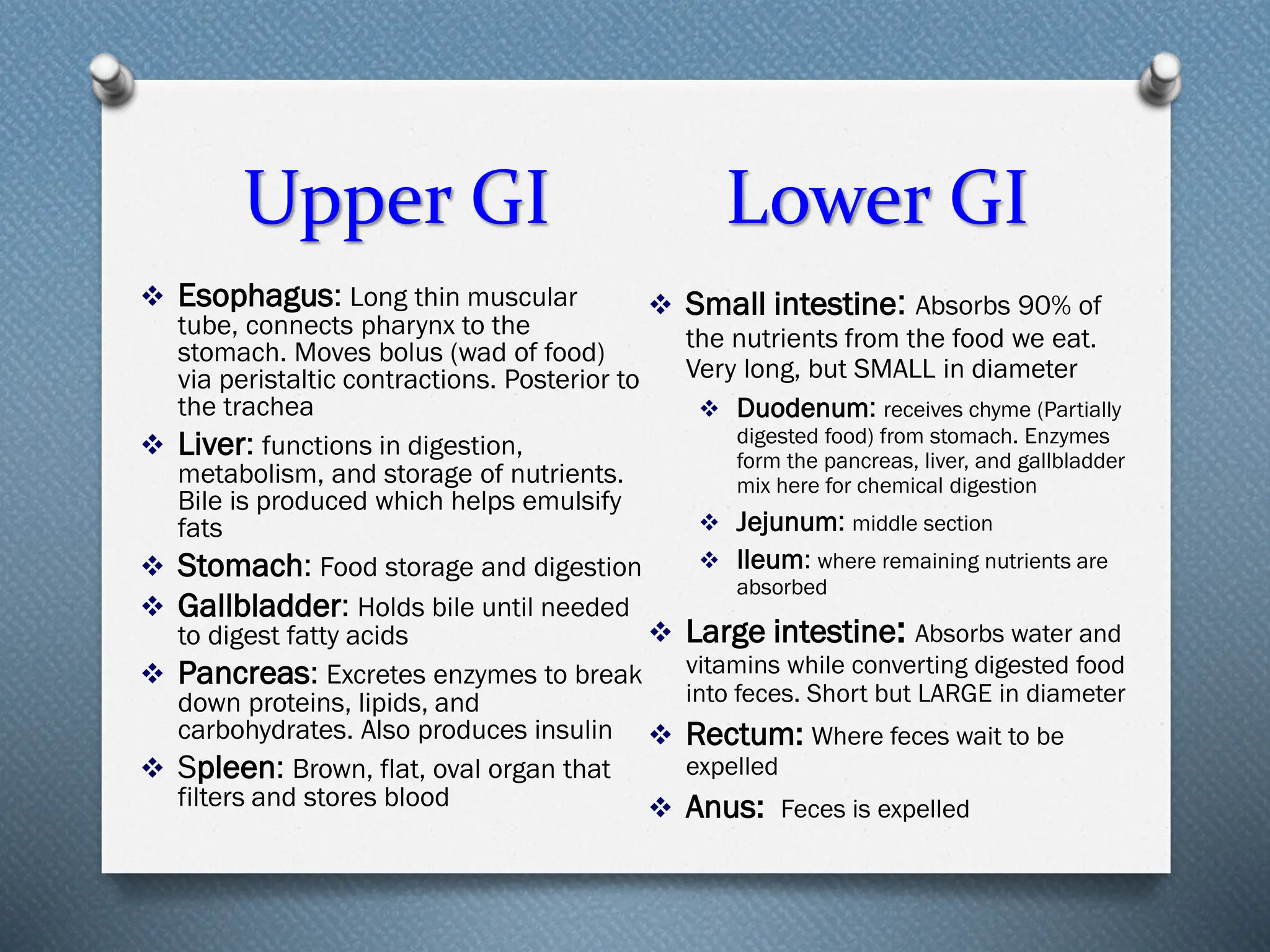
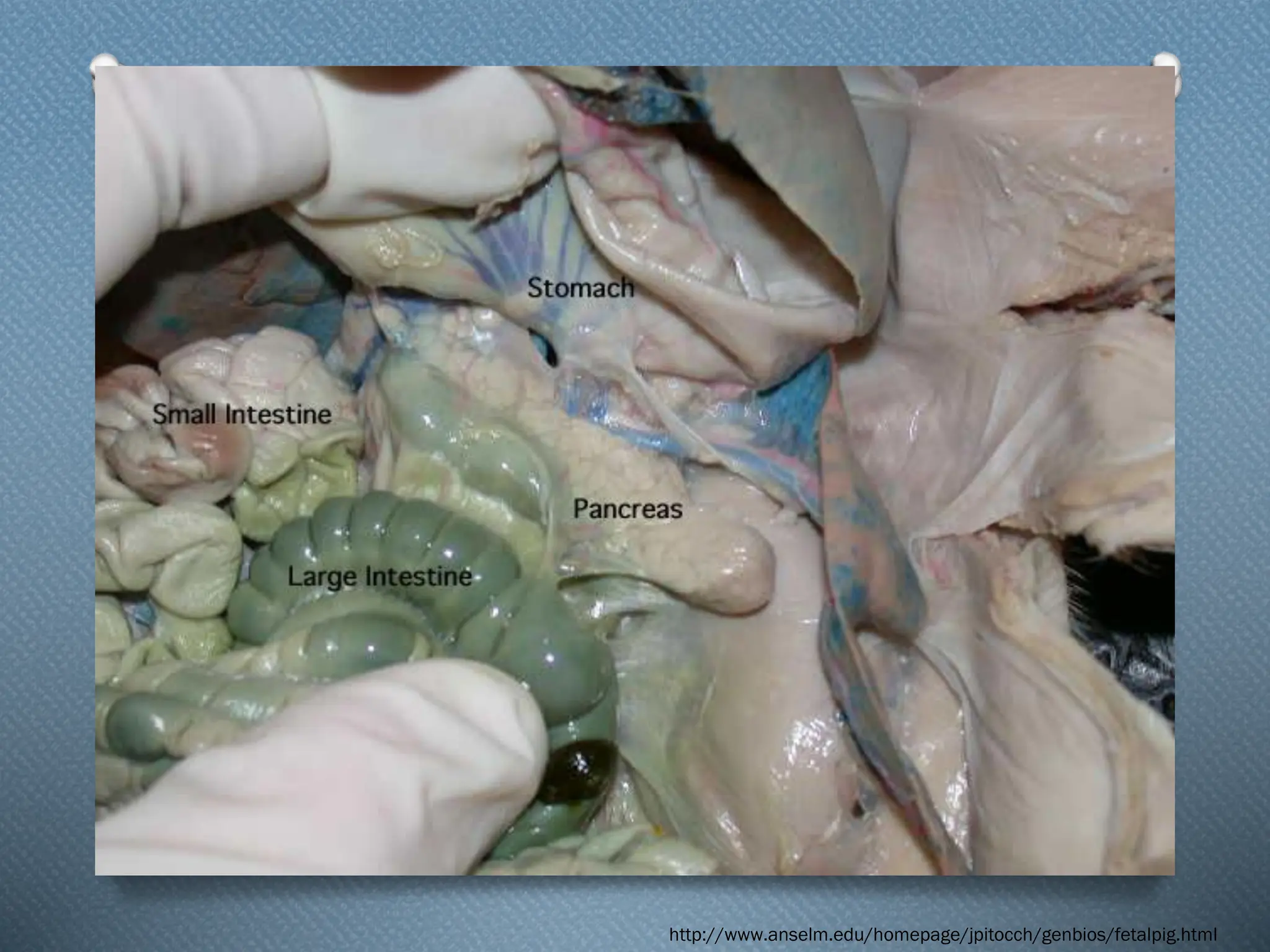
Upper GI
❖ Esophagus
❖ Liver
❖ Stomach
❖ Gallbladder
❖ Pancreas
❖ Spleen
http://www.clker.com/clipart-15593.html
Lower GI
❖ Small intestine
❖ Larger Intestine
❖ Rectum
❖ Anus
Goal: To get nutrients into the body via the breakdown of
food into smaller molecules and to excrete waste
14.
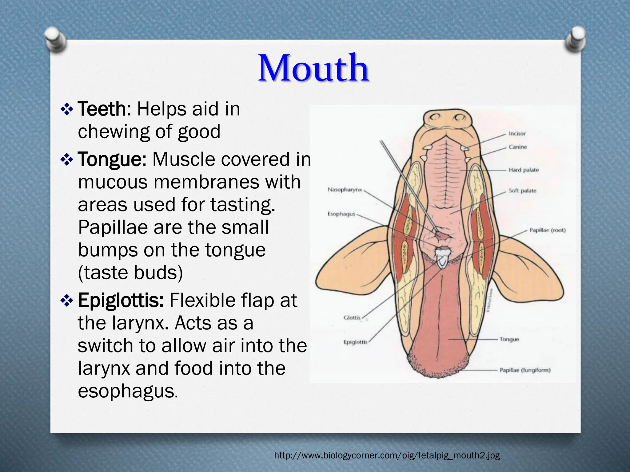
Mouth
❖ Teeth: Helpsaid in
chewing of good
❖ Tongue: Muscle covered in
mucous membranes with
areas used for tasting.
Papillae are the small
bumps on the tongue
(taste buds)
❖ Epiglottis: Flexible flap at
the larynx. Acts as a
switch to allow air into the
larynx and food into the
esophagus.
http://www.biologycorner.com/pig/fetalpig_mouth2.jpg
15.
Glands of theMouth
❖Parotid Gland :
Largest of the salivary
glands located anterior
and inferior to the ear.
❖Sublingual Gland:
One of the salivary
glands; located under
the floor of the mouth.
Mandibular Gland:
One of the salivary
glands inferior to the
mandible.
16.
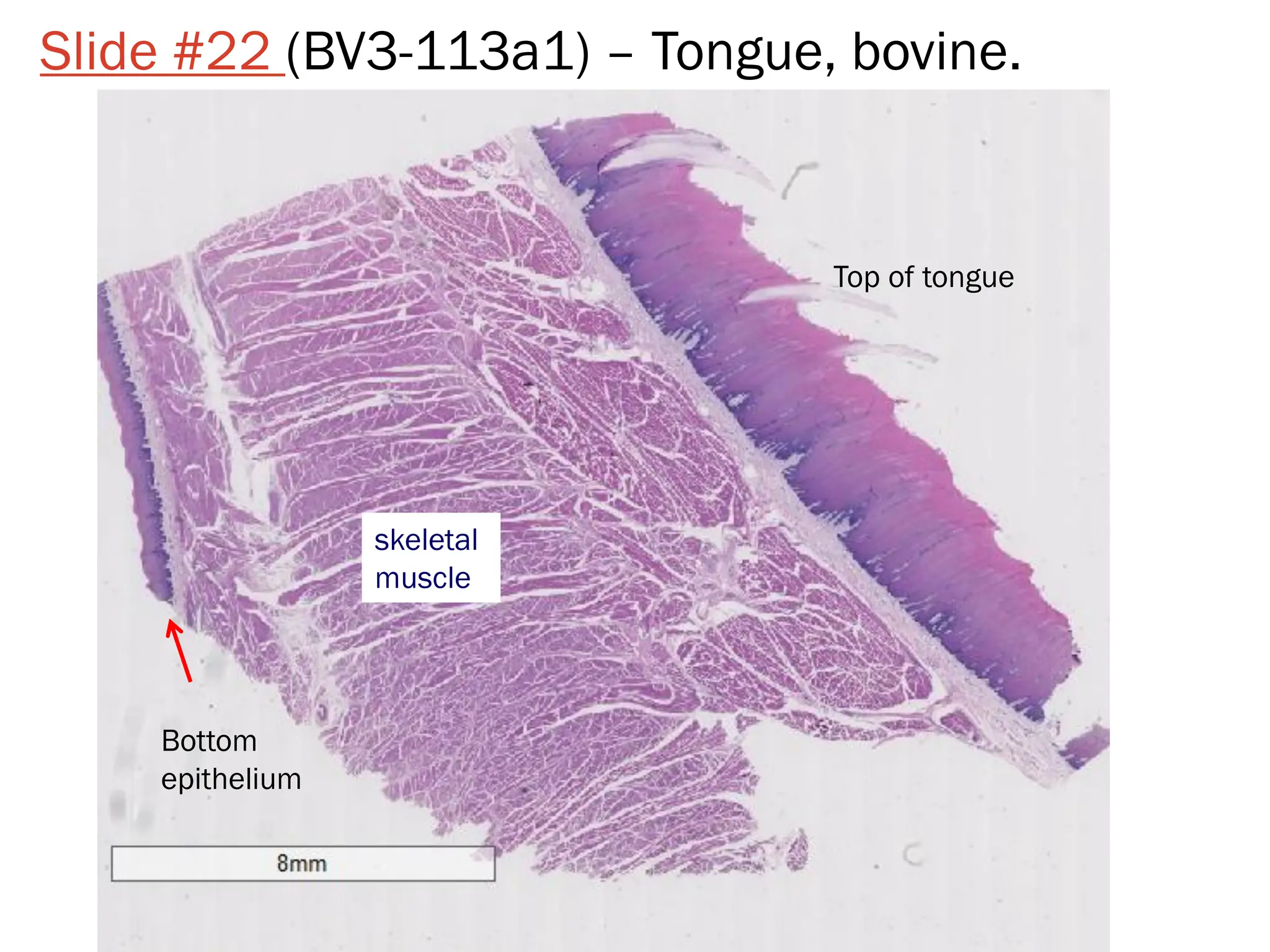
Slide #22 (BV3-113a1)– Tongue, bovine.
skeletal
muscle
Top of tongue
Bottom
epithelium
Upper GI LowerGI
❖ Esophagus: Long thin muscular
tube, connects pharynx to the
stomach. Moves bolus (wad of food)
via peristaltic contractions. Posterior to
the trachea
❖ Liver: functions in digestion,
metabolism, and storage of nutrients.
Bile is produced which helps emulsify
fats
❖ Stomach: Food storage and digestion
❖ Gallbladder: Holds bile until needed
to digest fatty acids
❖ Pancreas: Excretes enzymes to break
down proteins, lipids, and
carbohydrates. Also produces insulin
❖ Spleen: Brown, flat, oval organ that
filters and stores blood
❖ Small intestine: Absorbs 90% of
the nutrients from the food we eat.
Very long, but SMALL in diameter
❖ Duodenum: receives chyme (Partially
digested food) from stomach. Enzymes
form the pancreas, liver, and gallbladder
mix here for chemical digestion
❖ Jejunum: middle section
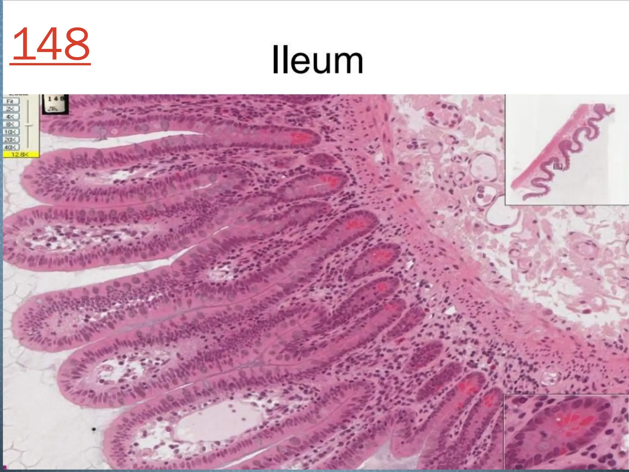
❖ Ileum: where remaining nutrients are
absorbed
❖ Large intestine: Absorbs water and
vitamins while converting digested food
into feces. Short but LARGE in diameter
❖ Rectum: Where feces wait to be
expelled
❖ Anus: Feces is expelled
Let’s take aCloser Look!
❖ Submandibular gland
❖ Esophagus
❖ Spleen
❖ Liver and Spleen
❖ Bile Duct and portal vein
❖ Gallbladder
❖ Pancreas
❖ Ileum
❖ Duodenum
❖ Liver
28.
Respiratory Tract
Goal: Toremove carbon dioxide from the blood and replace it
with oxygen
❖ Works in conjunction with the circulatory system
❖ Organs:
❖ Upper Tract
❖ Nose with nares
❖ Pharynx
❖ Larynx
❖ Lower Tract
❖ Trachea
❖ Lungs which contain the bronchi, bronchioles, and alveoli
❖ Diaphragm
29.
Nose
❖ Air entersthe respiratory
system through the paired
nares or nostrils
❖ Two nasal passages:
separated by bone
❖ Used for smell
❖ Hair and mucous
membranes clean out
particles
❖ Carbon dioxide is expired
through the nares
http://www.whitman.edu/academics/courses-of-study/biology/virtual-pig/respiratory-system
30.
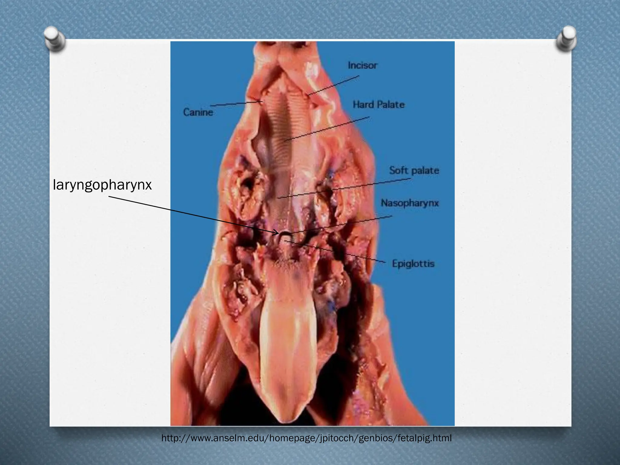
Pharynx and Epiglottis
❖Nasopharynx
❖ Above the soft palate
❖ Leads from the nose to the trachea for breathing
❖ Laryngopharynx
❖ Upper border of the glottis
❖ Helps guide food and air
❖ Epiglottis
❖ Small flap of cartilage
❖ Lies above the pharynx
❖ Allows for air to flow into larynx and trachea
❖ Directs food into the esophagus
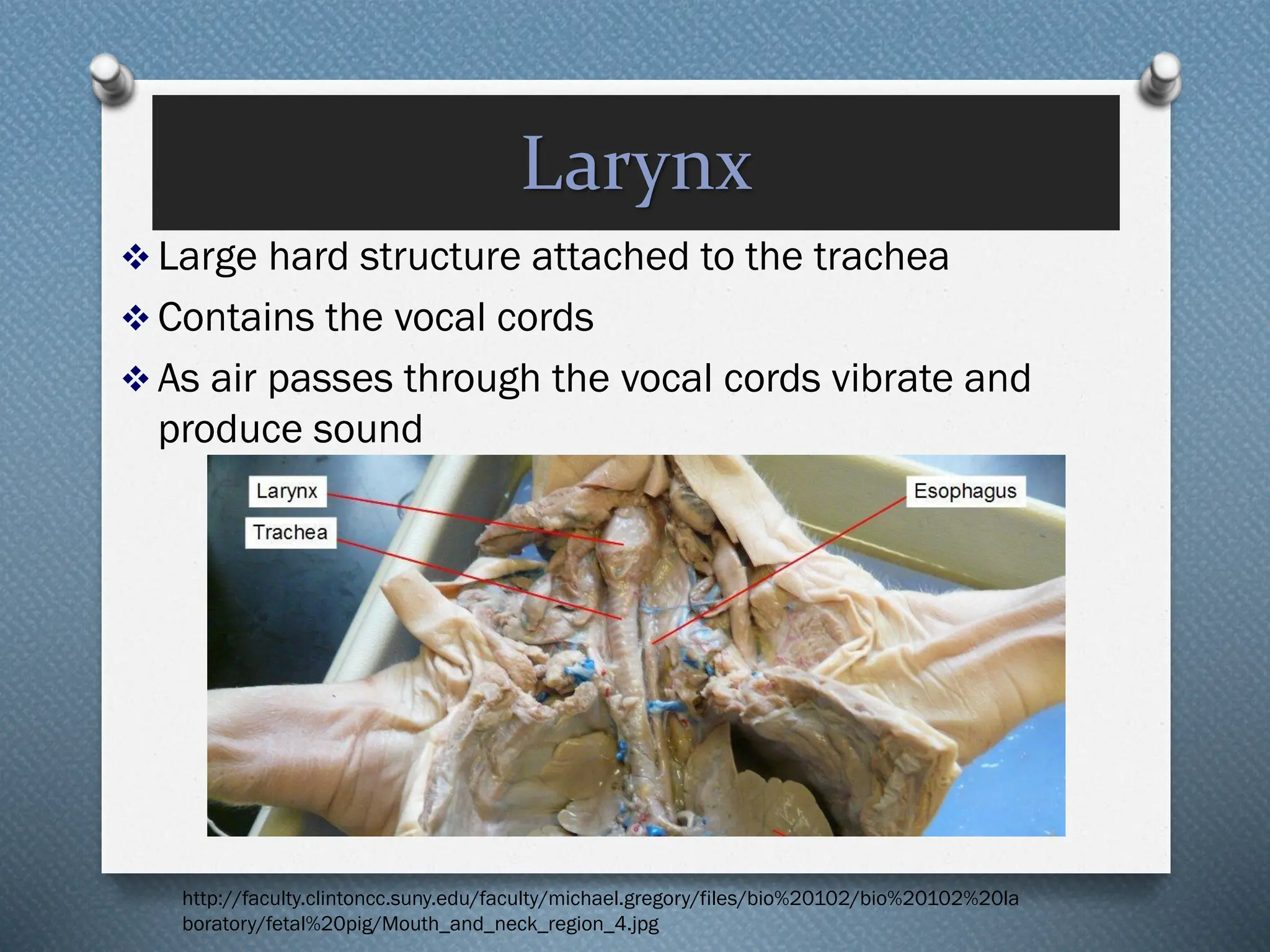
Larynx
❖ Large hardstructure attached to the trachea
❖ Contains the vocal cords
❖ As air passes through the vocal cords vibrate and
produce sound
http://faculty.clintoncc.suny.edu/faculty/michael.gregory/files/bio%20102/bio%20102%20la
boratory/fetal%20pig/Mouth_and_neck_region_4.jpg
33.
Trachea
❖ Windpipe
❖ Connectslarynx to lungs
❖ Contains cartilage rings
that prevent trachea
from collapsing
❖ Extends into the thoracic
cavity
❖ Branches into bronchi
❖ Allows air into the lungs
❖ Anterior to the
esophagus
https://www.google.com/search?q=trachea+fetal+pig&espv=2&biw=972&bih=854&source=lnms&tbm=isch&sa=X&ei=qAQ4VKTyO4_o8AHl4oC4Ag&ved=0CAYQ_A
UoAQ#facrc=_&imgdii=ZJMqXEEmRUJLqM%3A%3BLYjzE16FarDrrM%3BZJMqXEEmRUJLqM%3A&imgrc=ZJMqXEEmRUJLqM%253A%3BREGnqwdLFxjyuM%3Bhttp%2
53A%252F%252Fclassconnection.s3.amazonaws.com%252F244%252Fflashcards%252F2198244%252Fjpg%252Fpulmonary_trunk1352727288605.jpg%3Bhttp
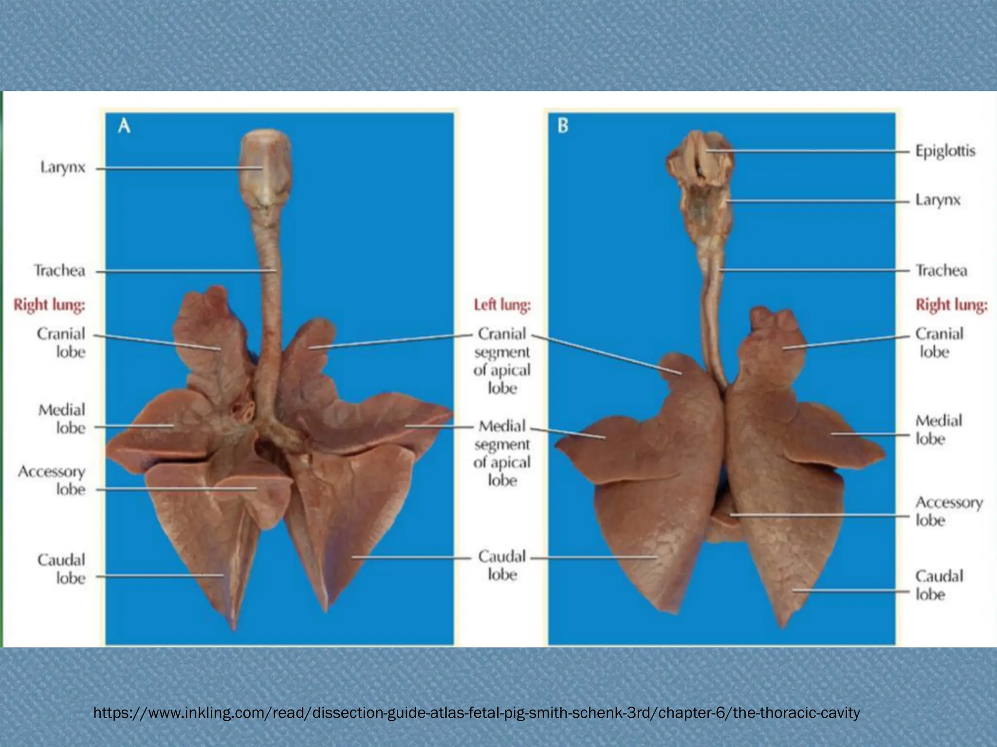
Lungs
❖ Branching ofthe trachea to bronchi to
lungs
❖ Located on either side of the heart
❖ The bronchi divide into bronchioles
❖ Bronchioles branch into alveolar sacs
❖ Alveolar sacs are bunches of alveoli
❖ Alveoli hold air and tightly bound in
blood vessels; allows for gas exchange
36.
Lungs continued
❖ Sectionedinto lobes
❖ Right lung is larger (4 lobes)
❖ cranial, medial, caudal, and accessory lobes
❖ Left lung is smaller because of space taken
up by the heart (2 lobes)
❖ apical and caudal lobe
Diaphragm
❖ Below thelungs
❖ Thin muscular sheet of tissue
❖ Only in mammals
❖ Expands to allow air in (diaphragm contracts)
❖ Compresses to expel air (diaphragm relaxes)
❖ Separates the chest and abdominal cavities
http://faculty.clintoncc.suny.edu/faculty/michael.gregory/files/bio%20102/bio%20102%20laboratory/fetal%20pig/Respiratory_system_5.jpg
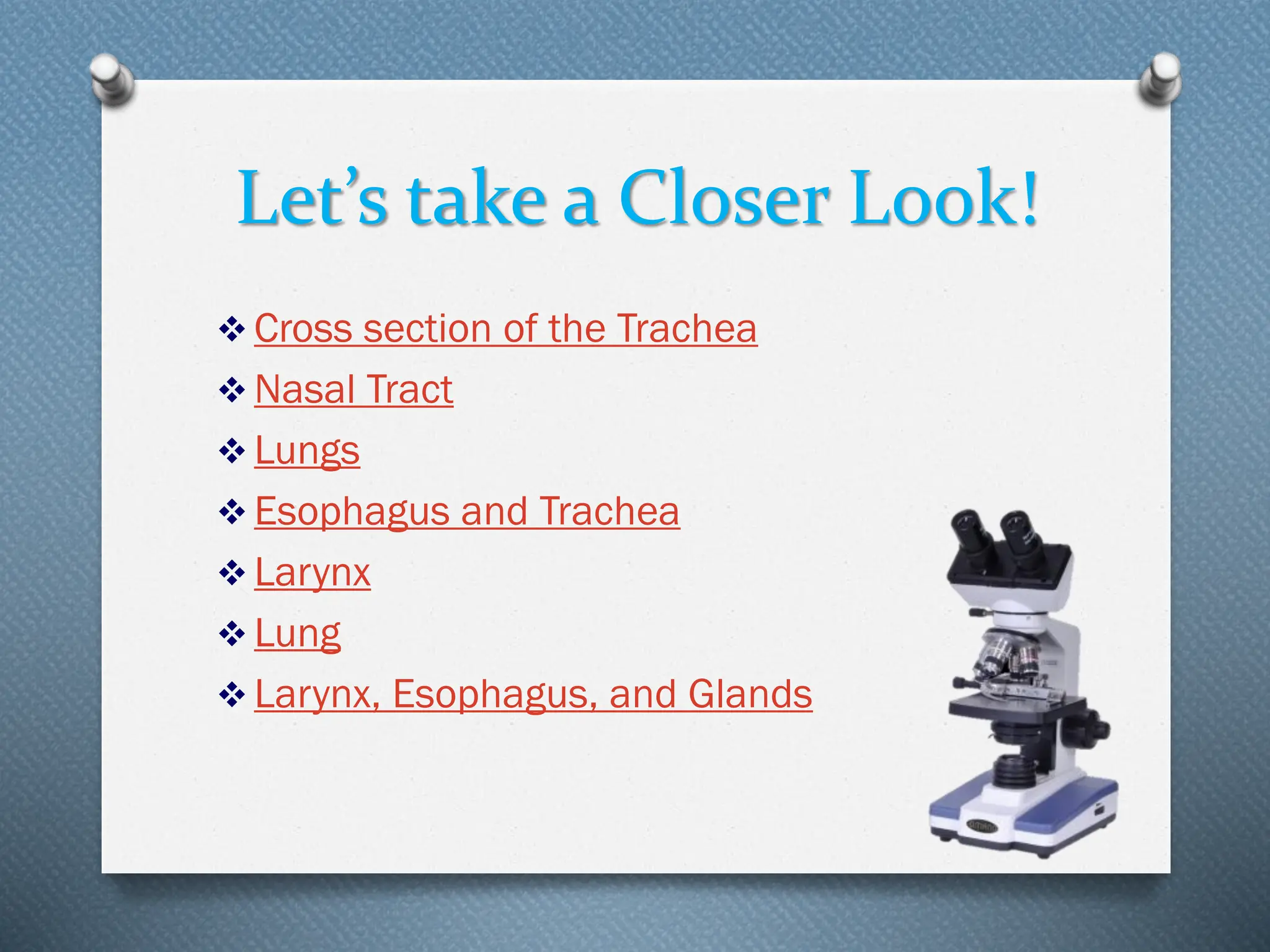
Let’s take aCloser Look!
❖ Cross section of the Trachea
❖ Nasal Tract
❖ Lungs
❖ Esophagus and Trachea
❖ Larynx
❖ Lung
❖ Larynx, Esophagus, and Glands
43.
Circulatory System
Allows blood,nutrients, oxygen and other gases, and
hormones to flow throughout the body
❖ Mammals (including humans)
❖ Double loop system
❖ 4 chambered heart
❖ Organs
❖ Heart
❖ Lungs
❖ Arteries
❖ Veins
http://www.ideacenter.org/contentmgr/showdetails.php/id/1113
44.
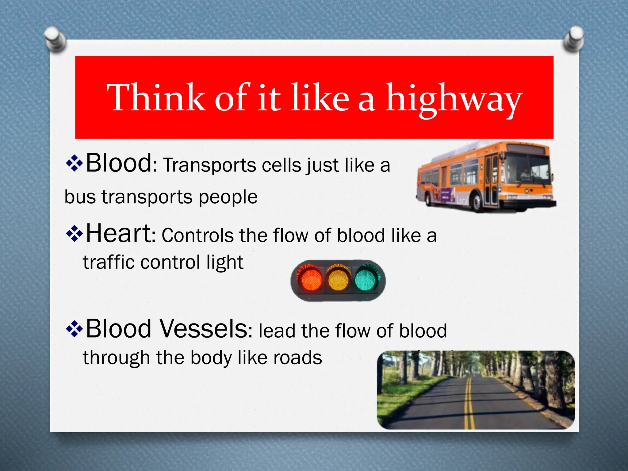
Think of itlike a highway
❖Blood: Transports cells just like a
bus transports people
❖Heart: Controls the flow of blood like a
traffic control light
❖Blood Vessels: lead the flow of blood
through the body like roads lead
45.
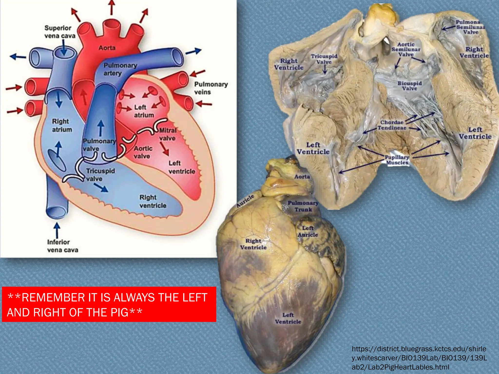
Heart
❖ Muscle usedto pump blood
❖ 4 chambers in mammals
❖ Right Atria
❖ Right Ventricle
❖ Left Atria
❖ Left Ventricle
❖ Atria pump blood to ventricles
❖ Ventricles pump blood out of heart and into circulatory
system
❖ Valves prevent backflow of blood
**REMEMBER IT ISALWAYS THE LEFT
AND RIGHT OF THE PIG**
https://district.bluegrass.kctcs.edu/shirle
y.whitescarver/BIO139Lab/BIO139/139L
ab2/Lab2PigHeartLables.html
50.
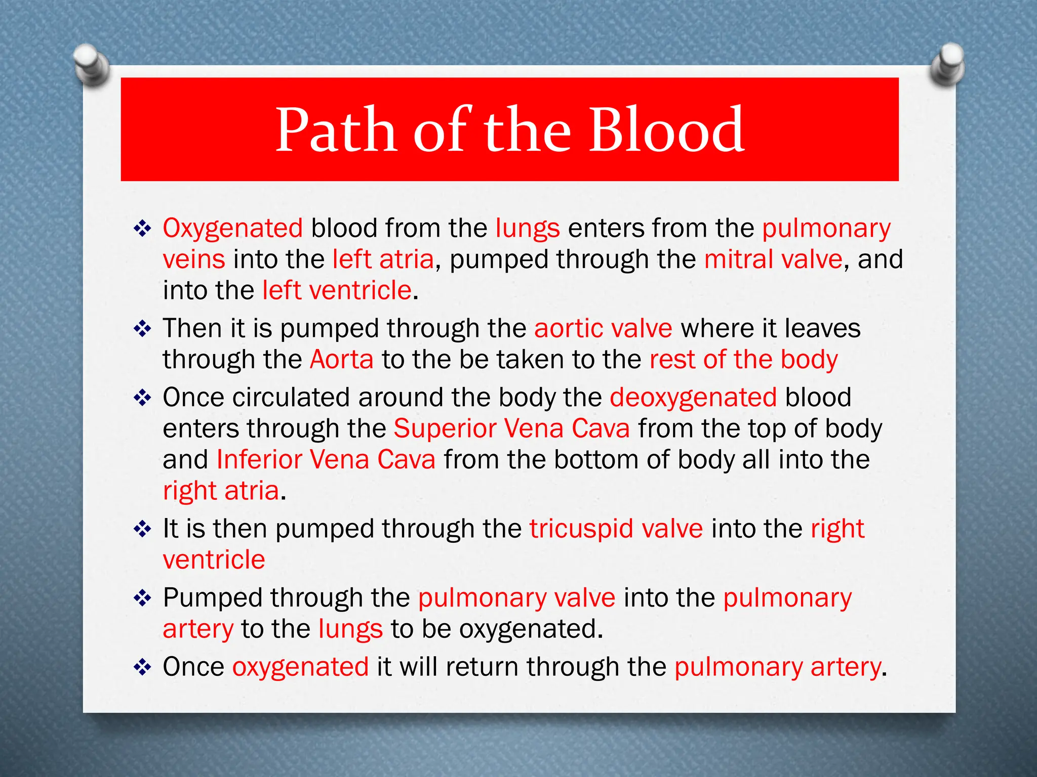
Path of theBlood
❖ Oxygenated blood from the lungs enters from the pulmonary
veins into the left atria, pumped through the mitral valve, and
into the left ventricle.
❖ Then it is pumped through the aortic valve where it leaves
through the Aorta to the be taken to the rest of the body
❖ Once circulated around the body the deoxygenated blood
enters through the Superior Vena Cava from the top of body
and Inferior Vena Cava from the bottom of body all into the
right atria.
❖ It is then pumped through the tricuspid valve into the right
ventricle
❖ Pumped through the pulmonary valve into the pulmonary
artery to the lungs to be oxygenated.
❖ Once oxygenated it will return through the pulmonary artery.
51.
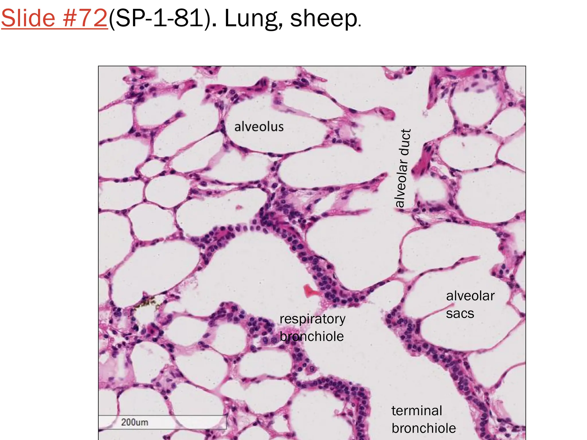
Lungs
❖ Provide oxygento the blood and remove
carbon dioxide from the blood
❖ Exchange of gases takes place in the
alveolar sacs
http://en.wikipedia.org/wiki/Pulmonary_circulation
52.
Blood
❖ Liquid thatcirculates in the blood vessels,
transporting oxygen, carbon dioxide, waste, and
hormones around the body.
❖ Blood contains
❖ (leukocytes)- fight against infections
❖ Red blood cells (erythrocytes) – transport oxygen
❖ Platelets (thrombocytes)- assist in clotting
❖ Plasma- liquid that suspends proteins and the solid
components of blood
White Blood Cells
53.
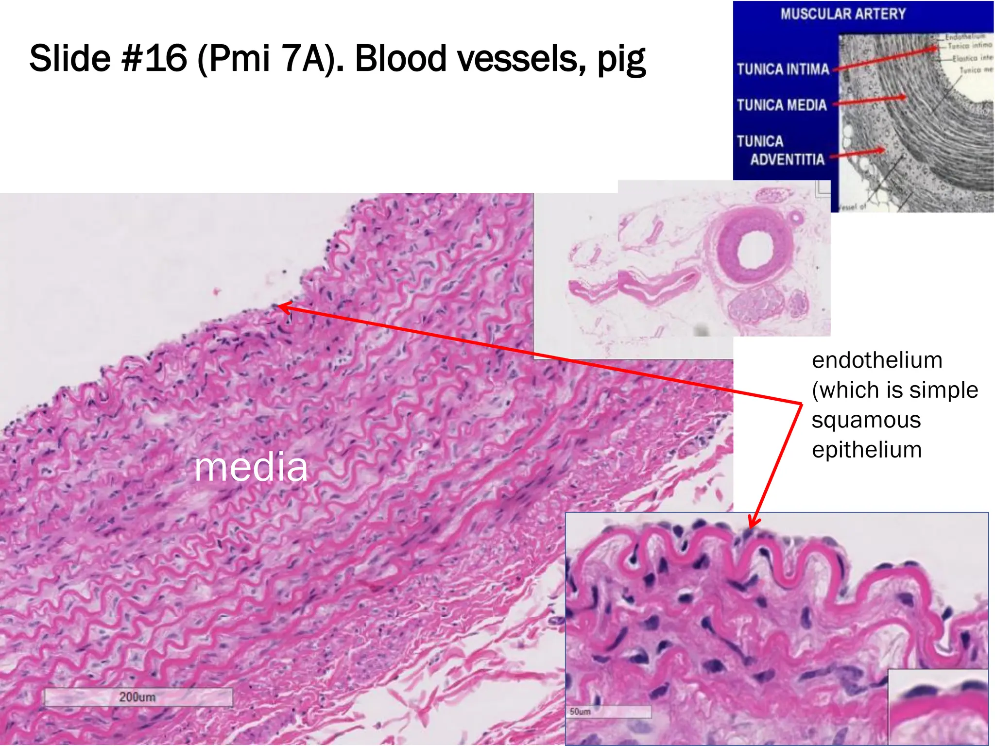
Blood Vessels
❖Veins- carryblood to the heart
❖Arteries- carry blood away from the heart
❖Capillaries – small pathway that
connects arteries to veins
❖Only one blood cell wide
https://www.studyblue.com/notes/note/n/emt-basic/deck/5473108
54.
Slide #16 (Pmi7A). Blood vessels, pig
endothelium
(which is simple
squamous
epithelium
media
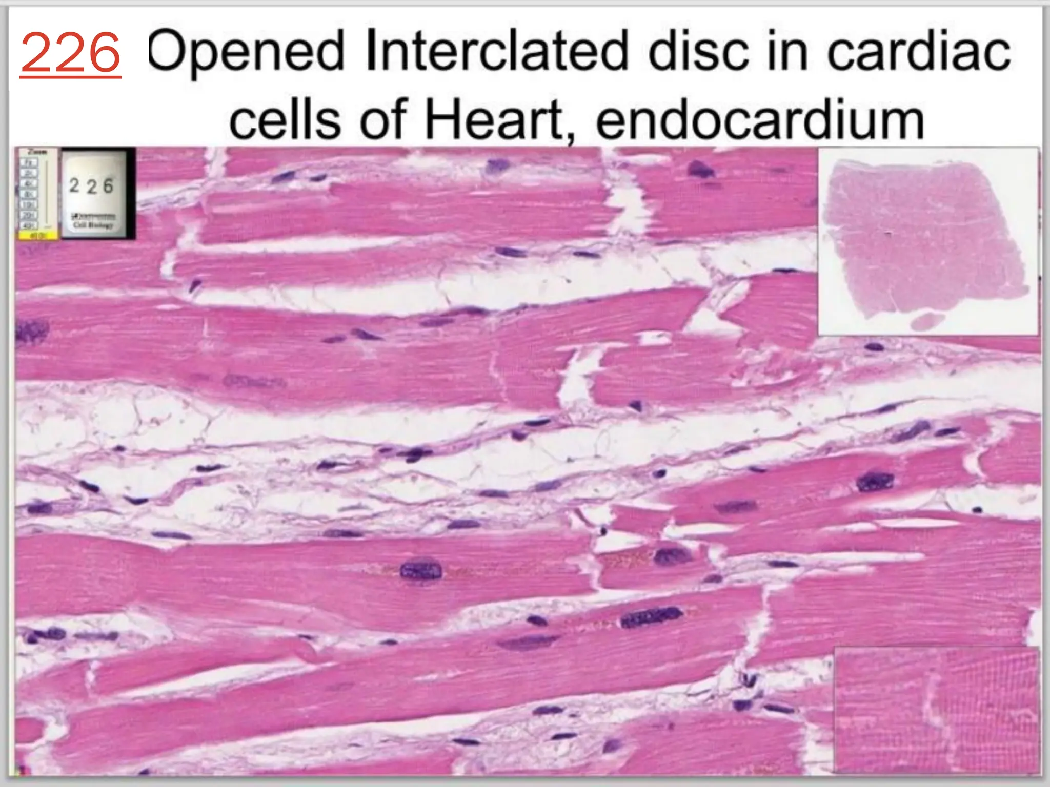
Let’s Take aCloser Look
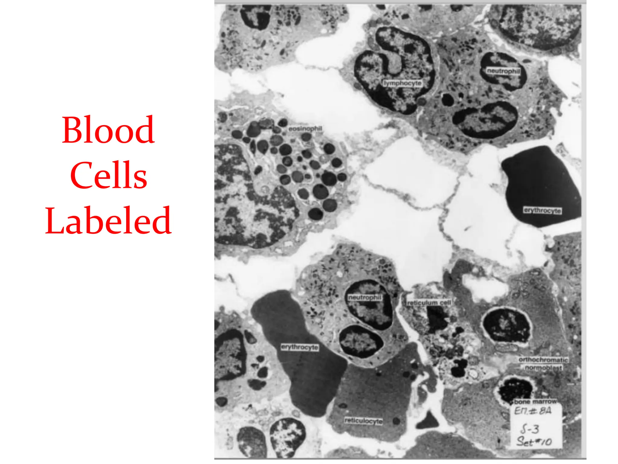
❖ Peripheral Blood Smear
❖ Red Bone Marrow Smear
❖ Heart, Epicardium
❖ Heart, Endocardium
❖ Blood 1
❖ Blood 2
❖ Blood 3
❖ Bone Marrow
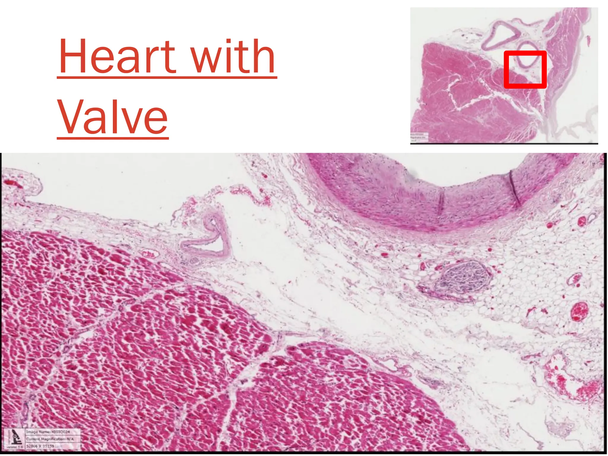
❖ Heart with Valve
❖ Heart Muscle
❖ Heart
http://www.microscope-microscope.org/basic/microscope-images/138-microscopes-lg.jpg
57.
Reproductive System
Expands thepopulation of a certain species
❖ Male and
female reproductive
tracts are different
❖ Males-produce
testosterone and sperm
❖ Females- produce
oocytes
58.
Male Repro Tract
❖Testes– produce sperm
❖Penis - deposits semen into female reproductive tract. Expels
urine from the body.
❖Cremaster muscle –pulls the testes
closer to the body to keep
warm
❖Scrotum -
houses the testes
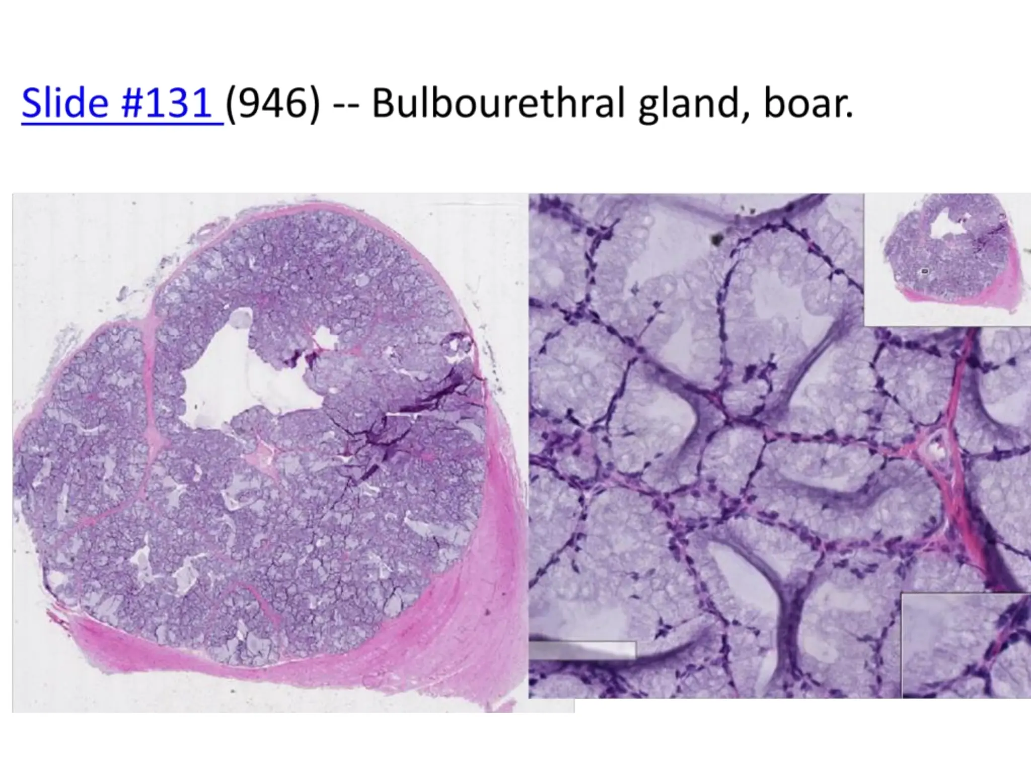
❖Bulbourethral Gland -
gives off seminal
fluid to urethra
59.
❖ Spermatic cord-
contains Vas
deferens,
spermatic artery
and vein, lymphatic
vessels, and leads
to the epididymis
❖ Epididymis- stores
sperm
❖ Seminiferous
tubules – sperm
produced
❖ Vas deferens-
transports sperm to
urethra
❖Urethra – receives seminal secretions form the testes. Drains excretory products from
the bladder
❖Prostate- secretes fluid that carries sperm
61.
Female Repro Tract
❖Ovaries- Produces oocytes (eggs)
❖ Oviduct- receives mature oocytes at
ovulation; site of fertilization
❖ Uterine Horns- Site of implantation and
embryonic development
❖ Vagina- receives penis during copulation;
serves as part of birth canal
❖ Urogenital Sinus- chamber in which the
vagina and urethra meet
63.
DEMO SLIDE BOX#208 – Cervix, sow.
complex folds of the luminal surface of the cervix.
Urinary System
Eliminates wastesfrom the body, regulates blood
volume and pressure, controls levels of electrolytes
and metabolites, and regulates blood pH
❖ Organs
❖ Adrenal Glands
❖ Kidneys
❖ Ureter
❖ Bladder
❖ Urethra
Adrenal
Gland
Kidney
Ureter
Bladder
http://memorize.com/urinary-system-pig-2/h4nsyh4ns
Urethra
67.
Adrenal Glands
❖ Siton top of the kidneys
❖ 2 total
❖ 2 Parts
❖ Adrenal Cortex- outer part that
produces hormones vital to life,
such as cortisol (metabolism) and
aldosterone (blood pressure)
❖ Adrenal Medulla- inner part that
produces nonessential hormones,
such as adrenaline (stress)
http://http://blog.lib.umn.edu/clar0841/psychblog/4/
68.
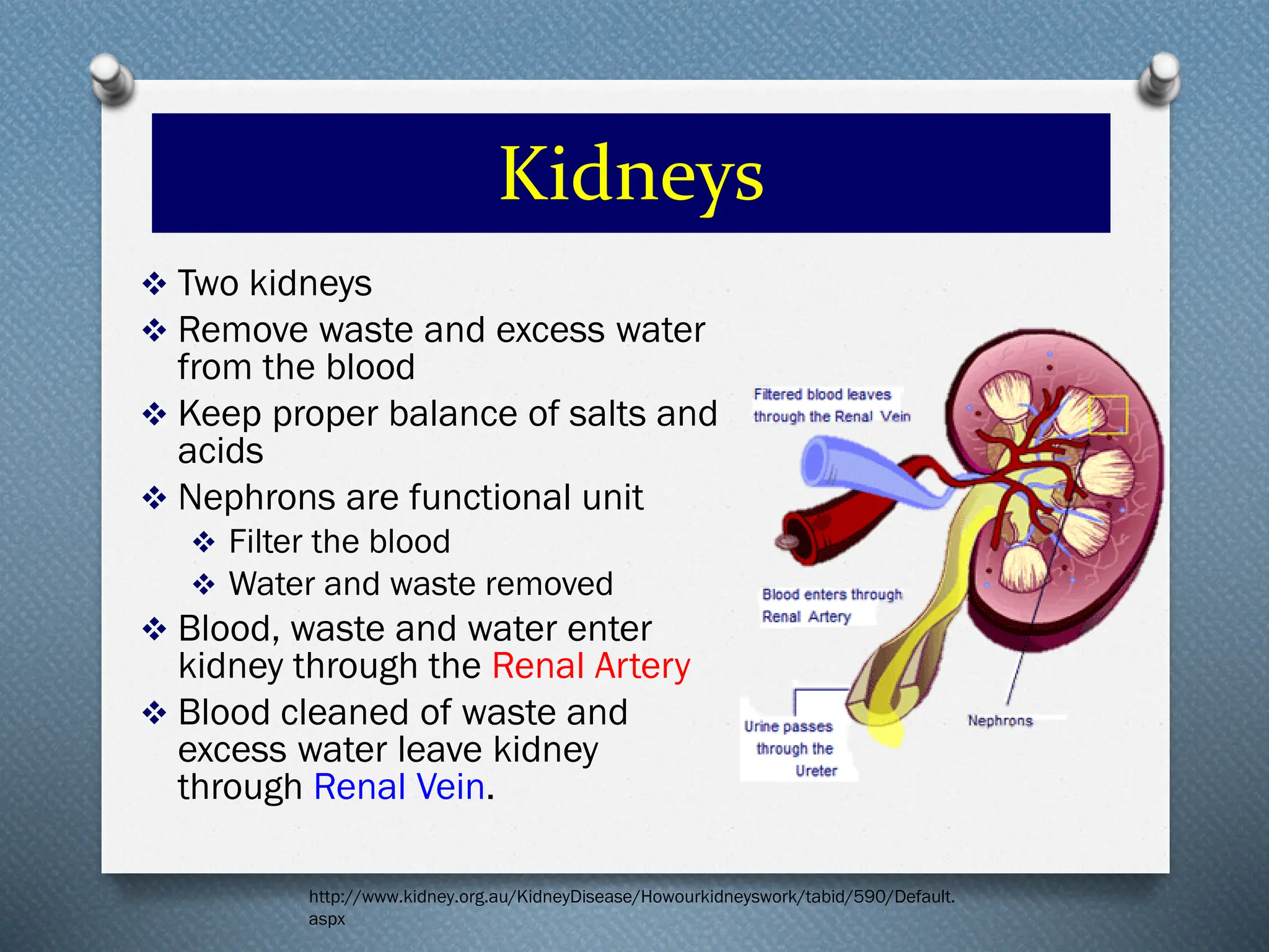
Kidneys
❖ Two kidneys
❖Remove waste and excess water
from the blood
❖ Keep proper balance of salts and
acids
❖ Nephrons are functional unit
❖ Filter the blood
❖ Water and waste removed
❖ Blood, waste and water enter
kidney through the Renal Artery
❖ Blood cleaned of waste and
excess water leave kidney
through Renal Vein.
http://www.kidney.org.au/KidneyDisease/Howourkidneyswork/tabid/590/Default.
aspx
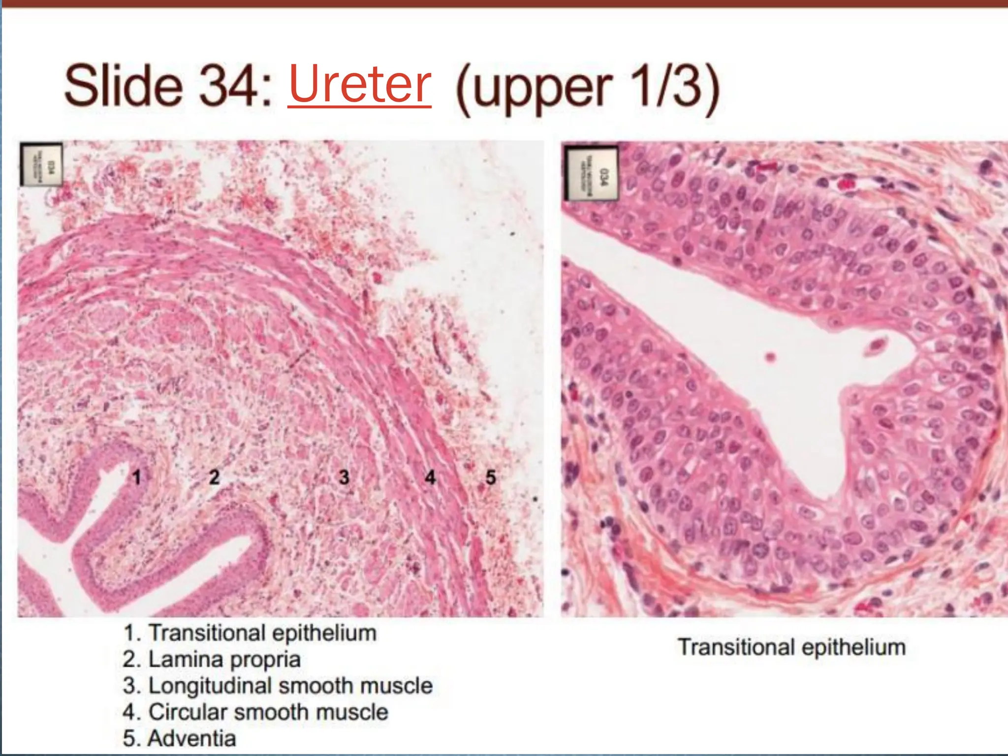
Ureter, Bladder, andUrethra
❖ Ureter
❖ One from each kidney
❖ Carries urine from
kidney to bladder
❖ Strong muscular tube
❖ Bladder
❖ Hollow organ
❖ Expands as it is filled
❖ Urethra
❖ Takes urine from the bladder to be eliminated
Who would have
thoughta pig would
be so closely related
to a human??!!
https://www.google.com/search?q=pig+and+farmer+friend+cartoon&espv=2&bi
w=1197&bih=753&source=lnms&tbm=isch&sa=X&ei=5khRVOqeA4upgwS7n4H
QBw&ved=0CAYQ_AUoAQ#tbm=isch&q=human+in+mud+cartoon&imgdii=_


![Human_Digestive_System[1].pptx by medical with us.pptx](https://cdn.slidesharecdn.com/ss_thumbnails/humandigestivesystem1-250517060919-d012c22d-thumbnail.jpg?width=640&height=640&fit=bounds)


