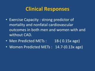
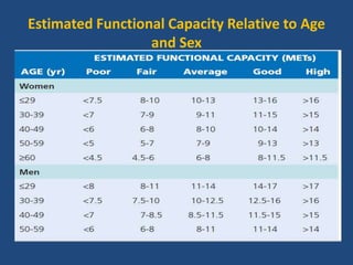
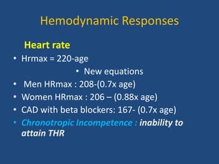
ECG round
Exercise Tolerance Test
by
Dr Galaleldin Ibrahim Ali
King Saud University Hospital
EP Devision
General indications for EXT
• Diagnosis of CAD
• Assessment of prognosis in CAD
• Evaluation of functional capacity
• Evaluation of therapy for CAD
outlines
• EXT physiology
• Technical aspects
• Contraindications
• EXT modalities and protocols
• Indications for terminations
• Pt monitoring during EXT
• Clinical response
• Diagnostic and prognostic values
• Prognostic scores
• guidelines
Physiology of exercising testing
• What is MET?
• What is the Vo2max?
• What is VO2 paek?
• MET = resting energy
expenditure
• I MET = 3.5ml O2/kg/min
• 5METs activity= 5 times resting
energy expenditure
• Vmax O2 is the peak oxygen uptake achieved
during performance of the highest level of
dynamic exercise
• cannot be exceeded despite increases in work
rate
• It is related to age, sex, heredity, exercise
habits, and cardiovascular status.
• Cardiac output can increase as much as four
to six times resting levels in the upright
position.
• Maximum cardiac output is the result of a
twofold to threefold increase in heart rate
from resting levels and an increase in stroke
volume.
• Stroke volume in healthy persons generally
plateaus at 50% to 60% of VmaxO2.
• VO2 peak =VO2 that is attained during
symptom-limited maximum exercise tolerated
• What you need to achieve during clinical EXT
Effects of Exercise on Myocardial Oxygen
Demand and Supply Relationships
• Myocardial ischemia occurs when the supply
of oxygenated blood to myocardial cells is
inadequate to meet demands.
• Exercise testing is performed to stress these
relationships and observe the physiologic
responses that ensue
Myocardial oxygen demand
Affected by:
• HR easiest to measure
• BP
• LV contractility
• LV wall stress
- LV cavity size
- LV pressure
- wall thickness
Physiologic responses to acute EXT
Myocardial oxygen supply
• Coronary blood flow increases during exercise
in response to neurohumoral stimulation(ß
receptors)
• In healthy persons during acute exercise,
coronary arteries dilate and coronary blood
flow rises in response to the increases in
myocardial oxygen demand
• a 50% to 70% reduction in luminal diameter
will impair peak reactive hyperemia
• whereas 90% or greater stenosis will reduce
resting flow.
• Atherosclerotic arteries often fail to dilate and
may actually constrict with exercise,
Technical aspect of EXT
• Patient assessment
History:
 symptoms
 risk factors for CAD
 Recent illness, hospitalization, or surgical
procedure
 Medication dose and schedule
 Ability to perform physical activity
Physical exam
• Pulse rate and regularity
• Resting blood pressure while sitting and standing
• Auscultation of the lungs, with specific attention
to uniformity of breath sounds in all areas
• Auscultation of the heart, particularly in patients
with heart failure or valvular disease
• Examination related to orthopedic, neurologic, or
other medical problems that might limit EXT
Contraindications for EXT
• Absolute Contraindications
• Acute myocardial infarction (within 2 days)
• High-risk unstable angina
• Uncontrolled cardiac arrhythmia with hemodynamic
compromise
• Active endocarditis
• Symptomatic severe aortic stenosis
• Decompensated heart failure
• Acute pulmonary embolism or pulmonary infarction
• Acute myocarditis or pericarditis
• Physical disability precluding safe and adequate testing
Relative Contraindications
• Known left main coronary artery stenosis
• Moderate aortic stenosis with uncertain relationship
to symptoms
• Tachyarrhythmias with uncontrolled ventricular rates
• Acquired complete heart block
• Hypertrophic cardiomyopathy with a severe resting
gradient
• Mental impairment with limited ability to cooperate
• From Fletcher GF, Ades PA, Kligfield P, et al: Exercise standards for testing and training: a
scientific statement from the American Heart Association. Circulation 128:873, 2013.
Exercise Test Modality and Protocols
Should be selected according to:
• the patient’s estimated functional capacity
based on
• age
• estimated physical fitness (history)
• underlying disease.
Types of protocols
• Stepped :- Work rate increments( stages),1-
2.5METs
• Ramp protocols :- designed with stages that
are no longer than 1 minute and for the
patient to attain peak effort within 8 to 12
minutes.
Submaximal EXT
• Has predetermined endpoint often defined as a
peak heart rate as HR =120 or 70%of THR or
5METs
• used after myocardial infarction pre discharge to
give prognostic information to guide
management.
• useful in the evaluation of a patient’s ability to
engage in daily activities after discharge .
• serve as a baseline for cardiac rehabilitative
exercise therapy
• Symptom-limited test: designed to continue
until the patient demonstrates signs and/or
symptoms necessitating termination of
exercise
Modalities of EXT
• Treadmill ( more physiological stress )
• Stationary Cycle.
• Arm Cycle Ergometry (alternative to former)
• Six-Minute Walk Test( not useful in deteming
CAD , used in a serial manner to evaluate
changes in exercise capacity and the response
to interventions
• Cardiopulmonary Exercise Testing
Indication for termination EXT
• Absolute Indications
• ST elevation (>1.0 mm) in leads without Q waves because of
previous MI (other than aVR, aVL, or V1)
• Drop in systolic BP of >10 mm Hg, despite an increase in
workload, when accompanied by any other evidence of
ischemia
• Moderate to severe angina
• Central nervous system symptoms (e.g., ataxia, dizziness, or
near-syncope)
• Signs of poor perfusion (cyanosis or pallor)
• Sustained ventricular tachycardia or other arrhythmia that
interferes with normal maintenance of cardiac output during
exercise
• Technical difficulties monitoring the ECG or systolic BP
• Patient’s request to stop
Relative Indications
• Marked ST displacement (>2 mm horizontal or
downsloping) in a patient with suspected ischemia
• Drop in systolic BP of >10 mm Hg (persistently below
baseline), despite an increase in workload, in the absence
of other evidence of ischemia
• Increasing chest pain Fatigue, shortness of breath,
wheezing, leg cramps, or claudication
• Arrhythmias other than sustained ventricular tachycardia,
including multifocal ectopy, ventricular triplets,
supraventricular tachycardia, atrioventricular heart block,
or bradyarrhythmias
• Exaggerated hypertensive response (systolic BP >250 mm
Hg and/or diastolic BP >115 mm Hg)
• Development of a BBB that cannot be distinguished from
ventricular tachycardia
Patient Monitoring During Exercise Testing
During the Exercise Period
• 12-lead ECG during the last minute of each
stage or at least every 3 min
• Blood pressure during the last minute of each
stage or at least every 3 min
• Symptom rating scales as appropriate for the
test indication and laboratory protocol
During the Recovery Period
• Monitoring for a minimum of 6 min after exercise in a sitting or
supine position or until near-baseline heart rate blood pressure,
ECG, and symptom measures are reached.
• A period of active cool down may be included in the recovery
period, particularly following high levels of exercise, to minimize the
postexercise hypotensive effects of venous pooling in the lower
extremities
• 12-lead ECG every minute
• Heart rate and blood pressure immediately after exercise and then
every 1 or 2 min thereafter until near-baseline measures are
reached
• Symptomatic ratings every minute as long as they persist after
exercise. Patients should be observed until all symptoms have
resolved or returned to baseline levels
Bruce protocol for treadmill testing
From American College of Sports Medicine Guidelines for Exercise
Testing and Prescription. 9th ed. Philadelphia, Lippincott,
Williams & Wilkins, 2013.
Clinical Responses
• Exercise Capacity : strong predictor of
mortality and nonfatal cardiovascular
outcomes in both men and women with and
without CAD.
• Men Predicted METs : 18-( 0.15x age)
• Women Predicted METs : 14.7-(0.13x age)
Estimated Functional Capacity Relative to Age
and Sex
Hemodynamic Responses
Heart rate
• Hrmax = 220-age
• New equations
• Men HRmax : 208-(0.7x age)
• Women HRmax : 206 – (0.88x age)
• CAD with beta blockers: 167- (0.7x age)
• Chronotropic Incompetence : inability to
attain THR
• Early Heart Rate Acceleration : seen in atrial
fibrillation, hypovolemia, anemia, and left
ventricular dysfunction (or deconditioning)
• Has prognostic value ( under investigation)
• Heart Rate Recovery : Abnormal HRR is
associated with an increase in all-cause
mortality in both asymptomatic individuals
and patients with established heart disease.
Definition of abnormal HRR
• less than 12 beats/min after 1 minute with
postexercise cool down,
• less than 18 beats/min after 1 minute with
immediate cessation of movement into either
the supine or sitting position,
• and less than 42 beats/min after 2 minutes
Blood Pressure
Exaggerated Systolic Pressure Response:
• defined as greater than 210 mm Hg in men
and greater than 190 mm Hg in women.
• may be indicative of the future development
of hypertension or adverse cardiac events.
• Exercise-Induced Hypotension : falling below
resting systolic pressure or 20 mm Hg fall after
an initial rise (severe multivessel CAD with left
ventricular dysfunction)
• Other causes:
• cardiomyopathy
• left ventricular outflow tract obstruction
• enhanced vagal tone
• hypovolemia
• antihypertensive medications
• arrhythmias.
• Low Systolic Pressure Peak : defined as a rise
to less than 140 mm Hg or a lower than 10
mm Hg rise overall.
• Ass. With severe CAD
• worse cardiovascular outcomes in persons
with and without known CAD and warrants
further evaluation.
• Recovery Systolic Pressure Response:
expressed as the ratio of 1-, 2-, or 3-minute
recovery pressure to peak exercise pressure.
• studies have shown a worse cardiovascular
prognosis when an abnormal recovery
pressure (e.g., peak-recovery ratio of ≥0.9) is
present.
• Double-Product Reserve : difference between
the peak and resting double product (HR-SBP)
• inversely related to cardiovascular events in
patients with and without known CAD
• It may have greater prognostic power than the
maximum heart rate, exercise capacity, and
HRR.
• Values lower than 10,000 warrant further
evaluation
ECG response
• ST-Segment Changes :
Sensitivity = 60- 70 %
Specificity = 70- 80 %
ECG patterns at rest and during EXT
usual criterion
• 1 mm or greater or 0.1 mV or greater of
horizontal or downsloping (i.e., <0.5 mV/sec)
ST-segment depression in three consecutive
beats.
• PQ point (not the TP )  isoelectric line
• point of ST-segment measurement is 60 to 80
milliseconds after the J point.
• The 60-millisecond post–J point criterion is
used at heart rates higher than 130 beats/min.
ACC/AHA Practice Guidelines on Exercise Testing: Pretest
Probability of Coronary Heart Disease by Age, Sex, and
symptoms
Diagnostic Value of the EXT for Identification of Coronary
Artery Disease
- Sensitivity and Specificity :- (68% - 70% respectively)
- Sensitivity influenced by:
- Disease severity
- Effort level
- Use of anti-ischemic
Specificity is affected by:
resting ECG pattren
LVHST-T abnormality
IVCD
drugs ( digoxin)
Prognostic Value of the Exercise
Electrocardiogram
Prognostic Variables :
• The strongest predictor of prognosis
derived from the exercise test is
exercise capacity. The weakest
predictor is ST-segment depression.
Prognostic Scores
1- Duke Treadmill Score.
ET –( 5xST D ) –(4x angina index )
 high risk < -11
 low risk > 5
 intermediate risk between -11 and 5
2- Sex-Specific Scores.
3- Cleveland Clinic Score
LM or MVD predictors
• early onset of ischemic ST segment depression,
ST segment depression of mm (0.20 mV) involving
five or more leads, or persisting minutes into
recovery,
• A failure to increase systolic blood pressure by 10
mm Hg,
• or a sustained decrease in systolic blood pressure
of 10 mm Hg or more,
• reproducible sustained or symptomatic
ventricular tachycardia
GUIDELINES
References
• BRAUNWALD’S heart disease of CVS
10th edition 2015
(with review more 65 references )
Thank you
for
attention

ECG Exercise Tolerance Test

  • 1.
    ECG round Exercise ToleranceTest by Dr Galaleldin Ibrahim Ali King Saud University Hospital EP Devision
  • 2.
    General indications forEXT • Diagnosis of CAD • Assessment of prognosis in CAD • Evaluation of functional capacity • Evaluation of therapy for CAD
  • 3.
    outlines • EXT physiology •Technical aspects • Contraindications • EXT modalities and protocols • Indications for terminations • Pt monitoring during EXT • Clinical response • Diagnostic and prognostic values • Prognostic scores • guidelines
  • 4.
    Physiology of exercisingtesting • What is MET? • What is the Vo2max? • What is VO2 paek?
  • 5.
    • MET =resting energy expenditure • I MET = 3.5ml O2/kg/min • 5METs activity= 5 times resting energy expenditure
  • 6.
    • Vmax O2is the peak oxygen uptake achieved during performance of the highest level of dynamic exercise • cannot be exceeded despite increases in work rate • It is related to age, sex, heredity, exercise habits, and cardiovascular status.
  • 7.
    • Cardiac outputcan increase as much as four to six times resting levels in the upright position. • Maximum cardiac output is the result of a twofold to threefold increase in heart rate from resting levels and an increase in stroke volume. • Stroke volume in healthy persons generally plateaus at 50% to 60% of VmaxO2.
  • 8.
    • VO2 peak=VO2 that is attained during symptom-limited maximum exercise tolerated • What you need to achieve during clinical EXT
  • 9.
    Effects of Exerciseon Myocardial Oxygen Demand and Supply Relationships • Myocardial ischemia occurs when the supply of oxygenated blood to myocardial cells is inadequate to meet demands. • Exercise testing is performed to stress these relationships and observe the physiologic responses that ensue
  • 10.
    Myocardial oxygen demand Affectedby: • HR easiest to measure • BP • LV contractility • LV wall stress - LV cavity size - LV pressure - wall thickness
  • 11.
  • 12.
    Myocardial oxygen supply •Coronary blood flow increases during exercise in response to neurohumoral stimulation(ß receptors) • In healthy persons during acute exercise, coronary arteries dilate and coronary blood flow rises in response to the increases in myocardial oxygen demand
  • 13.
    • a 50%to 70% reduction in luminal diameter will impair peak reactive hyperemia • whereas 90% or greater stenosis will reduce resting flow. • Atherosclerotic arteries often fail to dilate and may actually constrict with exercise,
  • 14.
    Technical aspect ofEXT • Patient assessment History:  symptoms  risk factors for CAD  Recent illness, hospitalization, or surgical procedure  Medication dose and schedule  Ability to perform physical activity
  • 15.
    Physical exam • Pulserate and regularity • Resting blood pressure while sitting and standing • Auscultation of the lungs, with specific attention to uniformity of breath sounds in all areas • Auscultation of the heart, particularly in patients with heart failure or valvular disease • Examination related to orthopedic, neurologic, or other medical problems that might limit EXT
  • 16.
    Contraindications for EXT •Absolute Contraindications • Acute myocardial infarction (within 2 days) • High-risk unstable angina • Uncontrolled cardiac arrhythmia with hemodynamic compromise • Active endocarditis • Symptomatic severe aortic stenosis • Decompensated heart failure • Acute pulmonary embolism or pulmonary infarction • Acute myocarditis or pericarditis • Physical disability precluding safe and adequate testing
  • 17.
    Relative Contraindications • Knownleft main coronary artery stenosis • Moderate aortic stenosis with uncertain relationship to symptoms • Tachyarrhythmias with uncontrolled ventricular rates • Acquired complete heart block • Hypertrophic cardiomyopathy with a severe resting gradient • Mental impairment with limited ability to cooperate • From Fletcher GF, Ades PA, Kligfield P, et al: Exercise standards for testing and training: a scientific statement from the American Heart Association. Circulation 128:873, 2013.
  • 18.
    Exercise Test Modalityand Protocols Should be selected according to: • the patient’s estimated functional capacity based on • age • estimated physical fitness (history) • underlying disease.
  • 19.
    Types of protocols •Stepped :- Work rate increments( stages),1- 2.5METs • Ramp protocols :- designed with stages that are no longer than 1 minute and for the patient to attain peak effort within 8 to 12 minutes.
  • 20.
    Submaximal EXT • Haspredetermined endpoint often defined as a peak heart rate as HR =120 or 70%of THR or 5METs • used after myocardial infarction pre discharge to give prognostic information to guide management. • useful in the evaluation of a patient’s ability to engage in daily activities after discharge . • serve as a baseline for cardiac rehabilitative exercise therapy
  • 21.
    • Symptom-limited test:designed to continue until the patient demonstrates signs and/or symptoms necessitating termination of exercise
  • 22.
    Modalities of EXT •Treadmill ( more physiological stress ) • Stationary Cycle. • Arm Cycle Ergometry (alternative to former) • Six-Minute Walk Test( not useful in deteming CAD , used in a serial manner to evaluate changes in exercise capacity and the response to interventions • Cardiopulmonary Exercise Testing
  • 23.
    Indication for terminationEXT • Absolute Indications • ST elevation (>1.0 mm) in leads without Q waves because of previous MI (other than aVR, aVL, or V1) • Drop in systolic BP of >10 mm Hg, despite an increase in workload, when accompanied by any other evidence of ischemia • Moderate to severe angina • Central nervous system symptoms (e.g., ataxia, dizziness, or near-syncope) • Signs of poor perfusion (cyanosis or pallor) • Sustained ventricular tachycardia or other arrhythmia that interferes with normal maintenance of cardiac output during exercise • Technical difficulties monitoring the ECG or systolic BP • Patient’s request to stop
  • 24.
    Relative Indications • MarkedST displacement (>2 mm horizontal or downsloping) in a patient with suspected ischemia • Drop in systolic BP of >10 mm Hg (persistently below baseline), despite an increase in workload, in the absence of other evidence of ischemia • Increasing chest pain Fatigue, shortness of breath, wheezing, leg cramps, or claudication • Arrhythmias other than sustained ventricular tachycardia, including multifocal ectopy, ventricular triplets, supraventricular tachycardia, atrioventricular heart block, or bradyarrhythmias • Exaggerated hypertensive response (systolic BP >250 mm Hg and/or diastolic BP >115 mm Hg) • Development of a BBB that cannot be distinguished from ventricular tachycardia
  • 25.
    Patient Monitoring DuringExercise Testing During the Exercise Period • 12-lead ECG during the last minute of each stage or at least every 3 min • Blood pressure during the last minute of each stage or at least every 3 min • Symptom rating scales as appropriate for the test indication and laboratory protocol
  • 26.
    During the RecoveryPeriod • Monitoring for a minimum of 6 min after exercise in a sitting or supine position or until near-baseline heart rate blood pressure, ECG, and symptom measures are reached. • A period of active cool down may be included in the recovery period, particularly following high levels of exercise, to minimize the postexercise hypotensive effects of venous pooling in the lower extremities • 12-lead ECG every minute • Heart rate and blood pressure immediately after exercise and then every 1 or 2 min thereafter until near-baseline measures are reached • Symptomatic ratings every minute as long as they persist after exercise. Patients should be observed until all symptoms have resolved or returned to baseline levels
  • 28.
    Bruce protocol fortreadmill testing From American College of Sports Medicine Guidelines for Exercise Testing and Prescription. 9th ed. Philadelphia, Lippincott, Williams & Wilkins, 2013.
  • 29.
    Clinical Responses • ExerciseCapacity : strong predictor of mortality and nonfatal cardiovascular outcomes in both men and women with and without CAD. • Men Predicted METs : 18-( 0.15x age) • Women Predicted METs : 14.7-(0.13x age)
  • 30.
    Estimated Functional CapacityRelative to Age and Sex
  • 31.
    Hemodynamic Responses Heart rate •Hrmax = 220-age • New equations • Men HRmax : 208-(0.7x age) • Women HRmax : 206 – (0.88x age) • CAD with beta blockers: 167- (0.7x age) • Chronotropic Incompetence : inability to attain THR
  • 32.
    • Early HeartRate Acceleration : seen in atrial fibrillation, hypovolemia, anemia, and left ventricular dysfunction (or deconditioning) • Has prognostic value ( under investigation) • Heart Rate Recovery : Abnormal HRR is associated with an increase in all-cause mortality in both asymptomatic individuals and patients with established heart disease.
  • 33.
    Definition of abnormalHRR • less than 12 beats/min after 1 minute with postexercise cool down, • less than 18 beats/min after 1 minute with immediate cessation of movement into either the supine or sitting position, • and less than 42 beats/min after 2 minutes
  • 34.
    Blood Pressure Exaggerated SystolicPressure Response: • defined as greater than 210 mm Hg in men and greater than 190 mm Hg in women. • may be indicative of the future development of hypertension or adverse cardiac events. • Exercise-Induced Hypotension : falling below resting systolic pressure or 20 mm Hg fall after an initial rise (severe multivessel CAD with left ventricular dysfunction)
  • 35.
    • Other causes: •cardiomyopathy • left ventricular outflow tract obstruction • enhanced vagal tone • hypovolemia • antihypertensive medications • arrhythmias.
  • 36.
    • Low SystolicPressure Peak : defined as a rise to less than 140 mm Hg or a lower than 10 mm Hg rise overall. • Ass. With severe CAD • worse cardiovascular outcomes in persons with and without known CAD and warrants further evaluation.
  • 37.
    • Recovery SystolicPressure Response: expressed as the ratio of 1-, 2-, or 3-minute recovery pressure to peak exercise pressure. • studies have shown a worse cardiovascular prognosis when an abnormal recovery pressure (e.g., peak-recovery ratio of ≥0.9) is present.
  • 38.
    • Double-Product Reserve: difference between the peak and resting double product (HR-SBP) • inversely related to cardiovascular events in patients with and without known CAD • It may have greater prognostic power than the maximum heart rate, exercise capacity, and HRR. • Values lower than 10,000 warrant further evaluation
  • 39.
    ECG response • ST-SegmentChanges : Sensitivity = 60- 70 % Specificity = 70- 80 %
  • 40.
    ECG patterns atrest and during EXT
  • 42.
    usual criterion • 1mm or greater or 0.1 mV or greater of horizontal or downsloping (i.e., <0.5 mV/sec) ST-segment depression in three consecutive beats. • PQ point (not the TP )  isoelectric line • point of ST-segment measurement is 60 to 80 milliseconds after the J point. • The 60-millisecond post–J point criterion is used at heart rates higher than 130 beats/min.
  • 43.
    ACC/AHA Practice Guidelineson Exercise Testing: Pretest Probability of Coronary Heart Disease by Age, Sex, and symptoms
  • 44.
    Diagnostic Value ofthe EXT for Identification of Coronary Artery Disease - Sensitivity and Specificity :- (68% - 70% respectively) - Sensitivity influenced by: - Disease severity - Effort level - Use of anti-ischemic Specificity is affected by: resting ECG pattren LVHST-T abnormality IVCD drugs ( digoxin)
  • 45.
    Prognostic Value ofthe Exercise Electrocardiogram Prognostic Variables : • The strongest predictor of prognosis derived from the exercise test is exercise capacity. The weakest predictor is ST-segment depression.
  • 46.
    Prognostic Scores 1- DukeTreadmill Score. ET –( 5xST D ) –(4x angina index )  high risk < -11  low risk > 5  intermediate risk between -11 and 5 2- Sex-Specific Scores. 3- Cleveland Clinic Score
  • 48.
    LM or MVDpredictors • early onset of ischemic ST segment depression, ST segment depression of mm (0.20 mV) involving five or more leads, or persisting minutes into recovery, • A failure to increase systolic blood pressure by 10 mm Hg, • or a sustained decrease in systolic blood pressure of 10 mm Hg or more, • reproducible sustained or symptomatic ventricular tachycardia
  • 49.
  • 53.
    References • BRAUNWALD’S heartdisease of CVS 10th edition 2015 (with review more 65 references )
  • 54.