2010-Era computer assisted microscopy- 4M microscopes
•Download as PPT, PDF•
0 likes•309 views

Small Size Virtual Slide
Recommended

Recommended
More Related Content
What's hot
What's hot (20)
Similar to 2010-Era computer assisted microscopy- 4M microscopes
Similar to 2010-Era computer assisted microscopy- 4M microscopes (20)
More from Unesco Telemedicine
More from Unesco Telemedicine (20)
Recently uploaded
Recently uploaded (20)
2010-Era computer assisted microscopy- 4M microscopes
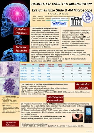
- 1. www.imprimelo.com O. Ferrer-Roca & F. Marcano UNESCO Chair of Telemedicine. Chair of Telemedicine of Telefonica. Faculty of Medicine. University of La Laguna. Tenerife 38071. Canary Islands. Spain. catai@teide.net. www.catai.net/blog Referencias: References: Gráficos y tablas / Graphs and tables Métodos: Methods: The FOV images had four times higher discrimination power in spite of the low sampling density. The ROI images, with a sampling density close to Shannon theory had six times higher discrimination power. SM OnScreen magnification FOV achieved 640x & ROI 3200x augments that could never been reached using analog microscopy. (1) Projection magnification onChip (PMoC) is essential to evaluate the system sampling capabilities, the so-called DR o digital resolution that influenced visibility and digital image quality with or without computer assisted techniques. (2) Differences OnScreen of RAW /RGB images were related to higher DR and the contrast enhancement & light gathering provided by superresolution algorithm. on the RAW images. (3) that SSVS are ideal for hand-held microscopes, 4M or even mobile phones with ad-on capture systems COMPUTER ASSISTED MICROSCOPY Era Small Size Slide & 4M Microscope Objetivos: Objetives: MIAD-Medical Image Analysis & Description Diagnostic Systems. Small Size Virtual Slides (SSVS) have two parts: (1) Low power Field of view digital images (FOV 4x) & (2) Regions of interest scanned at 20x (ROI 20x). Used in conjunction with Multi-Modal Miniature Microscopes (4M) are able to provide quality images capable to be diagnosed at distance. The paper described the technique to evaluate : (1) digital resolution (DR), (2)Visual Magnification (VM), (3)onScreen Magnification (SM) and (4)Useful magnification (US) in order to compare image quality and resolution for diagnostic purposes on computer assisted microscopes (CAM). The study was done on surgical pathology and cytological specimens comparing analog microscopic images versus digital SSVS images. The SSVS were obtained with an 8 megapixel camera, in JPEG2000 format using a superresolution algorithm of capture. The monochip was a color mosaic (R-G x G-B) with 2x2 pixel sensitivity. Signal to noise ratio (SNR) was 36,19dB. Obj(NA) VM Low UM 500xNA High UM 1000xNA CCD PMoC Zoom-out (fingerprint) 1:1 Zoom-in 4 x(0.13). 40x 65x 130x 3x 9x 294x 640x 20x(0.46). 200x 250x 460x 15x 46x 1467x 3200x 40x(0.95)* 400x 475x 950x 30x 92x 2933x 5800x 60x(0.95)* 600x 475x 950x 45x nt nt nt 100x(1.4)* 1000x 700x 1400x 75x nt nt nt Resultados: Results: Conclusiones: Conclusions: Cytology specimen. (urinary 53 yr old man). SSVS-RGB. A. FOV onScreen Zoom-in SM 640x. System: OR=2.12 μm & DR=3μm /px (one third of the Shannon theory). B. ROI onScreen . Zoom-in SM 3200x showing a tridimensional papillary structure. System: OR= 0.55μm OR & DR= 1μm /2px (half of the Shannon theory). Small size virtual slides in pathology : FERRER-ROCA, O.; MARCANO, F.; QUINTANA, J. J. (2008) Virchows Archiv 452: S9.