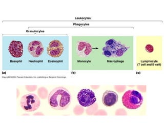
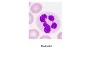
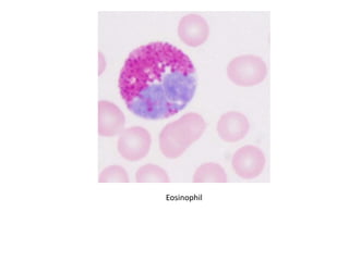
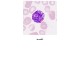
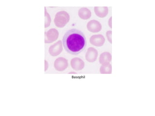
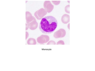
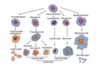
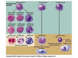
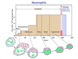
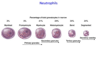
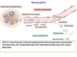
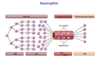
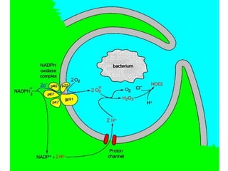
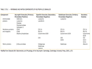
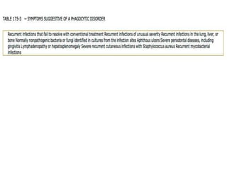
This document discusses the toxicology of leukocytes, including their components, functions, and toxic effects. It covers the kinetics, morphology, and interpretation of changes to neutrophils, eosinophils, basophils, monocytes, and lymphocytes. Important topics include mechanisms of neutrophilic leukocytosis, idiosyncratic toxic neutropenia, phagocyte function, the eosinophilia-myalgia syndrome, and causes of changes in the different leukocyte types.
Somatic cells are mainly epithelial cells that has been shed from the epithelial lining of the gland and White blood cells (leukocyte) has entered the mammary gland in response to injury or infection.
Lecturer notes for metabolic diseases in Cattle.which is benificial for student of BVSc& AH/DVM and MVsc student. It is My first presentation need your feedback for more presentation like this.
Interpretation of Canine Leukocyte Responses Pankaj Gaonkar
Interpreting a leukogram can be a challenging task. One has to interpret both the presence of as well as the degree of the abnormality. Evaluating the leukogram, including a total leukocyte count, a differential leukocyte count, absolute numbers of specific leukocytes and examination of morphology on a blood smear, can help identify abnormalities that may suggest specific diseases such as a viral or bacterial infection or even a neoplastic process.
Deploying Automated Workstreams and Computational Approaches for Generation of Toxicity Data Used for Hazard Identification, by Robert T. Dunn, II, Ph.D., DABT
Leukocytes constitute the cellular components of the innate and adaptive immune system and are critical for host defense. These cells mediate acute and chronic inflammation, modulate immune responses, and protect the host against numerous pathogens.
Disorders affecting leukocytes can be divided broadly into malignant disorders (tumors of leukocytes or their progenitors) and non-malignant disorders.
The malignant disorders are uncommon but clinically important entities
Non- malignant leukocyte disorders can involve any any of the leukocytes (neutrophils, eosinophils, basophils, monocytes, B cells, T cells, and natural killer cells)
but the disorders of greatest clinical relevance affect neutrophils; these will be our major focus.
inflammation is the body's immune system's response to an irritant. The irritant might be a germ, but it could also be a foreign object, such as a splinter in your finger.
Immunology is the study of the immune system and is a very important branch of the medical and biological sciences. The immune system protects us from infection through
The term necrosis is derived from a greek word nekros which means dead body.
Definition: Necrosis is defined as local or focal death of cells along with degradation of tissues by hydrolytic enzymes released from lysosome of the cell.
It is often associated with surrounding inflammatory reaction.
Necrosis is a series of morphological changes that follows cell death due to the irreversible cell injury/lethal cell injury/pathological cell injury.
1. Toxicology of the Leukon
Dr R B Cope BVSc BSc(Hon 1) PhD cGLPCP DABT ERT
2. Learning Objectives
• To understand the key basic functional concepts of the
leukon;
• To understand the fundamentals of the kinetics of the leukon;
• To understand and accurately interpret changes in the status
of the leukon;
• To understand and accurately interpret changes to leukon
morphology;
• To understand and accurately recognize important
toxicological effects on the leukon
3. Components of the Peripheral Blood Leukogram
• Consists of:
– Granulocytes
• Neutrophils
• Eosinophils
• Basophils
– Mononuclear cells
• T lymphocytes
• B lymphocytes
• Other lymphocytes
• Monocytes
17. Neutrophils
• Bone marrow storage pool
– Consists of metamyelocytes, bands and segmented N0
– Cells cannot replicate
– Cells are functionally mature despite the differences in
morphology
18. Neutrophils
• Bone marrow storage pool
– Normal transit time in the SP is 2 – 3 days but this can
shorten considerably on increased demand
– In health, ~ 80% of N0 are in bone marrow are in the
storage compartment – a bout a 5 day supply of N0 under
normal conditions
– Release from the SP is ordered – oldest cells (segmented
N0) are released first
– During increased peripheral demand for N0, younger cells
(bands, and in extreme cases metamyelocytes) are
released – this is called a “Left Shift”
21. Neutrophils
• Circulating and Marginal Pools
– N0 move more slowly in the post-capillary venules than
RBCs due to the presence of adhesion molecules on the N0
and on the endothelium of the post-capillary venules – this
population is referred to as the marginated pool
– In the axial or central blood flow of blood vessels make up
the circulating pool
– Net result is an uneven distribution of N0 in the
circulation: the circulating pool of unadhered N0 and the
marginal pool of N0 that are have adhered or are in the
process of adhering to the vascular endothelium in the
post-capillary venules
23. Neutrophils
• Circulating and Marginal Pools
– Average transit time of a N0 in the circulation is ~ 10 hours
under normal conditions
– N0 subsequently migrate into the tissues. The migration is
unidirectional (i.e. they do not re-enter the circulation)
– Some N0 are lost across mucosal surfaces and in secretions
24. Neutrophils
• Mechanisms of neutrophilic leukocytosis
(increase in WBC primarily due to an increase in N0)
– Increased stem cell recruitment
• Normal response to inctreased demand for N0
• Takes 3 – 5 days to have an effect
– Increased effective granulopoesis
• Due to an increased number cell divisions in the
development/maturation pool
• Also occurs due to decreased death of meylocytes in
the development/maturation pool
• Takes 2 – 3 days to have an effect
25. Neutrophils
• Mechanisms of neutrophilic leukocytosis
(increase in WBC primarily due to an increase in N0)
– Shortened marrow transit time
– Shift of mature N0 from the marginated pool in the peripheral
blood vessels to the circulating pool of mature N0. This results
in a neutrophilia without a left shift. This commonly occurs in
combination with a reduced number of lymphocytes
(lymphopenia) and a reduced number of eosinophis
(eosinopenia) in the peripheral blood. This response is referred
to as a “stress leukogram.” May be accompanied with
lymphopenia, and eosinopenia.
Stress leukograms are caused by adrenalin or cortisol release.
They are a common finding associated with exercise/excitement
handling and blood collection in many species.
The majority of stress leukograms are NORMAL
26.
27. Causes of neutropenia arranged
according to the compartment
with which the
pathophysiologically relevant
mechanism is linked. One
should begin the diagnostic
approach to a neutropenic
patient by seeking to identify
the pathophysiologically
relevant compartment.
Management of a neutropenic
patient whose neutrophil
production is reduced is
entirely different from that of a
neutropenic patient whose
production is normal and in
whom the rate of delivery to
the extravascular compartment
is normal or appropriately
increased in the context of
acute infections.
28.
29.
30. Idiosyncratic Toxic Neutropenia
• Rare but extremely important – potentially lethal
agranulocytosis (profound depletion of N0)
• Occurs sporadically within a population
• MOA is not the same as the pharmacologic properties of the
xenobiotic
• Preclinical tox studies rarely identify or predict this problem
• May or may not be dose-related
31. Idiosyncratic Toxic Neutropenia
• MOA
– Dose responsive disruption of N0 precursor cell division in
the bone marrow
– Non-dose responsive immune-mediated destruction of N0
or N0 precursors – common effect of many drugs, more
common in older patients, more common in
women, involves anti-N0 antibodies
• Typically involves a sudden decrease in circulating N0 (why?)
• Typically persists as long as the xenobiotic is present
• Greatly increases the risk of infection and sepsis if the
neutropenia is severe
33. Phagocyte Function
• Neutrophils and monocyte-macrophages are the
key phagocytes of the innate immune system
• Their principal innate immune role is to recognize
and eliminate microorganisms that make their
way past primary physical barriers, such as the
epithelium and body secretions that protect the
external and lining surfaces of the body.
• Monocyte/macrophages carry out sentinel duty
looking for microbes in healthy tissue and act as a
bridge between the innate and adaptive immune
systems
34. Phagocyte Function
• Neutrophils appear only in infected or damaged
tissue after being recruited by inflammatory
mediators released from activated macrophages
and endothelial cells or by chemical signals
released by invading microorganisms themselves
• After accumulation of these key immune cells at
sites of infection, the microbes are eliminated
through the process of phagocytosis, which is
defined as the engulfment, internalization, and
degradation of extracellular material.
35. Phagocyte Function
• Processes involved with N0:
– Receptor-mediated adherence to the blood vessel
endothelial wall (shift from the circulating pool to the
marginated pool of N0)
– Diapedesis – movement through the blood vessel wall
– Chemotaxis – movement up a chemical concentration of a
chemoattractant (endogenously produced and produced
by bacteria)
– Binding of the chemoattractant to the N0 cell membrane
activation phagocytosis degranulation/oxidative
burst/killing
36.
37.
38.
39.
40.
41.
42. Eosinophils
• Important in Type I (IgE-Mast Cell-histamine-mediated)
allergic reactions ( e.g. asthma)
• Important in the innate immune response to helminth
parasites
• Phagocytic and bacteriocidal capacity resemble that of N0
• Both circulating and marginated pools exist
• Peripheral blood eosinophilia is generally associated with:
– Helminth infections
– Allergic processes
– Neoplasia (classically mast cell tumors)
– Eosinophilic leukemias
– Some drugs (e.g. penicillin)
– Idiopathic
45. Eosinophilia-Myalgia Syndrome
• Incurable and sometimes fatal syndrome associated with
ingestion of L-tryptophan or substances metabolized to L-
tryptophan
• Resembles Spanish toxic oil syndrome due to contaminated
rapeseed oil
• Potentially due to a contaminant - 1,1-ethylidenebis (L-
tryptophan) (tryptophan dimer)
• Potentially due to a metabolite – 4-aminophenol
• Associated with:
– Eosinophilia
– Intense myalgia (muscular pain)
46. Basophils and Mast Cells
• Basophils and mast cells have different lineages but similar
functions
• Important in Type I hypersensitivity and allergy
• Effects are rate
• Basopenias
– Acute hypersensitivity reactions (early stages)
– Glucocorticoids/stress
– Hyperthyroidism
• Basophilias
– Chronic allergy/hypersensitivity reactions
– Diabetes mellitus
– Estrogen
– Hypothyroidism
– Iron deficiency
– Neoplasia
48. Monocytes
• Derived from bone marrow monoblast.
• Circulate in blood for 1 – 3 days and then move into the tissues
• ~3-8% of blood leukoctes
• Significant storage pool in spleen (~50% of total body monocytes) in the
Cords of Biltrothare produced by the bone marrow from hematopoietic
stem cell precursors called monoblasts. Monocytes circulate in the
bloodstream for about one to three days and then typically move into
tissues throughout the body.
• Following migration from blood, monocytes mature into either tissue
macrophages or dendritic cells.
49. Monocytes
• Form part of the immune system – 3 main functions:
– Antigen presentation
– Phagocytosis
– Cytokine production
• 3 classical types of types of monocytes in blood:
– the classical monocyte (CD14++ CD16- monocyte)
– Non classical monocytes (CD14+CD16++ monocyte)
– Intermediate cells (CD14++CD16+)
– Different classes represent different developmental stages:
Classical Intermediate non-classical
– After stimulation with microbial products the CD14+CD16++
monocytes produce high amounts of pro-inflammatory
cytokines
50. Monocytes
• Form part of the immune system – 3 main functions:
– Antigen presentation
– Phagocytosis
– Cytokine production
• Moncytosis – increase in circulating blood monocyte numbers
– Infection
– Recovery phase of neutropenia following infection
– Hyperadreocorticism
– Autoimmune reactions
– Neoplasia/leukemia
– Sarcoidosis
– Lipid storage diseases
52. Lymphocytes
• Circulating part of the adaptive immune system
• Lymphocytosis is a feature of infection or neoplasia
(leukemias)
• Causes of absolute lymphocytosis include:
– Acute viral infections
– Acute & chronic bacterial infections
– Some protozoal infections
– Leukemias
53. Lymphocytes
• Leukemias/lymphomas in mice
– Extremely common spontaneous finding
– Virtually all mouse strains contain endogeous MuLV
retrovirus (Type C) sequences
– Most laboratory mice do not have exogenous MuLV
because this is controlled because of SPF