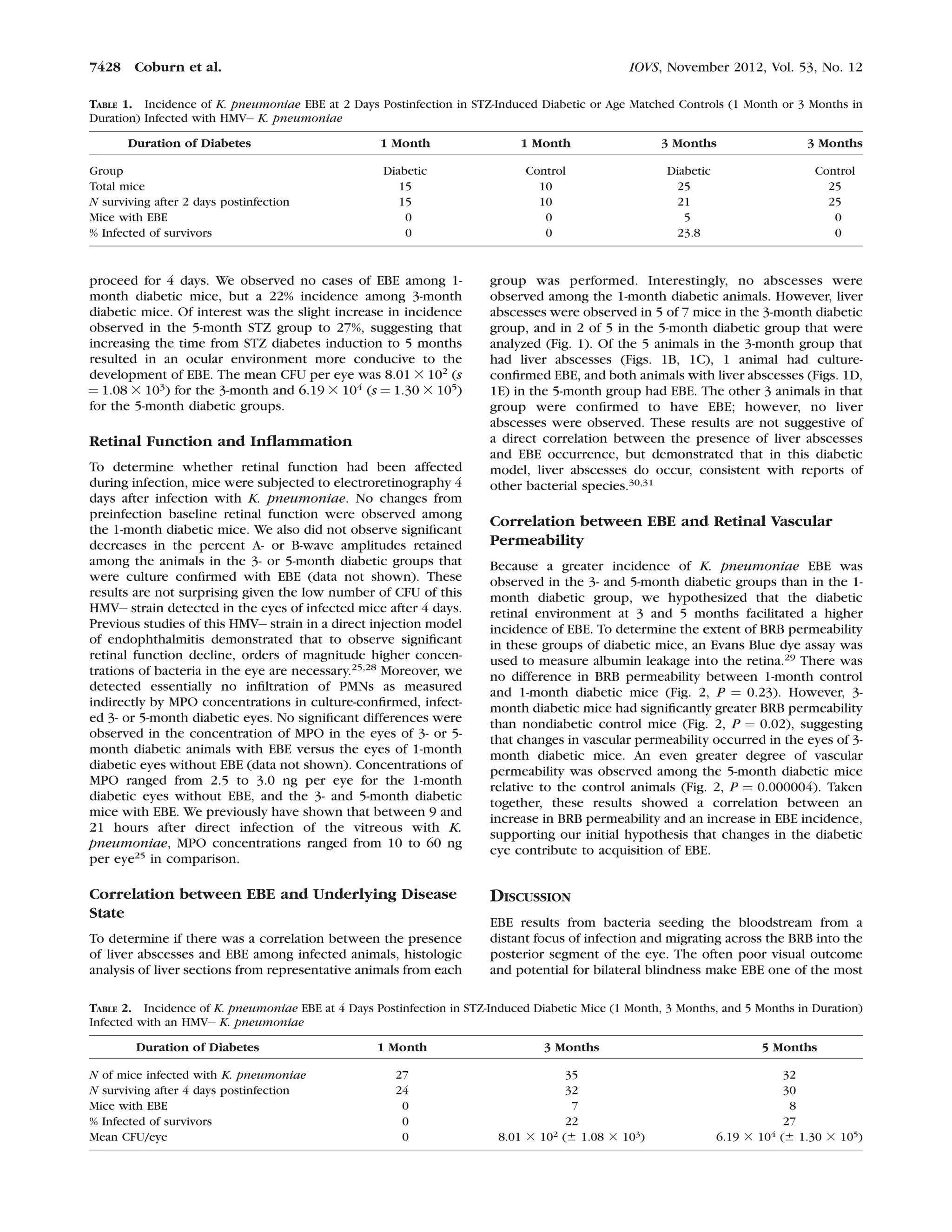
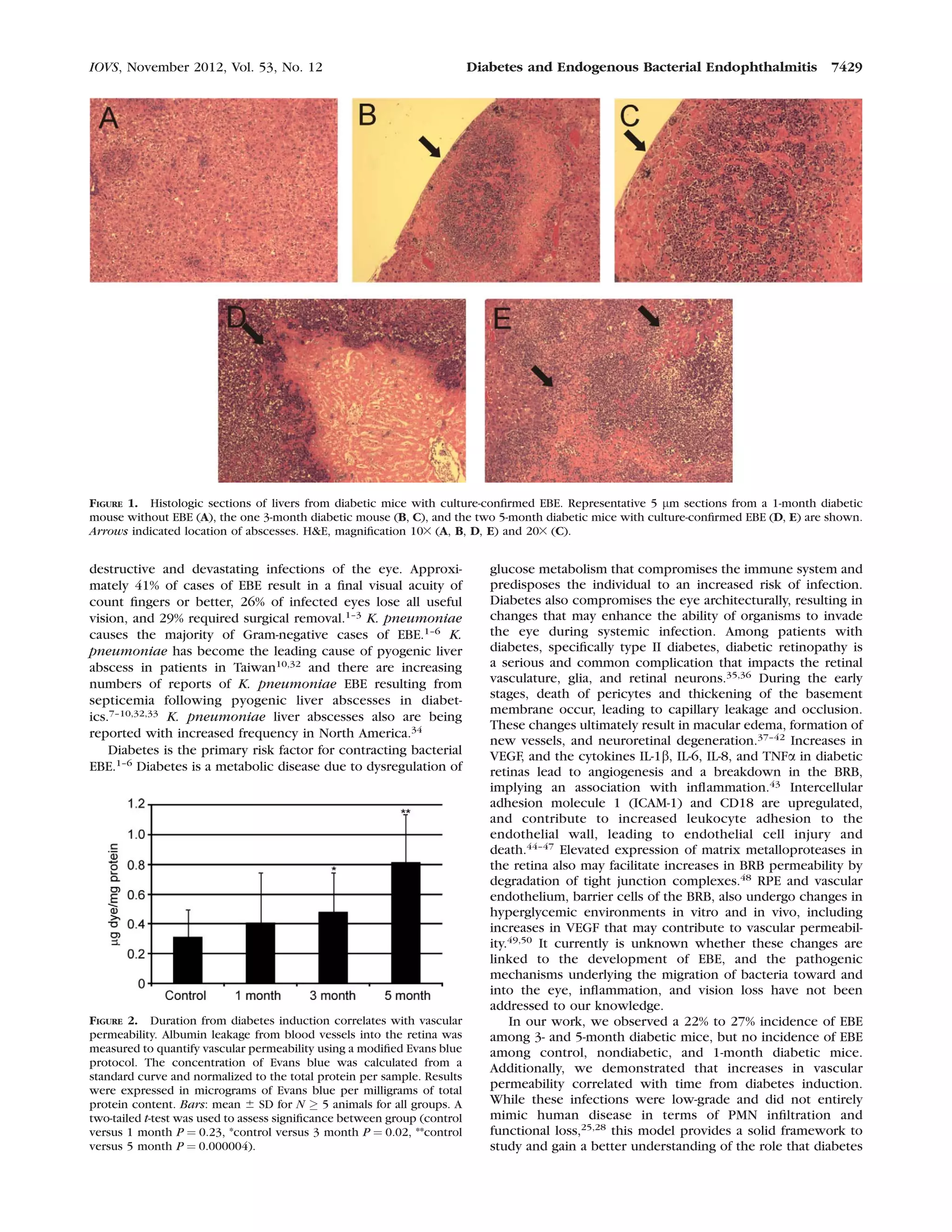
Immunology and Microbiology
The Diabetic Ocular Environment Facilitates the
Development of Endogenous Bacterial Endophthalmitis
Phillip S. Coburn,1
Brandt J. Wiskur,2
Elizabeth Christy,1
and Michelle C. Callegan1
PURPOSE. We tested the hypothesis that changes in the diabetic
ocular environment facilitate the development of endogenous
bacterial endophthalmitis (EBE).
METHODS. C57BL/6J mice were rendered diabetic with strep-
tozotocin (STZ) for 1, 3, or 5 months’ duration. Diabetic and
age-matched nondiabetic mice were tail vein–injected with 108
CFU of Klebsiella pneumoniae, a common cause of EBE in
diabetics. After either 2 or 4 days postinfection, the EBE
incidence was assessed by electroretinography, histology,
bacterial counts, and myeloperoxidase ELISAs. Blood-retinal
barrier (BRB) permeability in uninfected diabetic mice also was
determined.
RESULTS. No cases of EBE were observed among the 1-month
diabetic group. Extending the time from diabetes induction to
3 months resulted in a 23.8% EBE incidence after 2 days, and a
22% incidence after 4 days. The incidence of EBE increased to
27% in the 5-month diabetic group. Infected eyes had an
average 8.01 3 102
and 6.19 3 104
CFU/eye for the 3- and 5-
month diabetic groups, respectively. There was no significant
difference in BRB permeability between control and 1-month
uninfected diabetic mice. However, 3- and 5-month diabetic
mice had significantly greater BRB permeability than control
mice. These results suggested that increasing the time from
STZ diabetes induction to 3 and 5 months resulted in an ocular
environment more conducive to the development of EBE.
CONCLUSIONS. These results demonstrated a correlation between
an increase in BRB permeability and an increase in EBE
incidence, supporting the hypothesis that diabetic ocular
changes contribute to the development of EBE. (Invest
Ophthalmol Vis Sci. 2012;53:7426–7431) DOI:10.1167/
iovs.12-10661
Endogenous bacterial endophthalmitis (EBE) is a potentially
blinding form of intraocular infection resulting from
hematogenous spread of bacteria from a focus of infection
into the eye. A number of conditions predispose individuals to
EBE, including intravenous drug abuse and treatment with
immunosuppressive agents. However, the most common
predisposing risk factor is diabetes.1–6
During the course of
diabetes, permeability changes occur in the blood-retinal
barrier (BRB). Of particular interest is the role that the diabetic
ocular environment may have in the migration of bacteria from
the vasculature into the eye, resulting in EBE. At present, the
mechanisms underlying movement of bacteria across the BRB
and into the eye from the bloodstream are not well
understood.
One of the most common causes of EBE in diabetics is the
opportunistic pathogen Klebsiella pneumoniae, a Gram-
negative bacterium found in the environment and a constituent
of the gastrointestinal tract microbiota.1–6
K. pneumoniae is a
frequent cause of community-acquired and healthcare-associ-
ated infections, which include pneumonia, bacteremia, urinary
tract, and wound infections.7–10
K. pneumoniae is the most
prevalent bacteria isolated from pyogenic liver abscesses in the
United States, Trinidad, and Asian Pacific regions,11–16 with
diabetes as the major underlying condition associated with
these abscesses.17–23 Ten percent of pyogenic K. pneumoniae
liver abscess cases are complicated by EBE.8 The visual
outcome in patients with K. pneumoniae EBE is uniformly
poor, ranging from hand motion visualization to evisceration or
enucleation of the eye.1–3
The prevalence of cases of K.
pneumoniae EBE where diabetes is a predisposing risk factor
led to the hypothesis that changes that occur within the ocular
environment during diabetes contribute to the development of
K. pneumoniae EBE. To test this hypothesis, we developed a
streptozotocin (STZ)-induced diabetic mouse model of K.
pneumoniae EBE. In this report, we demonstrated that the
incidence of K. pneumoniae EBE was greater in STZ-induced
diabetic mice than in control mice, and that the degree of BRB
permeability may have facilitated a higher incidence of EBE.
The results demonstrated a correlation between the duration
of diabetes and the incidence of K. pneumoniae EBE, and also
suggested that increases in BRB permeability may contribute to
the enhanced incidence of K. pneumoniae EBE.
METHODS
Animals
Six-week-old C57BL/6J mice were acquired from the Jackson Labora-
tory and used in accordance with institutional guidelines and the ARVO
Statement for the Use of Animals in Ophthalmic and Vision Research.
Mice were allowed to acclimate to the environment for two weeks
before the establishment of diabetes. Mice were anesthetized with an
intramuscular injection of 85 mg ketamine/kg and 14 mg xylazine/kg
before tail-vein injections and electroretinography.
From the 1Department of Ophthalmology and the 2Oklahoma
Center for Neuroscience, The University of Oklahoma Health
Sciences Center, Oklahoma City, Oklahoma.
Presented in part at the annual meeting of the Association for
Research in Vision in Ophthalmology, Fort Lauderdale, Florida, May
2011.
Supported by a Lew R. Wasserman Award from Research to
Prevent Blindness (MCC), and in part by NIH Grants R01EY012985
(MCC), P30EY12191 (NIH CORE grant to Robert E. Anderson,
OUHSC), P20RR17702 (NCRR COBRE grant to Robert E. Anderson,
OUHSC), and an unrestricted grant to the Dean A. McGee Eye
Institute from Research to Prevent Blindness. The authors alone are
responsible for the content and writing of this paper.
Submitted for publication July 27, 2012; revised September 11,
2012; accepted September 22, 2012.
Disclosure: P.S. Coburn, None; B.J. Wiskur, None; E. Christy,
None; M.C. Callegan, None
Corresponding author: Michelle C. Callegan, Department of
Ophthalmology, DMEI PA-418, 608 Stanton L. Young Boulevard,
Oklahoma City, OK 73104; michelle-callegan@ouhsc.edu.
Investigative Ophthalmology & Visual Science, November 2012, Vol. 53, No. 12
7426 Copyright 2012 The Association for Research in Vision and Ophthalmology, Inc.
STZ Induction of Diabetes
To establish diabetes, 8-week-old male C57BL/6J mice were given a
series of 2 STZ injections at 100 mg/kg one week apart.24 STZ (Sigma-
Aldrich, St. Louis, MO) was solubilized in freshly prepared 10 mM
citrate buffer pH 4.5 and immediately injected intraperitoneally. Blood
glucose levels were measured two weeks following the second STZ
injection. All mice demonstrated blood glucose levels greater than or
equal to 300 mg/dl. Mice were allowed to proceed for either 1, 3, or 5
months, and were the same age at the time of infection. Control
animals were administered citrate buffer pH 4.5 only. Before injections
with K. pneumoniae or Evans Blue dye, blood glucose levels were
measured again to ensure a diabetic state.
Endogenous Bacterial Endophthalmitis Model
A clinical hypermucoviscosity (HMV) negative K. pneumoniae
endophthalmitis isolate25 was grown for 18 hours in brain heart
infusion media (BHI; Difco Laboratories, Detroit, MI), and subcultured
in prewarmed BHI to logarithmic phase. Bacteria then were
centrifuged and resuspended in PBS. To establish endogenous
endophthalmitis infections, all groups of mice were tail vein injected
with 108 CFU in 100 lL PBS. At 2 days postinfection, some eyes were
harvested for bacterial quantitation as described below. At 4 days
postinfection, retinal function of both eyes was assessed by
electroretinography. Both eyes from each mouse were harvested for
bacterial and polymorphonuclear leukocyte (PMN) quantitation or
histology.
Electroretinography
To determine the degree of retinal function 4 days after mice were
infected with K. pneumoniae, mice were dark adapted for at least 6
hours and then anesthetized as described above. Pupils were dilated
with 10% topical phenylephrine (Akorn, Inc., Lake Forest, IL) and
gold-wire electrodes were placed over both corneas with a drop of
GONAK (Hypromeliose Ophthalmic Demulscen Solution 2.5%;
Akorn, Inc.) to ensure maximum conductance. Scotopic ERGs were
recorded for both eyes using a UTAS-E3000 Electrodiagnostic System
(LKC Technologies, Inc., Gaithersburg, MD). The A-wave amplitude
was measured from the prestimulus baseline to the A-wave trough,
and the B-wave amplitude was measured from the A-wave trough to
the B-wave peak. Percent ERG retention was calculated relative to
mean baseline ERGs recorded from 6 eyes per group before
infection.26,27
Bacterial Quantitation
For each group of infected mice, eyes were selected at random for
bacterial quantitation. Briefly, the left and right eyes from each mouse
selected were enucleated, placed into separate tubes containing 400
ll of sterile PBS and 1.0 mm sterile glass beads (Biospec Products,
Inc., Bartlesville, OK), and homogenized for 60 seconds at 5,000
revolutions per minute (rpm) in a Mini-BeadBeater (Biospect
Products, Inc.). Eye homogenates were diluted serially and plated in
triplicate on brain heart infusion (BHI) agar and the CFU per eye
determined.26,27
PMN Quantitation
PMN leukocyte infiltration into eyes was assessed indirectly by
measurement of myeloperoxidase (MPO) concentrations in whole
eye homogenates. Eyes were enucleated and homogenized with glass
beads in PBS as described for bacterial quantitation. Homogenates were
diluted 1:4 and analyzed for MPO by ELISA according to manufacturer’s
instructions (Mouse MPO ELISA Test Kit; Cell Sciences, Canton,
MA).27,28
Histology
Because homogenization of eyes for bacterial quantitation precludes
subsequent histologic analysis on eyes that are confirmed culture
positive, eyes from each group were selected randomly and fixed in
Excalibur’s Alcoholic Z-Fix for 24 hours, and then embedded in
paraffin. Sections were stained with hematoxylin and eosin (H&E), and
tissue Gram stain. Livers were fixed in 10% neutral buffered formalin
for 24 hours, embedded in paraffin, and sections stained with
hematoxylin and eosin.26,27
Vascular Permeability
Albumin leakage from blood vessels into the retina was measured to
quantify vascular permeability using a modified Evans blue protocol.29
Mice were anesthetized as described above and 15 mg Evans Blue dye
(Sigma-Aldrich) per kg were injected into the tail vein. After two hours,
mice were perfused with 1% paraformaldehyde in citrate buffer (pH
4.2) for 1 minute, and retinas harvested and placed in 150 lL
formamide. Retinas were incubated at 708C for 18 hours to extract the
dye and centrifuged at 70,000 rpm (TLA 100.3 rotor; Beckman Coulter,
Fullerton, CA) for 20 minutes. The optical density at 620 nanometers
(OD620) of 100 lL of the supernatants was measured and the
concentration of Evans blue was calculated from a standard curve of
Evans Blue in formamide. The pellets then were solubilized in 1 mL of
0.2% SDS in PBS and the protein concentrations measured using a BCA
protein assay. The concentration of Evans Blue in each sample then
was normalized to the total protein per sample. Results were expressed
in micrograms of Evans blue/mg total protein content.
Statistics
All values represent the mean 6 SD of the number of eyes per group
indicated in the figure legends for each assay. Two-tailed, two-sample t-
tests were used for statistical comparisons between groups. A P value
of 0.05 was considered significant.
RESULTS
Diabetes and Incidence of K. pneumoniae EBE
To test the hypothesis that changes in the diabetic ocular
environment facilitated the development of K. pneumoniae
EBE, we used a mouse model of STZ -induced diabetes.24
Male
C57BL/6J mice were injected with 100 mg/kg STZ and diabetes
was allowed to progress for one month. One-month diabetic
and age-matched sham injected control mice were tail vein-
injected with 108 CFU of a clinical ocular isolate of K.
pneumoniae. After 2 days postinfection, the incidence of EBE
was evaluated. No difference was observed in the incidence of
EBE between the one-month diabetic and control mice (Table
1). We surmised that the changes in the architecture of the eye
observed during diabetes progression had not yet occurred
after one month, even though the mean blood glucose level of
these mice was >400 mg/dl. We sought to determine whether
extending the time from diabetes induction to 3 months might
facilitate the changes necessary for EBE development. After 2
days postinfection, we observed a 23.8% incidence of infection
among 3-month diabetic mice, while no cases of EBE were
observed in control mice (Table 1).
To determine whether extending the time from diabetes
induction to 5 months might result in greater incidence of EBE,
we repeated the above-described experiments and included a
5-month diabetic group. Mice in all groups were age-matched
at the time of infection and had similar blood glucose levels of
>300 mg/dl. As shown in Table 2, the incidence of K.
pneumoniae EBE among 1- and 3-month groups was similar to
that observed previously, even though infection was allowed to
IOVS, November 2012, Vol. 53, No. 12 Diabetes and Endogenous Bacterial Endophthalmitis 7427
proceed for 4 days. We observed no cases of EBE among 1-
month diabetic mice, but a 22% incidence among 3-month
diabetic mice. Of interest was the slight increase in incidence
observed in the 5-month STZ group to 27%, suggesting that
increasing the time from STZ diabetes induction to 5 months
resulted in an ocular environment more conducive to the
development of EBE. The mean CFU per eye was 8.01 3 102
(s
¼ 1.08 3 103) for the 3-month and 6.19 3 104 (s ¼ 1.30 3 105)
for the 5-month diabetic groups.
Retinal Function and Inflammation
To determine whether retinal function had been affected
during infection, mice were subjected to electroretinography 4
days after infection with K. pneumoniae. No changes from
preinfection baseline retinal function were observed among
the 1-month diabetic mice. We also did not observe significant
decreases in the percent A- or B-wave amplitudes retained
among the animals in the 3- or 5-month diabetic groups that
were culture confirmed with EBE (data not shown). These
results are not surprising given the low number of CFU of this
HMVÀ strain detected in the eyes of infected mice after 4 days.
Previous studies of this HMVÀ strain in a direct injection model
of endophthalmitis demonstrated that to observe significant
retinal function decline, orders of magnitude higher concen-
trations of bacteria in the eye are necessary.25,28
Moreover, we
detected essentially no infiltration of PMNs as measured
indirectly by MPO concentrations in culture-confirmed, infect-
ed 3- or 5-month diabetic eyes. No significant differences were
observed in the concentration of MPO in the eyes of 3- or 5-
month diabetic animals with EBE versus the eyes of 1-month
diabetic eyes without EBE (data not shown). Concentrations of
MPO ranged from 2.5 to 3.0 ng per eye for the 1-month
diabetic eyes without EBE, and the 3- and 5-month diabetic
mice with EBE. We previously have shown that between 9 and
21 hours after direct infection of the vitreous with K.
pneumoniae, MPO concentrations ranged from 10 to 60 ng
per eye25
in comparison.
Correlation between EBE and Underlying Disease
State
To determine if there was a correlation between the presence
of liver abscesses and EBE among infected animals, histologic
analysis of liver sections from representative animals from each
group was performed. Interestingly, no abscesses were
observed among the 1-month diabetic animals. However, liver
abscesses were observed in 5 of 7 mice in the 3-month diabetic
group, and in 2 of 5 in the 5-month diabetic group that were
analyzed (Fig. 1). Of the 5 animals in the 3-month group that
had liver abscesses (Figs. 1B, 1C), 1 animal had culture-
confirmed EBE, and both animals with liver abscesses (Figs. 1D,
1E) in the 5-month group had EBE. The other 3 animals in that
group were confirmed to have EBE; however, no liver
abscesses were observed. These results are not suggestive of
a direct correlation between the presence of liver abscesses
and EBE occurrence, but demonstrated that in this diabetic
model, liver abscesses do occur, consistent with reports of
other bacterial species.30,31
Correlation between EBE and Retinal Vascular
Permeability
Because a greater incidence of K. pneumoniae EBE was
observed in the 3- and 5-month diabetic groups than in the 1-
month diabetic group, we hypothesized that the diabetic
retinal environment at 3 and 5 months facilitated a higher
incidence of EBE. To determine the extent of BRB permeability
in these groups of diabetic mice, an Evans Blue dye assay was
used to measure albumin leakage into the retina.29 There was
no difference in BRB permeability between 1-month control
and 1-month diabetic mice (Fig. 2, P ¼ 0.23). However, 3-
month diabetic mice had significantly greater BRB permeability
than nondiabetic control mice (Fig. 2, P ¼ 0.02), suggesting
that changes in vascular permeability occurred in the eyes of 3-
month diabetic mice. An even greater degree of vascular
permeability was observed among the 5-month diabetic mice
relative to the control animals (Fig. 2, P ¼ 0.000004). Taken
together, these results showed a correlation between an
increase in BRB permeability and an increase in EBE incidence,
supporting our initial hypothesis that changes in the diabetic
eye contribute to acquisition of EBE.
DISCUSSION
EBE results from bacteria seeding the bloodstream from a
distant focus of infection and migrating across the BRB into the
posterior segment of the eye. The often poor visual outcome
and potential for bilateral blindness make EBE one of the most
TABLE 2. Incidence of K. pneumoniae EBE at 4 Days Postinfection in STZ-Induced Diabetic Mice (1 Month, 3 Months, and 5 Months in Duration)
Infected with an HMVÀ K. pneumoniae
Duration of Diabetes 1 Month 3 Months 5 Months
N of mice infected with K. pneumoniae 27 35 32
N surviving after 4 days postinfection 24 32 30
Mice with EBE 0 7 8
% Infected of survivors 0 22 27
Mean CFU/eye 0 8.01 3 102 (6 1.08 3 103) 6.19 3 104 (6 1.30 3 105)
TABLE 1. Incidence of K. pneumoniae EBE at 2 Days Postinfection in STZ-Induced Diabetic or Age Matched Controls (1 Month or 3 Months in
Duration) Infected with HMVÀ K. pneumoniae
Duration of Diabetes 1 Month 1 Month 3 Months 3 Months
Group Diabetic Control Diabetic Control
Total mice 15 10 25 25
N surviving after 2 days postinfection 15 10 21 25
Mice with EBE 0 0 5 0
% Infected of survivors 0 0 23.8 0
7428 Coburn et al. IOVS, November 2012, Vol. 53, No. 12
destructive and devastating infections of the eye. Approxi-
mately 41% of cases of EBE result in a final visual acuity of
count fingers or better, 26% of infected eyes lose all useful
vision, and 29% required surgical removal.1–3
K. pneumoniae
causes the majority of Gram-negative cases of EBE.1–6
K.
pneumoniae has become the leading cause of pyogenic liver
abscess in patients in Taiwan10,32
and there are increasing
numbers of reports of K. pneumoniae EBE resulting from
septicemia following pyogenic liver abscesses in diabet-
ics.7–10,32,33 K. pneumoniae liver abscesses also are being
reported with increased frequency in North America.34
Diabetes is the primary risk factor for contracting bacterial
EBE.1–6
Diabetes is a metabolic disease due to dysregulation of
glucose metabolism that compromises the immune system and
predisposes the individual to an increased risk of infection.
Diabetes also compromises the eye architecturally, resulting in
changes that may enhance the ability of organisms to invade
the eye during systemic infection. Among patients with
diabetes, specifically type II diabetes, diabetic retinopathy is
a serious and common complication that impacts the retinal
vasculature, glia, and retinal neurons.35,36 During the early
stages, death of pericytes and thickening of the basement
membrane occur, leading to capillary leakage and occlusion.
These changes ultimately result in macular edema, formation of
new vessels, and neuroretinal degeneration.37–42
Increases in
VEGF, and the cytokines IL-1b, IL-6, IL-8, and TNFa in diabetic
retinas lead to angiogenesis and a breakdown in the BRB,
implying an association with inflammation.43 Intercellular
adhesion molecule 1 (ICAM-1) and CD18 are upregulated,
and contribute to increased leukocyte adhesion to the
endothelial wall, leading to endothelial cell injury and
death.44–47
Elevated expression of matrix metalloproteases in
the retina also may facilitate increases in BRB permeability by
degradation of tight junction complexes.48 RPE and vascular
endothelium, barrier cells of the BRB, also undergo changes in
hyperglycemic environments in vitro and in vivo, including
increases in VEGF that may contribute to vascular permeabil-
ity.49,50
It currently is unknown whether these changes are
linked to the development of EBE, and the pathogenic
mechanisms underlying the migration of bacteria toward and
into the eye, inflammation, and vision loss have not been
addressed to our knowledge.
In our work, we observed a 22% to 27% incidence of EBE
among 3- and 5-month diabetic mice, but no incidence of EBE
among control, nondiabetic, and 1-month diabetic mice.
Additionally, we demonstrated that increases in vascular
permeability correlated with time from diabetes induction.
While these infections were low-grade and did not entirely
mimic human disease in terms of PMN infiltration and
functional loss,25,28
this model provides a solid framework to
study and gain a better understanding of the role that diabetes
FIGURE 1. Histologic sections of livers from diabetic mice with culture-confirmed EBE. Representative 5 lm sections from a 1-month diabetic
mouse without EBE (A), the one 3-month diabetic mouse (B, C), and the two 5-month diabetic mice with culture-confirmed EBE (D, E) are shown.
Arrows indicated location of abscesses. H&E, magnification 103 (A, B, D, E) and 203 (C).
FIGURE 2. Duration from diabetes induction correlates with vascular
permeability. Albumin leakage from blood vessels into the retina was
measured to quantify vascular permeability using a modified Evans blue
protocol. The concentration of Evans blue was calculated from a
standard curve and normalized to the total protein per sample. Results
were expressed in micrograms of Evans blue per milligrams of total
protein content. Bars: mean 6 SD for N ‡ 5 animals for all groups. A
two-tailed t-test was used to assess significance between group (control
versus 1 month P ¼ 0.23, *control versus 3 month P ¼ 0.02, **control
versus 5 month P ¼ 0.000004).
IOVS, November 2012, Vol. 53, No. 12 Diabetes and Endogenous Bacterial Endophthalmitis 7429
has in development of EBE, and of the interplay of host and
bacterial factors that contribute to this devastating disease. In
our model, we used an HMVÀ strain of K. pneumoniae to
establish the model and provide baseline data for future
comparisons with HMVþ K. pneumoniae. Our laboratory has
demonstrated in a mouse model of experimentally-induced
endophthalmitis, in which bacteria are introduced into the
vitreous by direct injection, that the HMV phenotype
contributes to pathogenesis. As reported by Wiskur et al., an
HMVþ strain caused significantly greater inflammation and
greater loss of visual function than an HMVÀ strain,25
the same
strain we used in our study. Further, the HMVþ strain was not
readily cleared from the eye in this model.25
More recently, our
group used the experimentally-induced endophthalmitis model
to compare directly a wild type HMVþ K. pneumoniae strain
with an isogenic mutant defective in magA, the gene primarily
responsible for the HMV phenotype.28
The studies showed
that mice infected with the magAÀ strain showed a
significantly improved visual outcome relative to mice infected
with the wildtype strain.28
Both of these studies clearly
established a role for the HMV phenotype in K. pneumoniae
endophthalmitis. As this direct injection model circumvents
systemic infection, it remains to be seen whether the HMV
phenotype is important for crossing the BRB and establishing
EBE. The HMV phenotype might enable bacteria to more
readily cross the BRB, adhere to structures within the eye, and/
or evade PMN clearance. Future studies in our laboratory will
address this hypothesis.
To our knowledge, no studies have examined whether
patients suffering from bacterial EBE had a history of diabetes-
induced vision changes or diabetic retinopathy before infec-
tion. However, if barrier function of the BRB were altered and
the immune response was unable to suppress transient
bacteremia, bacteria could circumvent the immune response,
reach the retinal vasculature, and gain access into the eye
through a permeable BRB. While we did not observe EBE in
nondiabetic mice, it might be possible that K. pneumoniae
contributed directly to increasing vascular permeability. It has
been shown that Neisseria meningitidis, especially unencap-
sulated strains, induce the upregulation of the vascular
adhesion molecules CD62E, ICAM-1, and VCAM-1, leading to
increased binding of leukocytes to the endothelium, and
ultimately vascular damage and permeability.51 While it has not
been shown that K. pneumoniae similarly induces upregula-
tion of vascular adhesion molecules, we cannot rule out the
possibility that K. pneumoniae contributes to the pathogen-
esis of EBE by this mechanism. We further acknowledge that
systemic infection with K. pneumoniae might elicit increases
in vascular permeability through the liberation of TNFa, as do
other encapsulated organisms.52,53 However, as stated, we
have not observed any incidence of EBE among control
animals, so it is unlikely that systemic infection alone caused
the permeability necessary for EBE development.
In addition to changes in BRB permeability during the
course of diabetes, suppression of the innate immune system
also contributes to an increased risk of infection, and
potentially to EBE pathogenesis. PMNs are necessary for
clearance of pathogens from the eye during acute bacterial
endophthalmitis.25–29 However, PMNs from diabetic mice and
humans display defective abilities to phagocytize and kill
bacteria, including Staphylococcus aureus and K. pneumo-
niae.54,55 The possibility arises that the inability of PMNs to
clear pathogens adequately from the bloodstream and the eye
also may contribute to the development of EBE. However, this
hypothesis has not been tested, and future studies will be
necessary to address the role of the innate immune system in
EBE pathogenesis.
To our knowledge, this is the first reported experimental
model of endogenous bacterial endophthalmitis. Using this
model, we have demonstrated a correlation between the
duration of diabetes and the incidence of K. pneumoniae EBE.
We also have shown that increases in retinal vascular
permeability correlate with the time from diabetes induction.
This model will prove useful in elucidating the mechanisms of
EBE development and will allow the role of bacterial virulence
traits to be dissected, as well as explore host factors and
components of the immune system that are involved in EBE
pathogenesis. Future studies will continue to explore the
relationship between changes in the diabetic ocular environ-
ment and the development of EBE.
Acknowledgments
Bo Novosad, Jonathan Hunt, Bradley Blackburn, and Nanette
Wheatley (OUHSC) provided technical assistance. Excalibur
Pathology (Moore, OK) and the DMEI/NEI Imaging Core Facility
(Oklahoma City, OK) assisted with preparation of eye and liver
histology, respectively.
References
1. Jackson TL, Eykyn SJ, Graham EM, Stanford MR. Endogenous bacterial
endophthalmitis: a 17-year prospective series and review of 267
reported cases. Surv Ophthalmol. 2003;48:403–423.
2. Greenwald MJ, Wohl LG, Sell CH. Metastatic bacterial endophthalmitis:
a contemporary reappraisal. Surv Ophthalmol. 1986;31:81–101.
3. Shammas HF. Endogenous E. coli endophthalmitis. Surv Ophthalmol.
1977;21:429–435.
4. Okada AA, Johnson RP, Liles WC, D’Amico DJ, Baker AS. Endogenous
bacterial endophthalmitis. Report of a ten-year retrospective study.
Ophthalmology. 1994;101:832–838.
5. Romero CF, Rai MK, Lowder CY, Adal KA. Endogenous endophthalmitis:
case report and brief review. Am Fam Physician. 1999;60:510–514.
6. Shrader SK, Band JD, Lauter CB, Murphy P. The clinical spectrum of
endophthalmitis: incidence, predisposing factors, and features influ-
encing outcome. J Infect Dis. 1990;162:115–120.
7. Fang CT, Chuang YP, Shun CT, Chang SC, Wang JT. A novel virulence
gene in Klebsiella pneumoniae strains causing primary liver abscess
and septic metastatic complications. J Exp Med. 2004;199:697–705.
8. Fung CP, Chang FY, Lee SC, et al. A global emerging disease of Klebsiella
pneumoniae liver abscess: is serotype K1 an important factor for
complicated endophthalmitis? Gut. 2002;50:420–424.
9. Chuang YP, Fang CT, Lai SY, Chang SC, Wang JT. Genetic determinants
of capsular serotype K1 of Klebsiella pneumoniae causing primary
pyogenic liver abscess. J Infect Dis. 2006;193:645–654.
10. Chang SC, Fang CT, Hsueh PR, Chen YC, Luh KT. Klebsiella pneumo-
niae isolates causing liver abscess in Taiwan. Diagn Microbiol Infect
Dis. 2000;37:279–284.
11. Lederman ER, Crum NF. Pyogenic liver abscess with a focus on
Klebsiella pneumoniae as a primary pathogen: an emerging disease
with unique clinical characteristics. Am J Gastroenterol. 2005;100:
322–331.
12. Rahimian J, Wilson T, Oram V, Holzman RS. Pyogenic liver abscess:
recent trends in etiology and mortality. Clin Infect Dis. 2004;39:1654–
1659.
13. Fang FC, Sandler N, Libby SJ. Liver abscess caused by magAþ
Klebsiella pneumoniae in North America. J Clin Microbiol. 2005;43:
991–992.
14. Lederman ER, Crum NF. Klebsiella liver abscess: a coast-to-coast
phenomenon. Clin Infect Dis. 2005;41:273.
15. Ko WC, Paterson DL, Sagnimeni AJ, et al. Community-acquired
Klebsiella pneumoniae bacteremia: global differences in clinical
patterns. Emerg Infect Dis. 2002;8:160–166.
7430 Coburn et al. IOVS, November 2012, Vol. 53, No. 12
16. Ohmori S, Shiraki K, Ito K, et al. Septic endophthalmitis and meningitis
associated with Klebsiella pneumoniae liver abscess. Hepatol Res.
2002;22:307–312.
17. Jardeleza MS, Chao-Chan C, Shalaby I, de la Cruz Z, Shen de F, Green
WR. Clinicopathologic and ultrastructural study of endogenous
Klebsiella pneumoniae endophthalmitis. Retina. 2005;25:657–662.
18. Scott IU, Matharoo N, Flynn HW Jr, Miller D. Endophthalmitis caused by
Klebsiella species. Am J Ophthalmol. 2004;138:662–663.
19. Chen YJ, Kuo HK, Wu PC, et al. 10-year comparison of endogenous
endophthalmitis outcomes: an east Asian experience with Klebsiella
pneumoniae infection. Retina. 2004;24:383–390.
20. Giobbia M, Scotton PG, Carniato A, et al. Community-acquired
Klebsiella pneumoniae bacteremia with meningitis and endophthalmi-
tis in Italy. Int J Infect Dis. 2003;7:234–235.
21. Ayinala SR, Vulpe M, Azaz M, Cohen H, Donelson SS, Lee M. Pyogenic
liver abscesses due to Klebsiella pneumoniae in a diabetic patient. J
Miss State Med Assoc. 2001;42:67–70.
22. Saccente M. Klebsiella pneumoniae liver abscess, endophthalmitis, and
meningitis in a man with newly recognized diabetes mellitus. Clin
Infect Dis. 1999;29:1570–1571.
23. Liao HR, Lee HW, Leu HS, Lin BJ, Juang CJ. Endogenous Klebsiella
pneumoniae endophthalmitis in diabetic patients. Can J Ophthalmol.
1992;27:143–147.
24. Rajala RV, Wiskur B, Tanito M, Callegan M, Rajala A. Diabetes reduces
autophosphorylation of retinal insulin receptor and increases protein-
tyrosine phosphatase-1B activity. Invest Ophthalmol Vis Sci. 2009;50:
1033–1040.
25. Wiskur BJ, Hunt JJ, Callegan MC. Hypermucoviscosity as a virulence
factor in experimental Klebsiella pneumoniae endophthalmitis. Invest
Ophthalmol Vis Sci. 2008;49:4931–4938.
26. Ramadan RT, Ramirez R, Novosad BD, Callegan MC. Acute inflamma-
tion and loss of retinal architecture and function during experimental
Bacillus endophthalmitis. Curr Eye Res. 2006;31:955–965.
27. Ramadan RT, Moyer AL, Callegan MC. A role for tumor necrosis factor-
alpha in experimental Bacillus cereus endophthalmitis pathogenesis.
Invest Ophthalmol Vis Sci. 2008;49:4482–4489.
28. Hunt JJ, Wang JT, Callegan MC. Contribution of mucoviscosity
associated gene A (magA) to virulence in experimental Klebsiella
pneumoniae endophthalmitis. Invest Ophthalmol Vis Sci. 2011;52:
6860–6866.
29. Moyer AL, Ramadan RT, Novosad BD, Astley R, Callegan MC. Bacillus
cereus-induced permeability of the blood-ocular barrier during
experimental endophthalmitis. Invest Ophthalmol Vis Sci. 2009;50:
3783–3793.
30. Nielsen OL, Iburg T, Aalbaek B, et al. A pig model of acute
Staphylococcus aureus induced pyemia. Acta Vet Scand. 2009;51:14.
31. Carter PB. Pathogenecity of Yersinia enterocolitica for mice. Infect
Immun. 1975;11:164–170.
32. Wang JH, Liu YC, Lee SS, et al. Primary liver abscess due to Klebsiella
pneumoniae in Taiwan. Clin Infect Dis. 1998;26:1434–1438.
33. Chen YJ, Kuo HK, Wu PC, et al. 10-year comparison of endogenous
endophthalmitis outcomes: an east Asian experience with Klebsiella
pneumoniae infection. Retina. 2004;24:383–390.
34. Pope JV, Teich DL, Clardy P, McGillicuddy DC. Klebsiella pneumoniae
liver abscess: an emerging problem in North America. J Emerg Med.
2011;41:103–105.
35. Engler C, Krogsaa B, Lund-Andersen H. Blood-retina barrier permeabil-
ity and its relation to the progression of diabetic retinopathy in type 1
diabetics. An 8-year follow-up study. Graefes Arch Clin Exp Ophthal-
mol. 1991;229:442–446.
36. Aiello LP, Gardner TW, King GL, et al. Diabetic retinopathy. Diabetes
Care. 1998;21:143–156.
37. Fong DS, Aiello L, Gardner TW, et al. American Diabetes Association:
diabetic retinopathy. Diabetes Care. 2003;26:226–229.
38. Neely KA, Gardner TW. Ocular neovascularization: clarifying complex
interactions. Am J Pathol. 1998;153:665–670.
39. Qaum T, Xu Q, Joussen AM, et al. VEGF-initiated blood-retinal barrier
breakdown in early diabetes. Invest Ophthalmol Vis Sci. 2001;42:
2408–2413.
40. Takeda M, Mori F, Yoshida A, et al. Constitutive nitric oxide synthase is
associated with retinal vascular permeability in early diabetic rats.
Diabetologia. 2001;44:1043–1050.
41. Asnaghi V, Gerhardinger C, Hoehn T, Adeboje A, Lorenzi M. A role for
the polyol pathway in the early neuroretinal apoptosis and glial changes
induced by diabetes in the rat. Diabetes. 2003;52:506–511.
42. Martin PM, Roon P, Van Ells TK, Ganapathy V, Smith SB. Death of retinal
neurons in streptozotocin induced diabetic mice. Invest Ophthalmol
Vis Sci. 2004;45:3330–3336.
43. Jo DH, Kim JH, Kim JH. How to overcome retinal neuropathy: the fight
against angiogenesis-related blindness. Arch Pharm Res. 2010;33:
1557–1565.
44. Schroder S, Palinski W, Schmid-Schonbein GW. Activated monocytes
and granulocytes, capillary nonperfusion, and neovascularization in
diabetic retinopathy. Am J Pathol. 1991;139:81–100.
45. Miyamoto K, Hiroshiba N, Tsujikawa A, Ogura Y. In vivo demonstration
of increased leukocyte entrapment in retinal microcirculation of
diabetic rats. Invest Ophthalmol Vis Sci. 1998;39:2190–2194.
46. Miyamoto K, Khosrof S, Bursell SE, et al. Prevention of leukostasis and
vascular leakage in streptozotocin-induced diabetic retinopathy via
intercellular adhesion molecule-1 inhibition. Proc Natl Acad Sci U S A.
1999;96:10836–10841.
47. Funatsu H, Yamashita H, Sakata K, et al. Vitreous levels of vascular
endothelial growth factor and intercellular adhesion molecule 1 are
related to diabetic macular edema. Ophthalmology. 2005;112:806–816.
48. Giebel SJ, Menicucci G, McGuire PG, Das A. Matrix metalloproteinases
in early diabetic retinopathy and their role in alteration of the blood-
retinal barrier. Lab Invest. 2005;85:597–607.
49. Amrite AC, Ayalasomayajula SP, Cheruvu NP, Kompella UB. Single
periocular injection of celecoxib-PLGA microparticles inhibits diabetes-
induced elevations in retinal PGE2, VEGF, and vascular leakage. Invest
Ophthalmol Vis Sci. 2006;47:1149–1160.
50. Losso JN, Truax RE, Richard G. trans-resveratrol inhibits hyperglycemia-
induced inflammation and connexin downregulation in retinal pigment
epithelial cells. J Agric Food Chem. 2010;58:8246–8252.
51. Dixon GL, Heyderman RS, Kotovicz K, et al. Endothelial adhesion
molecule expression and its inhibition by recombinant bactericidal/
permeability-increasing protein are influenced by the capsulation and
lipooligosaccharide structure of Neisseria meningitidis. Infect Immun.
1999;67:5626–5633.
52. Simpson SQ, Singh R, Bice DE. Heat-killed pneumococci and
pneumococcal capsular polysaccharides stimulate tumor necrosis
factor-alpha production by murine macrophages. Am J Respir Cell
Mol Biol. 1994;10:284–289.
53. Vallejo JG, Baker CJ, Edwards MS. Roles of the bacterial cell wall and
capsule in induction of tumor necrosis factor alpha by type III group B
streptococci. Infect Immun. 1996;64:5042–5046.
54. Lin J, Siu L, Fung C, et al. Impaired phagocytosis of capsular serotypes
K1 or K2 Klebsiella pneumoniae in type 2 diabetes mellitus patients
with poor glycemic control. J Clin Endocrinol Metab. 2006;91:3084–
3087.
55. Park S, Rich J, Hanses F, Lee J. Defects in innate immunity predispose
C57BL/6J-Leprdb/Leprdb mice to infection by Staphylococcus aureus.
Infect Immun. 2009;77:1008–1014.
IOVS, November 2012, Vol. 53, No. 12 Diabetes and Endogenous Bacterial Endophthalmitis 7431

Endogenous Bacterial Endophthalmitis

  • 1.
    Immunology and Microbiology TheDiabetic Ocular Environment Facilitates the Development of Endogenous Bacterial Endophthalmitis Phillip S. Coburn,1 Brandt J. Wiskur,2 Elizabeth Christy,1 and Michelle C. Callegan1 PURPOSE. We tested the hypothesis that changes in the diabetic ocular environment facilitate the development of endogenous bacterial endophthalmitis (EBE). METHODS. C57BL/6J mice were rendered diabetic with strep- tozotocin (STZ) for 1, 3, or 5 months’ duration. Diabetic and age-matched nondiabetic mice were tail vein–injected with 108 CFU of Klebsiella pneumoniae, a common cause of EBE in diabetics. After either 2 or 4 days postinfection, the EBE incidence was assessed by electroretinography, histology, bacterial counts, and myeloperoxidase ELISAs. Blood-retinal barrier (BRB) permeability in uninfected diabetic mice also was determined. RESULTS. No cases of EBE were observed among the 1-month diabetic group. Extending the time from diabetes induction to 3 months resulted in a 23.8% EBE incidence after 2 days, and a 22% incidence after 4 days. The incidence of EBE increased to 27% in the 5-month diabetic group. Infected eyes had an average 8.01 3 102 and 6.19 3 104 CFU/eye for the 3- and 5- month diabetic groups, respectively. There was no significant difference in BRB permeability between control and 1-month uninfected diabetic mice. However, 3- and 5-month diabetic mice had significantly greater BRB permeability than control mice. These results suggested that increasing the time from STZ diabetes induction to 3 and 5 months resulted in an ocular environment more conducive to the development of EBE. CONCLUSIONS. These results demonstrated a correlation between an increase in BRB permeability and an increase in EBE incidence, supporting the hypothesis that diabetic ocular changes contribute to the development of EBE. (Invest Ophthalmol Vis Sci. 2012;53:7426–7431) DOI:10.1167/ iovs.12-10661 Endogenous bacterial endophthalmitis (EBE) is a potentially blinding form of intraocular infection resulting from hematogenous spread of bacteria from a focus of infection into the eye. A number of conditions predispose individuals to EBE, including intravenous drug abuse and treatment with immunosuppressive agents. However, the most common predisposing risk factor is diabetes.1–6 During the course of diabetes, permeability changes occur in the blood-retinal barrier (BRB). Of particular interest is the role that the diabetic ocular environment may have in the migration of bacteria from the vasculature into the eye, resulting in EBE. At present, the mechanisms underlying movement of bacteria across the BRB and into the eye from the bloodstream are not well understood. One of the most common causes of EBE in diabetics is the opportunistic pathogen Klebsiella pneumoniae, a Gram- negative bacterium found in the environment and a constituent of the gastrointestinal tract microbiota.1–6 K. pneumoniae is a frequent cause of community-acquired and healthcare-associ- ated infections, which include pneumonia, bacteremia, urinary tract, and wound infections.7–10 K. pneumoniae is the most prevalent bacteria isolated from pyogenic liver abscesses in the United States, Trinidad, and Asian Pacific regions,11–16 with diabetes as the major underlying condition associated with these abscesses.17–23 Ten percent of pyogenic K. pneumoniae liver abscess cases are complicated by EBE.8 The visual outcome in patients with K. pneumoniae EBE is uniformly poor, ranging from hand motion visualization to evisceration or enucleation of the eye.1–3 The prevalence of cases of K. pneumoniae EBE where diabetes is a predisposing risk factor led to the hypothesis that changes that occur within the ocular environment during diabetes contribute to the development of K. pneumoniae EBE. To test this hypothesis, we developed a streptozotocin (STZ)-induced diabetic mouse model of K. pneumoniae EBE. In this report, we demonstrated that the incidence of K. pneumoniae EBE was greater in STZ-induced diabetic mice than in control mice, and that the degree of BRB permeability may have facilitated a higher incidence of EBE. The results demonstrated a correlation between the duration of diabetes and the incidence of K. pneumoniae EBE, and also suggested that increases in BRB permeability may contribute to the enhanced incidence of K. pneumoniae EBE. METHODS Animals Six-week-old C57BL/6J mice were acquired from the Jackson Labora- tory and used in accordance with institutional guidelines and the ARVO Statement for the Use of Animals in Ophthalmic and Vision Research. Mice were allowed to acclimate to the environment for two weeks before the establishment of diabetes. Mice were anesthetized with an intramuscular injection of 85 mg ketamine/kg and 14 mg xylazine/kg before tail-vein injections and electroretinography. From the 1Department of Ophthalmology and the 2Oklahoma Center for Neuroscience, The University of Oklahoma Health Sciences Center, Oklahoma City, Oklahoma. Presented in part at the annual meeting of the Association for Research in Vision in Ophthalmology, Fort Lauderdale, Florida, May 2011. Supported by a Lew R. Wasserman Award from Research to Prevent Blindness (MCC), and in part by NIH Grants R01EY012985 (MCC), P30EY12191 (NIH CORE grant to Robert E. Anderson, OUHSC), P20RR17702 (NCRR COBRE grant to Robert E. Anderson, OUHSC), and an unrestricted grant to the Dean A. McGee Eye Institute from Research to Prevent Blindness. The authors alone are responsible for the content and writing of this paper. Submitted for publication July 27, 2012; revised September 11, 2012; accepted September 22, 2012. Disclosure: P.S. Coburn, None; B.J. Wiskur, None; E. Christy, None; M.C. Callegan, None Corresponding author: Michelle C. Callegan, Department of Ophthalmology, DMEI PA-418, 608 Stanton L. Young Boulevard, Oklahoma City, OK 73104; michelle-callegan@ouhsc.edu. Investigative Ophthalmology & Visual Science, November 2012, Vol. 53, No. 12 7426 Copyright 2012 The Association for Research in Vision and Ophthalmology, Inc.
  • 2.
    STZ Induction ofDiabetes To establish diabetes, 8-week-old male C57BL/6J mice were given a series of 2 STZ injections at 100 mg/kg one week apart.24 STZ (Sigma- Aldrich, St. Louis, MO) was solubilized in freshly prepared 10 mM citrate buffer pH 4.5 and immediately injected intraperitoneally. Blood glucose levels were measured two weeks following the second STZ injection. All mice demonstrated blood glucose levels greater than or equal to 300 mg/dl. Mice were allowed to proceed for either 1, 3, or 5 months, and were the same age at the time of infection. Control animals were administered citrate buffer pH 4.5 only. Before injections with K. pneumoniae or Evans Blue dye, blood glucose levels were measured again to ensure a diabetic state. Endogenous Bacterial Endophthalmitis Model A clinical hypermucoviscosity (HMV) negative K. pneumoniae endophthalmitis isolate25 was grown for 18 hours in brain heart infusion media (BHI; Difco Laboratories, Detroit, MI), and subcultured in prewarmed BHI to logarithmic phase. Bacteria then were centrifuged and resuspended in PBS. To establish endogenous endophthalmitis infections, all groups of mice were tail vein injected with 108 CFU in 100 lL PBS. At 2 days postinfection, some eyes were harvested for bacterial quantitation as described below. At 4 days postinfection, retinal function of both eyes was assessed by electroretinography. Both eyes from each mouse were harvested for bacterial and polymorphonuclear leukocyte (PMN) quantitation or histology. Electroretinography To determine the degree of retinal function 4 days after mice were infected with K. pneumoniae, mice were dark adapted for at least 6 hours and then anesthetized as described above. Pupils were dilated with 10% topical phenylephrine (Akorn, Inc., Lake Forest, IL) and gold-wire electrodes were placed over both corneas with a drop of GONAK (Hypromeliose Ophthalmic Demulscen Solution 2.5%; Akorn, Inc.) to ensure maximum conductance. Scotopic ERGs were recorded for both eyes using a UTAS-E3000 Electrodiagnostic System (LKC Technologies, Inc., Gaithersburg, MD). The A-wave amplitude was measured from the prestimulus baseline to the A-wave trough, and the B-wave amplitude was measured from the A-wave trough to the B-wave peak. Percent ERG retention was calculated relative to mean baseline ERGs recorded from 6 eyes per group before infection.26,27 Bacterial Quantitation For each group of infected mice, eyes were selected at random for bacterial quantitation. Briefly, the left and right eyes from each mouse selected were enucleated, placed into separate tubes containing 400 ll of sterile PBS and 1.0 mm sterile glass beads (Biospec Products, Inc., Bartlesville, OK), and homogenized for 60 seconds at 5,000 revolutions per minute (rpm) in a Mini-BeadBeater (Biospect Products, Inc.). Eye homogenates were diluted serially and plated in triplicate on brain heart infusion (BHI) agar and the CFU per eye determined.26,27 PMN Quantitation PMN leukocyte infiltration into eyes was assessed indirectly by measurement of myeloperoxidase (MPO) concentrations in whole eye homogenates. Eyes were enucleated and homogenized with glass beads in PBS as described for bacterial quantitation. Homogenates were diluted 1:4 and analyzed for MPO by ELISA according to manufacturer’s instructions (Mouse MPO ELISA Test Kit; Cell Sciences, Canton, MA).27,28 Histology Because homogenization of eyes for bacterial quantitation precludes subsequent histologic analysis on eyes that are confirmed culture positive, eyes from each group were selected randomly and fixed in Excalibur’s Alcoholic Z-Fix for 24 hours, and then embedded in paraffin. Sections were stained with hematoxylin and eosin (H&E), and tissue Gram stain. Livers were fixed in 10% neutral buffered formalin for 24 hours, embedded in paraffin, and sections stained with hematoxylin and eosin.26,27 Vascular Permeability Albumin leakage from blood vessels into the retina was measured to quantify vascular permeability using a modified Evans blue protocol.29 Mice were anesthetized as described above and 15 mg Evans Blue dye (Sigma-Aldrich) per kg were injected into the tail vein. After two hours, mice were perfused with 1% paraformaldehyde in citrate buffer (pH 4.2) for 1 minute, and retinas harvested and placed in 150 lL formamide. Retinas were incubated at 708C for 18 hours to extract the dye and centrifuged at 70,000 rpm (TLA 100.3 rotor; Beckman Coulter, Fullerton, CA) for 20 minutes. The optical density at 620 nanometers (OD620) of 100 lL of the supernatants was measured and the concentration of Evans blue was calculated from a standard curve of Evans Blue in formamide. The pellets then were solubilized in 1 mL of 0.2% SDS in PBS and the protein concentrations measured using a BCA protein assay. The concentration of Evans Blue in each sample then was normalized to the total protein per sample. Results were expressed in micrograms of Evans blue/mg total protein content. Statistics All values represent the mean 6 SD of the number of eyes per group indicated in the figure legends for each assay. Two-tailed, two-sample t- tests were used for statistical comparisons between groups. A P value of 0.05 was considered significant. RESULTS Diabetes and Incidence of K. pneumoniae EBE To test the hypothesis that changes in the diabetic ocular environment facilitated the development of K. pneumoniae EBE, we used a mouse model of STZ -induced diabetes.24 Male C57BL/6J mice were injected with 100 mg/kg STZ and diabetes was allowed to progress for one month. One-month diabetic and age-matched sham injected control mice were tail vein- injected with 108 CFU of a clinical ocular isolate of K. pneumoniae. After 2 days postinfection, the incidence of EBE was evaluated. No difference was observed in the incidence of EBE between the one-month diabetic and control mice (Table 1). We surmised that the changes in the architecture of the eye observed during diabetes progression had not yet occurred after one month, even though the mean blood glucose level of these mice was >400 mg/dl. We sought to determine whether extending the time from diabetes induction to 3 months might facilitate the changes necessary for EBE development. After 2 days postinfection, we observed a 23.8% incidence of infection among 3-month diabetic mice, while no cases of EBE were observed in control mice (Table 1). To determine whether extending the time from diabetes induction to 5 months might result in greater incidence of EBE, we repeated the above-described experiments and included a 5-month diabetic group. Mice in all groups were age-matched at the time of infection and had similar blood glucose levels of >300 mg/dl. As shown in Table 2, the incidence of K. pneumoniae EBE among 1- and 3-month groups was similar to that observed previously, even though infection was allowed to IOVS, November 2012, Vol. 53, No. 12 Diabetes and Endogenous Bacterial Endophthalmitis 7427
  • 3.
    proceed for 4days. We observed no cases of EBE among 1- month diabetic mice, but a 22% incidence among 3-month diabetic mice. Of interest was the slight increase in incidence observed in the 5-month STZ group to 27%, suggesting that increasing the time from STZ diabetes induction to 5 months resulted in an ocular environment more conducive to the development of EBE. The mean CFU per eye was 8.01 3 102 (s ¼ 1.08 3 103) for the 3-month and 6.19 3 104 (s ¼ 1.30 3 105) for the 5-month diabetic groups. Retinal Function and Inflammation To determine whether retinal function had been affected during infection, mice were subjected to electroretinography 4 days after infection with K. pneumoniae. No changes from preinfection baseline retinal function were observed among the 1-month diabetic mice. We also did not observe significant decreases in the percent A- or B-wave amplitudes retained among the animals in the 3- or 5-month diabetic groups that were culture confirmed with EBE (data not shown). These results are not surprising given the low number of CFU of this HMVÀ strain detected in the eyes of infected mice after 4 days. Previous studies of this HMVÀ strain in a direct injection model of endophthalmitis demonstrated that to observe significant retinal function decline, orders of magnitude higher concen- trations of bacteria in the eye are necessary.25,28 Moreover, we detected essentially no infiltration of PMNs as measured indirectly by MPO concentrations in culture-confirmed, infect- ed 3- or 5-month diabetic eyes. No significant differences were observed in the concentration of MPO in the eyes of 3- or 5- month diabetic animals with EBE versus the eyes of 1-month diabetic eyes without EBE (data not shown). Concentrations of MPO ranged from 2.5 to 3.0 ng per eye for the 1-month diabetic eyes without EBE, and the 3- and 5-month diabetic mice with EBE. We previously have shown that between 9 and 21 hours after direct infection of the vitreous with K. pneumoniae, MPO concentrations ranged from 10 to 60 ng per eye25 in comparison. Correlation between EBE and Underlying Disease State To determine if there was a correlation between the presence of liver abscesses and EBE among infected animals, histologic analysis of liver sections from representative animals from each group was performed. Interestingly, no abscesses were observed among the 1-month diabetic animals. However, liver abscesses were observed in 5 of 7 mice in the 3-month diabetic group, and in 2 of 5 in the 5-month diabetic group that were analyzed (Fig. 1). Of the 5 animals in the 3-month group that had liver abscesses (Figs. 1B, 1C), 1 animal had culture- confirmed EBE, and both animals with liver abscesses (Figs. 1D, 1E) in the 5-month group had EBE. The other 3 animals in that group were confirmed to have EBE; however, no liver abscesses were observed. These results are not suggestive of a direct correlation between the presence of liver abscesses and EBE occurrence, but demonstrated that in this diabetic model, liver abscesses do occur, consistent with reports of other bacterial species.30,31 Correlation between EBE and Retinal Vascular Permeability Because a greater incidence of K. pneumoniae EBE was observed in the 3- and 5-month diabetic groups than in the 1- month diabetic group, we hypothesized that the diabetic retinal environment at 3 and 5 months facilitated a higher incidence of EBE. To determine the extent of BRB permeability in these groups of diabetic mice, an Evans Blue dye assay was used to measure albumin leakage into the retina.29 There was no difference in BRB permeability between 1-month control and 1-month diabetic mice (Fig. 2, P ¼ 0.23). However, 3- month diabetic mice had significantly greater BRB permeability than nondiabetic control mice (Fig. 2, P ¼ 0.02), suggesting that changes in vascular permeability occurred in the eyes of 3- month diabetic mice. An even greater degree of vascular permeability was observed among the 5-month diabetic mice relative to the control animals (Fig. 2, P ¼ 0.000004). Taken together, these results showed a correlation between an increase in BRB permeability and an increase in EBE incidence, supporting our initial hypothesis that changes in the diabetic eye contribute to acquisition of EBE. DISCUSSION EBE results from bacteria seeding the bloodstream from a distant focus of infection and migrating across the BRB into the posterior segment of the eye. The often poor visual outcome and potential for bilateral blindness make EBE one of the most TABLE 2. Incidence of K. pneumoniae EBE at 4 Days Postinfection in STZ-Induced Diabetic Mice (1 Month, 3 Months, and 5 Months in Duration) Infected with an HMVÀ K. pneumoniae Duration of Diabetes 1 Month 3 Months 5 Months N of mice infected with K. pneumoniae 27 35 32 N surviving after 4 days postinfection 24 32 30 Mice with EBE 0 7 8 % Infected of survivors 0 22 27 Mean CFU/eye 0 8.01 3 102 (6 1.08 3 103) 6.19 3 104 (6 1.30 3 105) TABLE 1. Incidence of K. pneumoniae EBE at 2 Days Postinfection in STZ-Induced Diabetic or Age Matched Controls (1 Month or 3 Months in Duration) Infected with HMVÀ K. pneumoniae Duration of Diabetes 1 Month 1 Month 3 Months 3 Months Group Diabetic Control Diabetic Control Total mice 15 10 25 25 N surviving after 2 days postinfection 15 10 21 25 Mice with EBE 0 0 5 0 % Infected of survivors 0 0 23.8 0 7428 Coburn et al. IOVS, November 2012, Vol. 53, No. 12
  • 4.
    destructive and devastatinginfections of the eye. Approxi- mately 41% of cases of EBE result in a final visual acuity of count fingers or better, 26% of infected eyes lose all useful vision, and 29% required surgical removal.1–3 K. pneumoniae causes the majority of Gram-negative cases of EBE.1–6 K. pneumoniae has become the leading cause of pyogenic liver abscess in patients in Taiwan10,32 and there are increasing numbers of reports of K. pneumoniae EBE resulting from septicemia following pyogenic liver abscesses in diabet- ics.7–10,32,33 K. pneumoniae liver abscesses also are being reported with increased frequency in North America.34 Diabetes is the primary risk factor for contracting bacterial EBE.1–6 Diabetes is a metabolic disease due to dysregulation of glucose metabolism that compromises the immune system and predisposes the individual to an increased risk of infection. Diabetes also compromises the eye architecturally, resulting in changes that may enhance the ability of organisms to invade the eye during systemic infection. Among patients with diabetes, specifically type II diabetes, diabetic retinopathy is a serious and common complication that impacts the retinal vasculature, glia, and retinal neurons.35,36 During the early stages, death of pericytes and thickening of the basement membrane occur, leading to capillary leakage and occlusion. These changes ultimately result in macular edema, formation of new vessels, and neuroretinal degeneration.37–42 Increases in VEGF, and the cytokines IL-1b, IL-6, IL-8, and TNFa in diabetic retinas lead to angiogenesis and a breakdown in the BRB, implying an association with inflammation.43 Intercellular adhesion molecule 1 (ICAM-1) and CD18 are upregulated, and contribute to increased leukocyte adhesion to the endothelial wall, leading to endothelial cell injury and death.44–47 Elevated expression of matrix metalloproteases in the retina also may facilitate increases in BRB permeability by degradation of tight junction complexes.48 RPE and vascular endothelium, barrier cells of the BRB, also undergo changes in hyperglycemic environments in vitro and in vivo, including increases in VEGF that may contribute to vascular permeabil- ity.49,50 It currently is unknown whether these changes are linked to the development of EBE, and the pathogenic mechanisms underlying the migration of bacteria toward and into the eye, inflammation, and vision loss have not been addressed to our knowledge. In our work, we observed a 22% to 27% incidence of EBE among 3- and 5-month diabetic mice, but no incidence of EBE among control, nondiabetic, and 1-month diabetic mice. Additionally, we demonstrated that increases in vascular permeability correlated with time from diabetes induction. While these infections were low-grade and did not entirely mimic human disease in terms of PMN infiltration and functional loss,25,28 this model provides a solid framework to study and gain a better understanding of the role that diabetes FIGURE 1. Histologic sections of livers from diabetic mice with culture-confirmed EBE. Representative 5 lm sections from a 1-month diabetic mouse without EBE (A), the one 3-month diabetic mouse (B, C), and the two 5-month diabetic mice with culture-confirmed EBE (D, E) are shown. Arrows indicated location of abscesses. H&E, magnification 103 (A, B, D, E) and 203 (C). FIGURE 2. Duration from diabetes induction correlates with vascular permeability. Albumin leakage from blood vessels into the retina was measured to quantify vascular permeability using a modified Evans blue protocol. The concentration of Evans blue was calculated from a standard curve and normalized to the total protein per sample. Results were expressed in micrograms of Evans blue per milligrams of total protein content. Bars: mean 6 SD for N ‡ 5 animals for all groups. A two-tailed t-test was used to assess significance between group (control versus 1 month P ¼ 0.23, *control versus 3 month P ¼ 0.02, **control versus 5 month P ¼ 0.000004). IOVS, November 2012, Vol. 53, No. 12 Diabetes and Endogenous Bacterial Endophthalmitis 7429
  • 5.
    has in developmentof EBE, and of the interplay of host and bacterial factors that contribute to this devastating disease. In our model, we used an HMVÀ strain of K. pneumoniae to establish the model and provide baseline data for future comparisons with HMVþ K. pneumoniae. Our laboratory has demonstrated in a mouse model of experimentally-induced endophthalmitis, in which bacteria are introduced into the vitreous by direct injection, that the HMV phenotype contributes to pathogenesis. As reported by Wiskur et al., an HMVþ strain caused significantly greater inflammation and greater loss of visual function than an HMVÀ strain,25 the same strain we used in our study. Further, the HMVþ strain was not readily cleared from the eye in this model.25 More recently, our group used the experimentally-induced endophthalmitis model to compare directly a wild type HMVþ K. pneumoniae strain with an isogenic mutant defective in magA, the gene primarily responsible for the HMV phenotype.28 The studies showed that mice infected with the magAÀ strain showed a significantly improved visual outcome relative to mice infected with the wildtype strain.28 Both of these studies clearly established a role for the HMV phenotype in K. pneumoniae endophthalmitis. As this direct injection model circumvents systemic infection, it remains to be seen whether the HMV phenotype is important for crossing the BRB and establishing EBE. The HMV phenotype might enable bacteria to more readily cross the BRB, adhere to structures within the eye, and/ or evade PMN clearance. Future studies in our laboratory will address this hypothesis. To our knowledge, no studies have examined whether patients suffering from bacterial EBE had a history of diabetes- induced vision changes or diabetic retinopathy before infec- tion. However, if barrier function of the BRB were altered and the immune response was unable to suppress transient bacteremia, bacteria could circumvent the immune response, reach the retinal vasculature, and gain access into the eye through a permeable BRB. While we did not observe EBE in nondiabetic mice, it might be possible that K. pneumoniae contributed directly to increasing vascular permeability. It has been shown that Neisseria meningitidis, especially unencap- sulated strains, induce the upregulation of the vascular adhesion molecules CD62E, ICAM-1, and VCAM-1, leading to increased binding of leukocytes to the endothelium, and ultimately vascular damage and permeability.51 While it has not been shown that K. pneumoniae similarly induces upregula- tion of vascular adhesion molecules, we cannot rule out the possibility that K. pneumoniae contributes to the pathogen- esis of EBE by this mechanism. We further acknowledge that systemic infection with K. pneumoniae might elicit increases in vascular permeability through the liberation of TNFa, as do other encapsulated organisms.52,53 However, as stated, we have not observed any incidence of EBE among control animals, so it is unlikely that systemic infection alone caused the permeability necessary for EBE development. In addition to changes in BRB permeability during the course of diabetes, suppression of the innate immune system also contributes to an increased risk of infection, and potentially to EBE pathogenesis. PMNs are necessary for clearance of pathogens from the eye during acute bacterial endophthalmitis.25–29 However, PMNs from diabetic mice and humans display defective abilities to phagocytize and kill bacteria, including Staphylococcus aureus and K. pneumo- niae.54,55 The possibility arises that the inability of PMNs to clear pathogens adequately from the bloodstream and the eye also may contribute to the development of EBE. However, this hypothesis has not been tested, and future studies will be necessary to address the role of the innate immune system in EBE pathogenesis. To our knowledge, this is the first reported experimental model of endogenous bacterial endophthalmitis. Using this model, we have demonstrated a correlation between the duration of diabetes and the incidence of K. pneumoniae EBE. We also have shown that increases in retinal vascular permeability correlate with the time from diabetes induction. This model will prove useful in elucidating the mechanisms of EBE development and will allow the role of bacterial virulence traits to be dissected, as well as explore host factors and components of the immune system that are involved in EBE pathogenesis. Future studies will continue to explore the relationship between changes in the diabetic ocular environ- ment and the development of EBE. Acknowledgments Bo Novosad, Jonathan Hunt, Bradley Blackburn, and Nanette Wheatley (OUHSC) provided technical assistance. Excalibur Pathology (Moore, OK) and the DMEI/NEI Imaging Core Facility (Oklahoma City, OK) assisted with preparation of eye and liver histology, respectively. References 1. Jackson TL, Eykyn SJ, Graham EM, Stanford MR. Endogenous bacterial endophthalmitis: a 17-year prospective series and review of 267 reported cases. Surv Ophthalmol. 2003;48:403–423. 2. Greenwald MJ, Wohl LG, Sell CH. Metastatic bacterial endophthalmitis: a contemporary reappraisal. Surv Ophthalmol. 1986;31:81–101. 3. Shammas HF. Endogenous E. coli endophthalmitis. Surv Ophthalmol. 1977;21:429–435. 4. Okada AA, Johnson RP, Liles WC, D’Amico DJ, Baker AS. Endogenous bacterial endophthalmitis. Report of a ten-year retrospective study. Ophthalmology. 1994;101:832–838. 5. Romero CF, Rai MK, Lowder CY, Adal KA. Endogenous endophthalmitis: case report and brief review. Am Fam Physician. 1999;60:510–514. 6. Shrader SK, Band JD, Lauter CB, Murphy P. The clinical spectrum of endophthalmitis: incidence, predisposing factors, and features influ- encing outcome. J Infect Dis. 1990;162:115–120. 7. Fang CT, Chuang YP, Shun CT, Chang SC, Wang JT. A novel virulence gene in Klebsiella pneumoniae strains causing primary liver abscess and septic metastatic complications. J Exp Med. 2004;199:697–705. 8. Fung CP, Chang FY, Lee SC, et al. A global emerging disease of Klebsiella pneumoniae liver abscess: is serotype K1 an important factor for complicated endophthalmitis? Gut. 2002;50:420–424. 9. Chuang YP, Fang CT, Lai SY, Chang SC, Wang JT. Genetic determinants of capsular serotype K1 of Klebsiella pneumoniae causing primary pyogenic liver abscess. J Infect Dis. 2006;193:645–654. 10. Chang SC, Fang CT, Hsueh PR, Chen YC, Luh KT. Klebsiella pneumo- niae isolates causing liver abscess in Taiwan. Diagn Microbiol Infect Dis. 2000;37:279–284. 11. Lederman ER, Crum NF. Pyogenic liver abscess with a focus on Klebsiella pneumoniae as a primary pathogen: an emerging disease with unique clinical characteristics. Am J Gastroenterol. 2005;100: 322–331. 12. Rahimian J, Wilson T, Oram V, Holzman RS. Pyogenic liver abscess: recent trends in etiology and mortality. Clin Infect Dis. 2004;39:1654– 1659. 13. Fang FC, Sandler N, Libby SJ. Liver abscess caused by magAþ Klebsiella pneumoniae in North America. J Clin Microbiol. 2005;43: 991–992. 14. Lederman ER, Crum NF. Klebsiella liver abscess: a coast-to-coast phenomenon. Clin Infect Dis. 2005;41:273. 15. Ko WC, Paterson DL, Sagnimeni AJ, et al. Community-acquired Klebsiella pneumoniae bacteremia: global differences in clinical patterns. Emerg Infect Dis. 2002;8:160–166. 7430 Coburn et al. IOVS, November 2012, Vol. 53, No. 12
  • 6.
    16. Ohmori S,Shiraki K, Ito K, et al. Septic endophthalmitis and meningitis associated with Klebsiella pneumoniae liver abscess. Hepatol Res. 2002;22:307–312. 17. Jardeleza MS, Chao-Chan C, Shalaby I, de la Cruz Z, Shen de F, Green WR. Clinicopathologic and ultrastructural study of endogenous Klebsiella pneumoniae endophthalmitis. Retina. 2005;25:657–662. 18. Scott IU, Matharoo N, Flynn HW Jr, Miller D. Endophthalmitis caused by Klebsiella species. Am J Ophthalmol. 2004;138:662–663. 19. Chen YJ, Kuo HK, Wu PC, et al. 10-year comparison of endogenous endophthalmitis outcomes: an east Asian experience with Klebsiella pneumoniae infection. Retina. 2004;24:383–390. 20. Giobbia M, Scotton PG, Carniato A, et al. Community-acquired Klebsiella pneumoniae bacteremia with meningitis and endophthalmi- tis in Italy. Int J Infect Dis. 2003;7:234–235. 21. Ayinala SR, Vulpe M, Azaz M, Cohen H, Donelson SS, Lee M. Pyogenic liver abscesses due to Klebsiella pneumoniae in a diabetic patient. J Miss State Med Assoc. 2001;42:67–70. 22. Saccente M. Klebsiella pneumoniae liver abscess, endophthalmitis, and meningitis in a man with newly recognized diabetes mellitus. Clin Infect Dis. 1999;29:1570–1571. 23. Liao HR, Lee HW, Leu HS, Lin BJ, Juang CJ. Endogenous Klebsiella pneumoniae endophthalmitis in diabetic patients. Can J Ophthalmol. 1992;27:143–147. 24. Rajala RV, Wiskur B, Tanito M, Callegan M, Rajala A. Diabetes reduces autophosphorylation of retinal insulin receptor and increases protein- tyrosine phosphatase-1B activity. Invest Ophthalmol Vis Sci. 2009;50: 1033–1040. 25. Wiskur BJ, Hunt JJ, Callegan MC. Hypermucoviscosity as a virulence factor in experimental Klebsiella pneumoniae endophthalmitis. Invest Ophthalmol Vis Sci. 2008;49:4931–4938. 26. Ramadan RT, Ramirez R, Novosad BD, Callegan MC. Acute inflamma- tion and loss of retinal architecture and function during experimental Bacillus endophthalmitis. Curr Eye Res. 2006;31:955–965. 27. Ramadan RT, Moyer AL, Callegan MC. A role for tumor necrosis factor- alpha in experimental Bacillus cereus endophthalmitis pathogenesis. Invest Ophthalmol Vis Sci. 2008;49:4482–4489. 28. Hunt JJ, Wang JT, Callegan MC. Contribution of mucoviscosity associated gene A (magA) to virulence in experimental Klebsiella pneumoniae endophthalmitis. Invest Ophthalmol Vis Sci. 2011;52: 6860–6866. 29. Moyer AL, Ramadan RT, Novosad BD, Astley R, Callegan MC. Bacillus cereus-induced permeability of the blood-ocular barrier during experimental endophthalmitis. Invest Ophthalmol Vis Sci. 2009;50: 3783–3793. 30. Nielsen OL, Iburg T, Aalbaek B, et al. A pig model of acute Staphylococcus aureus induced pyemia. Acta Vet Scand. 2009;51:14. 31. Carter PB. Pathogenecity of Yersinia enterocolitica for mice. Infect Immun. 1975;11:164–170. 32. Wang JH, Liu YC, Lee SS, et al. Primary liver abscess due to Klebsiella pneumoniae in Taiwan. Clin Infect Dis. 1998;26:1434–1438. 33. Chen YJ, Kuo HK, Wu PC, et al. 10-year comparison of endogenous endophthalmitis outcomes: an east Asian experience with Klebsiella pneumoniae infection. Retina. 2004;24:383–390. 34. Pope JV, Teich DL, Clardy P, McGillicuddy DC. Klebsiella pneumoniae liver abscess: an emerging problem in North America. J Emerg Med. 2011;41:103–105. 35. Engler C, Krogsaa B, Lund-Andersen H. Blood-retina barrier permeabil- ity and its relation to the progression of diabetic retinopathy in type 1 diabetics. An 8-year follow-up study. Graefes Arch Clin Exp Ophthal- mol. 1991;229:442–446. 36. Aiello LP, Gardner TW, King GL, et al. Diabetic retinopathy. Diabetes Care. 1998;21:143–156. 37. Fong DS, Aiello L, Gardner TW, et al. American Diabetes Association: diabetic retinopathy. Diabetes Care. 2003;26:226–229. 38. Neely KA, Gardner TW. Ocular neovascularization: clarifying complex interactions. Am J Pathol. 1998;153:665–670. 39. Qaum T, Xu Q, Joussen AM, et al. VEGF-initiated blood-retinal barrier breakdown in early diabetes. Invest Ophthalmol Vis Sci. 2001;42: 2408–2413. 40. Takeda M, Mori F, Yoshida A, et al. Constitutive nitric oxide synthase is associated with retinal vascular permeability in early diabetic rats. Diabetologia. 2001;44:1043–1050. 41. Asnaghi V, Gerhardinger C, Hoehn T, Adeboje A, Lorenzi M. A role for the polyol pathway in the early neuroretinal apoptosis and glial changes induced by diabetes in the rat. Diabetes. 2003;52:506–511. 42. Martin PM, Roon P, Van Ells TK, Ganapathy V, Smith SB. Death of retinal neurons in streptozotocin induced diabetic mice. Invest Ophthalmol Vis Sci. 2004;45:3330–3336. 43. Jo DH, Kim JH, Kim JH. How to overcome retinal neuropathy: the fight against angiogenesis-related blindness. Arch Pharm Res. 2010;33: 1557–1565. 44. Schroder S, Palinski W, Schmid-Schonbein GW. Activated monocytes and granulocytes, capillary nonperfusion, and neovascularization in diabetic retinopathy. Am J Pathol. 1991;139:81–100. 45. Miyamoto K, Hiroshiba N, Tsujikawa A, Ogura Y. In vivo demonstration of increased leukocyte entrapment in retinal microcirculation of diabetic rats. Invest Ophthalmol Vis Sci. 1998;39:2190–2194. 46. Miyamoto K, Khosrof S, Bursell SE, et al. Prevention of leukostasis and vascular leakage in streptozotocin-induced diabetic retinopathy via intercellular adhesion molecule-1 inhibition. Proc Natl Acad Sci U S A. 1999;96:10836–10841. 47. Funatsu H, Yamashita H, Sakata K, et al. Vitreous levels of vascular endothelial growth factor and intercellular adhesion molecule 1 are related to diabetic macular edema. Ophthalmology. 2005;112:806–816. 48. Giebel SJ, Menicucci G, McGuire PG, Das A. Matrix metalloproteinases in early diabetic retinopathy and their role in alteration of the blood- retinal barrier. Lab Invest. 2005;85:597–607. 49. Amrite AC, Ayalasomayajula SP, Cheruvu NP, Kompella UB. Single periocular injection of celecoxib-PLGA microparticles inhibits diabetes- induced elevations in retinal PGE2, VEGF, and vascular leakage. Invest Ophthalmol Vis Sci. 2006;47:1149–1160. 50. Losso JN, Truax RE, Richard G. trans-resveratrol inhibits hyperglycemia- induced inflammation and connexin downregulation in retinal pigment epithelial cells. J Agric Food Chem. 2010;58:8246–8252. 51. Dixon GL, Heyderman RS, Kotovicz K, et al. Endothelial adhesion molecule expression and its inhibition by recombinant bactericidal/ permeability-increasing protein are influenced by the capsulation and lipooligosaccharide structure of Neisseria meningitidis. Infect Immun. 1999;67:5626–5633. 52. Simpson SQ, Singh R, Bice DE. Heat-killed pneumococci and pneumococcal capsular polysaccharides stimulate tumor necrosis factor-alpha production by murine macrophages. Am J Respir Cell Mol Biol. 1994;10:284–289. 53. Vallejo JG, Baker CJ, Edwards MS. Roles of the bacterial cell wall and capsule in induction of tumor necrosis factor alpha by type III group B streptococci. Infect Immun. 1996;64:5042–5046. 54. Lin J, Siu L, Fung C, et al. Impaired phagocytosis of capsular serotypes K1 or K2 Klebsiella pneumoniae in type 2 diabetes mellitus patients with poor glycemic control. J Clin Endocrinol Metab. 2006;91:3084– 3087. 55. Park S, Rich J, Hanses F, Lee J. Defects in innate immunity predispose C57BL/6J-Leprdb/Leprdb mice to infection by Staphylococcus aureus. Infect Immun. 2009;77:1008–1014. IOVS, November 2012, Vol. 53, No. 12 Diabetes and Endogenous Bacterial Endophthalmitis 7431