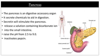
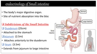
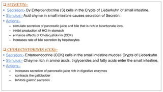
DR. HARI SINGH GOUR CENTRAL UNIVERSITY SAGAR (M.P)
2018-2019
ENDOCRINOLOGY
Hormonal control of digestion-from Oesophagus to intestine
(feed to digestion)
SUBMITTED BY :- NAGENDRA SAHU (MSc 1 sem / DEPARTMENT OF ZOOLOGY)
SUBMITTED TO:- DR. RASHMI SHRIVASTAVA (DEPARTMENT OF ZOOLOGY)
Hormonal control of digestion -from oesophagus to intestine (feed to digestion)
contents
 Hormonal control of digestion in digestive parts:-
 Digestive organs:- Accessory digestive organs:-
• Oral cavity 1.Salivary gland
• Phrynx 2.Liver/gall bladder
• Oesophagus 3.Pancrease
• Stomach
• Intestine
Oral cavity
 Boundaries are:
• lips (anteriorly)
• cheeks (laterally)
• palate (superiorly)
 Important in:
• Ingest food
• Saliva ph-7 (moisten and soften food)
• Tongue mixes food with saliva and rolls food
into bolus before swallowing
• mastication (chewing): mechanical digestion
• secretion of saliva for digestion
(amylase; digests starch)
Tongue and Teeth
Tongue
• (upper part of tongue covered
with papillae taste receptor)
• filiform papillae
• fungiform papillae
• circumvallate papillae
Teeth
• mechanical breakdown
• – Incisors
• – Canines
• – Molars
• Primary or deciduous teeth 20
• Secondary or permanent teeth 32
Salivary gland
 major salivary glands :-
• parotid glands - produce a serous, watery
secretion.
• submaxillary (mandibular) glands –
produce a serous and mucous secretion.
• Sublingual glands - secrete a saliva
mucous in character.
• Mixture of mucus and serous fluids Helps
to form a food bolus
• Contains salivary amylase to begin starch
digestion
pharynx
 Pharynx
• It is the passageway from the nose and mouth to the oesophagus and respiratory
tract
Nasopharynx
Oropharynx
Laryngopharynx
Serves as a passageway for air and food
• Food is propelled to the oesophagus by two
muscle layers:-
Longitudinal inner layer
Circular outer layer
• Food movement is by alternating contractions of the muscle layers (peristalsis)
oesophagus
• The oesophagus is a( 25 cm) passageway from the pharynx to stomach
Contains two sphincters: upper and lower:-
 Upper sphincter is skeletal (voluntary),
 lower sphincter is smooth muscle (involuntary)
• The oesophagus is posterior to the larynx and
trachea in the neck region and upper thorax.
• It travels on the right side of the descending aorta,
passes through the diaphragm, and connects with the
stomach.
Stomach
• Located on the left side of the abdominal cavity Food enters at the cardio
oesophageal sphincter.
• Site where food is churned into Chyme
Protein digestion begins.
• The stomach stores food, and mixes and
mechanically and chemically digests it
• The stomach also secretes digestive juices
pepsin: digests protein
• hydrochloric acid (acidic pH, required for
pepsin activity, and to kill ingested bacteria)
• Mucus: protects the stomach wall.
Histology and endocrine of stomach
 Cell types:
• Chief cells: produce pepsinogen (inactive
precursor to pepsin)
• Parietal cells: produce HCL(Hydrochloric acid)
“Endocrine” cells:
• G cells: gastrin
• D cells: somatostatin (paracrine)
• Enterochromaffin-like cells: histamine
(paracrine)
 Gastric hormones:-
 GASTRIN:-
 Secretion:- By enteroendocrine (G) cells in gastric pits of the mucosa.
 Stimulus-: Stomach distention and acid pH of chyme causes Gastrin.
 Action:-
1. increases HCL production in stomach
2. contract lower esophageal sphincter
3. relaxes pyloric sphincter
Somatostatin:
 Secretion:- By enteroendocrine (D) cells in gastric pits of the mucosa in the pylorus.
 Stimulus:- continuously released, overridden by Gastrin and nerves
 Action:-
• Inhibition of Gastrin production
liver and gallbladder
• Largest gland
• Located on the right side under
the diaphragm
• Consists of four lobes
(left and right, caudate , and quadrate)
• Connected to the gall bladder via
the common hepatic duct
• Detoxifies drugs and alcohol
• Degrades hormones
• Produce cholesterol, blood proteins .
 Bile:-
• Produced by cells in the liver
Composition:-
• Bile salts
• Bile pigment (mostly bilirubin
from the breakdown of haemoglobin)
• Cholesterol
• Phospholipids
• Electrolytes
 Gall Bladder:-
• Sac found in hollow fossa of liver
• Stores bile from the liver
by way of the cystic duct
• Gallstones can cause blockages
Pancreas
• The pancreas is an digestive accessory organ
• It secrete chemicals to aid in digestion.
• Secretin will stimulate the pancreas.
• release a solution containing bicarbonate ion
• into the small intestine.
• raise the pH from 2.5 to 9.0.
• inactivates pepsin.
Endocrinology of pancreas
• The acinar portion of pancreas has exocrine function .
• Endocrine portion consists of islets of Langerhans.
 Insulin:-
• hetero dimeric polypeptide .
• Glucose level more than 70mg/dl stimulates insulin synthesis , primarily by
enhancing protein translation & processing .
• Produced by beta cells.
• The major organs involved in insulin metabolism are liver , kidney & the
placenta .
 Glucagon:-
• synthesized as precursor molecule.
• Glucagon is inactivated by liver
enzyme removes 1st 2 amino acids.
• Produced by alpha cells.
 Somatostatin:-
• Produced by delta cells.
• It inhibit secretion of insulin and
glucagon.
endocrinology of Small intestine
• The body’s major digestive organ.
• Site of nutrient absorption into the blood.
 Subdivisions of the Small Intestine
 Duodenum (25cm)
• Attached to the stomach
Jejunum (2.5m)
• Attaches anteriorly to the duodenum
 Ileum (3.5m)
• Extends from jejunum to large intestine
 SECRETIN:-
 Secretion:- By Enteroendocrine (S) cells in the Crypts of Lieberkuhn of small intestine.
 Stimulus:- Acid chyme in small intestine causes secretion of Secretin:
 Actions:-
– stimulate secretion of pancreatic juice and bile that is rich in bicarbonate ions.
– inhibit production of HCl in stomach
– enhance effects of Cholecystokinin (CCK)
– Increases rate of bile secretion by hepatocytes
 CHOLECYSTOKININ (CCK):-
 Secretion:- Enteroendocrine (CCK) cells in the small intestine mucosa Crypts of Lieberkuhn
 Stimulus:- Chayme rich in amino acids, triglycerides and fatty acids enter the small intestine.
 Actions:-
– increases secretion of pancreatic juice rich in digestive enzymes
– contracts the gallbladder
– Inhibits gastric secretion .
Gastric Inhibitory Peptide (GIP):-
 Secretion:- Enteroendocrine cells in the small intestine mucosa Crypts of Lieberkuhn
 Stimulus:- Chayme rich in triglycerides, fatty acids, and glucose enter the small
intestine.
 Actions:-
– Stimulates release of insulin by beta cells
– Inhibits gastric secretion and motility
– Stimulates lipogenesis by adipose tissue
•REFRENCE:-
1. CLASS NOTES.
2. MEDICAL PHYSIOLOGY. Viz GUYTON AND HALL.
3. WIKIPEIDA.
4. YOUTUBE.
Endocrinology

Endocrinology

  • 1.
    DR. HARI SINGHGOUR CENTRAL UNIVERSITY SAGAR (M.P) 2018-2019 ENDOCRINOLOGY Hormonal control of digestion-from Oesophagus to intestine (feed to digestion) SUBMITTED BY :- NAGENDRA SAHU (MSc 1 sem / DEPARTMENT OF ZOOLOGY) SUBMITTED TO:- DR. RASHMI SHRIVASTAVA (DEPARTMENT OF ZOOLOGY)
  • 2.
    Hormonal control ofdigestion -from oesophagus to intestine (feed to digestion)
  • 3.
    contents  Hormonal controlof digestion in digestive parts:-  Digestive organs:- Accessory digestive organs:- • Oral cavity 1.Salivary gland • Phrynx 2.Liver/gall bladder • Oesophagus 3.Pancrease • Stomach • Intestine
  • 4.
    Oral cavity  Boundariesare: • lips (anteriorly) • cheeks (laterally) • palate (superiorly)  Important in: • Ingest food • Saliva ph-7 (moisten and soften food) • Tongue mixes food with saliva and rolls food into bolus before swallowing • mastication (chewing): mechanical digestion • secretion of saliva for digestion (amylase; digests starch)
  • 5.
    Tongue and Teeth Tongue •(upper part of tongue covered with papillae taste receptor) • filiform papillae • fungiform papillae • circumvallate papillae Teeth • mechanical breakdown • – Incisors • – Canines • – Molars • Primary or deciduous teeth 20 • Secondary or permanent teeth 32
  • 6.
    Salivary gland  majorsalivary glands :- • parotid glands - produce a serous, watery secretion. • submaxillary (mandibular) glands – produce a serous and mucous secretion. • Sublingual glands - secrete a saliva mucous in character. • Mixture of mucus and serous fluids Helps to form a food bolus • Contains salivary amylase to begin starch digestion
  • 7.
    pharynx  Pharynx • Itis the passageway from the nose and mouth to the oesophagus and respiratory tract Nasopharynx Oropharynx Laryngopharynx Serves as a passageway for air and food • Food is propelled to the oesophagus by two muscle layers:- Longitudinal inner layer Circular outer layer • Food movement is by alternating contractions of the muscle layers (peristalsis)
  • 8.
    oesophagus • The oesophagusis a( 25 cm) passageway from the pharynx to stomach Contains two sphincters: upper and lower:-  Upper sphincter is skeletal (voluntary),  lower sphincter is smooth muscle (involuntary) • The oesophagus is posterior to the larynx and trachea in the neck region and upper thorax. • It travels on the right side of the descending aorta, passes through the diaphragm, and connects with the stomach.
  • 9.
    Stomach • Located onthe left side of the abdominal cavity Food enters at the cardio oesophageal sphincter. • Site where food is churned into Chyme Protein digestion begins. • The stomach stores food, and mixes and mechanically and chemically digests it • The stomach also secretes digestive juices pepsin: digests protein • hydrochloric acid (acidic pH, required for pepsin activity, and to kill ingested bacteria) • Mucus: protects the stomach wall.
  • 10.
    Histology and endocrineof stomach  Cell types: • Chief cells: produce pepsinogen (inactive precursor to pepsin) • Parietal cells: produce HCL(Hydrochloric acid) “Endocrine” cells: • G cells: gastrin • D cells: somatostatin (paracrine) • Enterochromaffin-like cells: histamine (paracrine)
  • 11.
     Gastric hormones:- GASTRIN:-  Secretion:- By enteroendocrine (G) cells in gastric pits of the mucosa.  Stimulus-: Stomach distention and acid pH of chyme causes Gastrin.  Action:- 1. increases HCL production in stomach 2. contract lower esophageal sphincter 3. relaxes pyloric sphincter Somatostatin:  Secretion:- By enteroendocrine (D) cells in gastric pits of the mucosa in the pylorus.  Stimulus:- continuously released, overridden by Gastrin and nerves  Action:- • Inhibition of Gastrin production
  • 12.
    liver and gallbladder •Largest gland • Located on the right side under the diaphragm • Consists of four lobes (left and right, caudate , and quadrate) • Connected to the gall bladder via the common hepatic duct • Detoxifies drugs and alcohol • Degrades hormones • Produce cholesterol, blood proteins .
  • 13.
     Bile:- • Producedby cells in the liver Composition:- • Bile salts • Bile pigment (mostly bilirubin from the breakdown of haemoglobin) • Cholesterol • Phospholipids • Electrolytes  Gall Bladder:- • Sac found in hollow fossa of liver • Stores bile from the liver by way of the cystic duct • Gallstones can cause blockages
  • 14.
    Pancreas • The pancreasis an digestive accessory organ • It secrete chemicals to aid in digestion. • Secretin will stimulate the pancreas. • release a solution containing bicarbonate ion • into the small intestine. • raise the pH from 2.5 to 9.0. • inactivates pepsin.
  • 15.
    Endocrinology of pancreas •The acinar portion of pancreas has exocrine function . • Endocrine portion consists of islets of Langerhans.  Insulin:- • hetero dimeric polypeptide . • Glucose level more than 70mg/dl stimulates insulin synthesis , primarily by enhancing protein translation & processing . • Produced by beta cells. • The major organs involved in insulin metabolism are liver , kidney & the placenta .
  • 16.
     Glucagon:- • synthesizedas precursor molecule. • Glucagon is inactivated by liver enzyme removes 1st 2 amino acids. • Produced by alpha cells.  Somatostatin:- • Produced by delta cells. • It inhibit secretion of insulin and glucagon.
  • 17.
    endocrinology of Smallintestine • The body’s major digestive organ. • Site of nutrient absorption into the blood.  Subdivisions of the Small Intestine  Duodenum (25cm) • Attached to the stomach Jejunum (2.5m) • Attaches anteriorly to the duodenum  Ileum (3.5m) • Extends from jejunum to large intestine
  • 18.
     SECRETIN:-  Secretion:-By Enteroendocrine (S) cells in the Crypts of Lieberkuhn of small intestine.  Stimulus:- Acid chyme in small intestine causes secretion of Secretin:  Actions:- – stimulate secretion of pancreatic juice and bile that is rich in bicarbonate ions. – inhibit production of HCl in stomach – enhance effects of Cholecystokinin (CCK) – Increases rate of bile secretion by hepatocytes  CHOLECYSTOKININ (CCK):-  Secretion:- Enteroendocrine (CCK) cells in the small intestine mucosa Crypts of Lieberkuhn  Stimulus:- Chayme rich in amino acids, triglycerides and fatty acids enter the small intestine.  Actions:- – increases secretion of pancreatic juice rich in digestive enzymes – contracts the gallbladder – Inhibits gastric secretion .
  • 19.
    Gastric Inhibitory Peptide(GIP):-  Secretion:- Enteroendocrine cells in the small intestine mucosa Crypts of Lieberkuhn  Stimulus:- Chayme rich in triglycerides, fatty acids, and glucose enter the small intestine.  Actions:- – Stimulates release of insulin by beta cells – Inhibits gastric secretion and motility – Stimulates lipogenesis by adipose tissue
  • 20.
    •REFRENCE:- 1. CLASS NOTES. 2.MEDICAL PHYSIOLOGY. Viz GUYTON AND HALL. 3. WIKIPEIDA. 4. YOUTUBE.