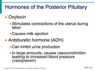
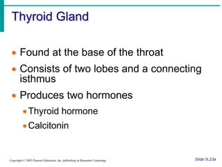
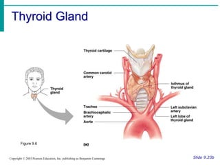
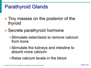
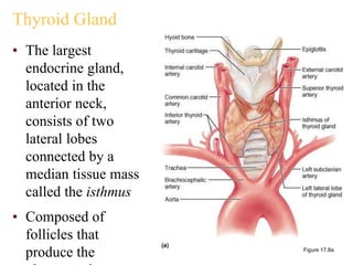
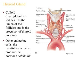
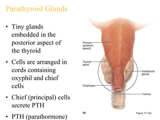
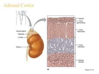
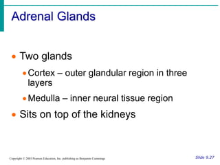
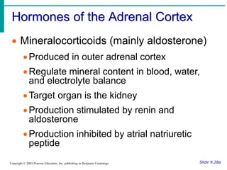
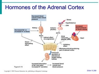
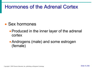
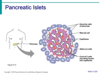
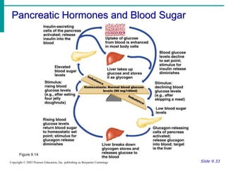
Hormones of the Posterior Pituitary
Slide 9.22
Copyright © 2003 Pearson Education, Inc. publishing as Benjamin Cummings
 Oxytocin
Stimulates contractions of the uterus during
labor
Causes milk ejection
 Antidiuretic hormone (ADH)
Can inhibit urine production
In large amounts, causes vasoconstriction
leading to increased blood pressure
(vasopressin)
Hormones of the Posterior Pituitary
Slide 9.22b
Copyright © 2003 Pearson Education, Inc. publishing as Benjamin Cummings
Figure 9.5
Thyroid Gland
Slide 9.23a
Copyright © 2003 Pearson Education, Inc. publishing as Benjamin Cummings
 Found at the base of the throat
 Consists of two lobes and a connecting
isthmus
 Produces two hormones
Thyroid hormone
Calcitonin
Thyroid Gland
Slide 9.23b
Copyright © 2003 Pearson Education, Inc. publishing as Benjamin Cummings
Figure 9.6
Thyroid Hormone
Slide 9.24
Copyright © 2003 Pearson Education, Inc. publishing as Benjamin Cummings
 Major metabolic hormone
 Composed of two active iodine-
containing hormones
Thyroxine (T4) – secreted by thyroid
follicles
Triiodothyronine (T3) – conversion of T4 at
target tissues
Calcitonin
Slide 9.25
Copyright © 2003 Pearson Education, Inc. publishing as Benjamin Cummings
 Decreases blood
calcium levels by
causing its
deposition on bone
 Antagonistic to
parathyroid
hormone
 Produced by C
(parafollicular)
cells
Figure 9.9
Parathyroid Glands
Slide 9.26
Copyright © 2003 Pearson Education, Inc. publishing as Benjamin Cummings
 Tiny masses on the posterior of the
thyroid
 Secrete parathyroid hormone
Stimulate osterclasts to remove calcium
from bone
Stimulate the kidneys and intestine to
absorb more calcium
Raise calcium levels in the blood
Thyroid Gland
• The largest
endocrine gland,
located in the
anterior neck,
consists of two
lateral lobes
connected by a
median tissue mass
called the isthmus
• Composed of
follicles that
produce the Figure 17.8a
Thyroid Gland
• Colloid
(thyroglobulin +
iodine) fills the
lumen of the
follicles and is the
precursor of thyroid
hormone
• Other endocrine
cells, the
parafollicular cells,
produce the
hormone calcitonin
Figure 17.8a
Thyroid Hormone (TH)
• Thyroid hormone – the body’s major metabolic
hormone
• Consists of two closely-related iodine-containing
compounds
• T4 – thyroxine; has two tyrosine molecules plus four
bound iodine atoms
• T3 – triiodothyronine; has two tyrosines with three
bound iodine atoms
Effects of Thyroid Hormone
• TH is concerned with:
• Glucose oxidation
• Increasing metabolic rate
• Heat production
• TH plays a role in:
• Maintaining blood pressure
• Regulating tissue growth
• Developing skeletal and nervous systems
• Maturation and reproductive capabilities
Transport and Regulation of TH
• T4 and T3 bind to thyroxine-binding globulins
(TBGs) produced by the liver
• Both bind to target receptors, but T3 is ten times
more active than T4
• Peripheral tissues convert T4 to T3
• Mechanisms of activity are similar to steroids
• Regulation is by negative feedback
• Hypothalamic thyrotropin-releasing hormone
(TRH) can overcome the negative feedback
Synthesis of Thyroid Hormone
• Thyroglobulin is synthesized and discharged into
the lumen
• Iodides (I–) are actively taken into the cell, oxidized
to iodine (I2), and released into the lumen
• Iodine attaches to tyrosine, mediated by peroxidase
enzymes, forming T1 (monoiodotyrosine, or MIT),
and T2 (diiodotyrosine, or DIT)
• Iodinated tyrosines link together to form T3 and T4
• Colloid is then endocytosed and combined with a
lysosome, where T3 and T4 are cleaved and diffuse
Synthesis of Thyroid Hormone
Figure 17.9
Calcitonin
• A peptide hormone produced by the parafollicular,
or C, cells
• Lowers blood calcium levels in children
• Antagonist to parathyroid hormone (PTH)
• Calcitonin targets the skeleton, where it:
• Inhibits osteoclast activity and thus bone resorption and
release of calcium from the bone matrix
• Stimulates calcium uptake and incorporation into the
bone matrix
• Regulated by a humoral (calcium ion concentration
Parathyroid Glands
• Tiny glands
embedded in the
posterior aspect of
the thyroid
• Cells are arranged in
cords containing
oxyphil and chief
cells
• Chief (principal) cells
secrete PTH
• PTH (parathormone)
Figure 17.10a
Effects of Parathyroid Hormone
• PTH release increases Ca2+
in the blood as it:
• Stimulates osteoclasts to
digest bone matrix
• Enhances the reabsorption
of Ca2+ and the secretion of
phosphate by the kidneys
• Increases absorption of Ca2+
by intestinal mucosal cells
• Rising Ca2+ in the blood
inhibits PTH release
Figure 17.11
Adrenal (Suprarenal) Glands
• Adrenal glands – paired, pyramid-shaped organs
atop the kidneys
• Structurally and functionally, they are two glands
in one
• Adrenal medulla – nervous tissue that acts as part of the
SNS
• Adrenal cortex – glandular tissue derived from
embryonic mesoderm
Adrenal Cortex
• Synthesizes and releases steroid hormones called
corticosteroids
• Different corticosteriods are produced in each of
the three layers
• Zona glomerulosa – mineralocorticoids (chiefly
aldosterone)
• Zona fasciculata – glucocorticoids (chiefly cortisol)
• Zona reticularis – gonadocorticoids (chiefly androgens)
Adrenal Cortex
Figure 17.12
Adrenal Glands
Slide 9.27
Copyright © 2003 Pearson Education, Inc. publishing as Benjamin Cummings
 Two glands
Cortex – outer glandular region in three
layers
Medulla – inner neural tissue region
 Sits on top of the kidneys
Hormones of the Adrenal Cortex
Slide 9.28a
Copyright © 2003 Pearson Education, Inc. publishing as Benjamin Cummings
 Mineralocorticoids (mainly aldosterone)
Produced in outer adrenal cortex
Regulate mineral content in blood, water,
and electrolyte balance
Target organ is the kidney
Production stimulated by renin and
aldosterone
Production inhibited by atrial natriuretic
peptide
Hormones of the Adrenal Cortex
Slide 9.28b
Copyright © 2003 Pearson Education, Inc. publishing as Benjamin Cummings
Figure 9.10
Hormones of the Adrenal Cortex
Slide 9.29a
Copyright © 2003 Pearson Education, Inc. publishing as Benjamin Cummings
 Glucocorticoids (including cortisone and
cortisol)
Produced in the middle layer of the adrenal
cortex
Promote normal cell metabolism
Help resist long-term stressors
Released in response to increased blood
levels of ACTH
Hormones of the Adrenal Cortex
Slide 9.29b
Copyright © 2003 Pearson Education, Inc. publishing as Benjamin Cummings
 Sex hormones
Produced in the inner layer of the adrenal
cortex
Androgens (male) and some estrogen
(female)
Hormones of the Adrenal Medulla
Slide 9.30
Copyright © 2003 Pearson Education, Inc. publishing as Benjamin Cummings
 Produces two similar hormones
(catecholamines)
Epinephrine
Norepinephrine
 These hormones prepare the body to
deal with short-term stress
Pancreatic Islets
Slide 9.32a
Copyright © 2003 Pearson Education, Inc. publishing as Benjamin Cummings
 The pancreas is a mixed gland
 The islets of the pancreas produce
hormones
Insulin – allows glucose to cross plasma
membranes into cells from beta cells
Glucagon – allows glucose to enter the
blood from alpha cells
These hormones are antagonists that
maintain blood sugar homeostasis
Pancreatic Islets
Slide 9.32b
Copyright © 2003 Pearson Education, Inc. publishing as Benjamin Cummings
Figure 9.13
Pancreatic Hormones and Blood Sugar
Slide 9.33
Copyright © 2003 Pearson Education, Inc. publishing as Benjamin Cummings
Figure 9.14
Pineal Gland
Slide 9.34
Copyright © 2003 Pearson Education, Inc. publishing as Benjamin Cummings
 Found on the third ventricle of the brain
 Secretes melatonin
Helps establish the body’s wake and sleep
cycles
May have other as-yet-unsubstantiated
functions
Thymus
Slide 9.35
Copyright © 2003 Pearson Education, Inc. publishing as Benjamin Cummings
 Located posterior to the sternum
 Largest in infants and children
 Produces thymosin
Matures some types of white blood cells
Important in developing the immune system
Hormones of the Ovaries
Slide 9.36
Copyright © 2003 Pearson Education, Inc. publishing as Benjamin Cummings
 Estrogens
 Produced by Graafian follicles or the placenta
 Stimulates the development of secondary
female characteristics
 Matures female reproductive organs
 Helps prepare the uterus to receive a fertilized
egg
 Helps maintain pregnancy
 Prepares the breasts to produce milk
Hormones of the Ovaries
Slide 9.37
Copyright © 2003 Pearson Education, Inc. publishing as Benjamin Cummings
 Progesterone
Produced by the corpus luteum
Acts with estrogen to bring about the
menstrual cycle
Helps in the implantation of an embryo in
the uterus
Hormones of the Testes
Slide 9.38
Copyright © 2003 Pearson Education, Inc. publishing as Benjamin Cummings
 Interstitial cells of testes are hormone-
producing
 Produce several androgens
 Testosterone is the most important
androgen
Responsible for adult male secondary sex
characteristics
Promotes growth and maturation of male
reproductive system
Required for sperm cell production
Other Hormone-Producing Tissues
and Organs
Slide 9.39
Copyright © 2003 Pearson Education, Inc. publishing as Benjamin Cummings
 Parts of the small intestine
 Parts of the stomach
 Kidneys
 Heart
 Many other areas have scattered
endocrine cells
Endocrine Function of the Placenta
Slide 9.40
Copyright © 2003 Pearson Education, Inc. publishing as Benjamin Cummings
 Produces hormones that maintain the
pregnancy
 Some hormones play a part in the
delivery of the baby
 Produces HCG in addition to estrogen,
progesterone, and other hormones
Developmental Aspects of the
Endocrine System
Slide 9.41
Copyright © 2003 Pearson Education, Inc. publishing as Benjamin Cummings
 Most endocrine organs operate smoothly
until old age
 Menopause is brought about by lack of
efficiency of the ovaries
 Problems associated with reduced estrogen
are common
 Growth hormone production declines with age
 Many endocrine glands decrease output with
age

EndoB'.ppt

  • 2.
    Hormones of thePosterior Pituitary Slide 9.22 Copyright © 2003 Pearson Education, Inc. publishing as Benjamin Cummings  Oxytocin Stimulates contractions of the uterus during labor Causes milk ejection  Antidiuretic hormone (ADH) Can inhibit urine production In large amounts, causes vasoconstriction leading to increased blood pressure (vasopressin)
  • 3.
    Hormones of thePosterior Pituitary Slide 9.22b Copyright © 2003 Pearson Education, Inc. publishing as Benjamin Cummings Figure 9.5
  • 4.
    Thyroid Gland Slide 9.23a Copyright© 2003 Pearson Education, Inc. publishing as Benjamin Cummings  Found at the base of the throat  Consists of two lobes and a connecting isthmus  Produces two hormones Thyroid hormone Calcitonin
  • 5.
    Thyroid Gland Slide 9.23b Copyright© 2003 Pearson Education, Inc. publishing as Benjamin Cummings Figure 9.6
  • 6.
    Thyroid Hormone Slide 9.24 Copyright© 2003 Pearson Education, Inc. publishing as Benjamin Cummings  Major metabolic hormone  Composed of two active iodine- containing hormones Thyroxine (T4) – secreted by thyroid follicles Triiodothyronine (T3) – conversion of T4 at target tissues
  • 7.
    Calcitonin Slide 9.25 Copyright ©2003 Pearson Education, Inc. publishing as Benjamin Cummings  Decreases blood calcium levels by causing its deposition on bone  Antagonistic to parathyroid hormone  Produced by C (parafollicular) cells Figure 9.9
  • 8.
    Parathyroid Glands Slide 9.26 Copyright© 2003 Pearson Education, Inc. publishing as Benjamin Cummings  Tiny masses on the posterior of the thyroid  Secrete parathyroid hormone Stimulate osterclasts to remove calcium from bone Stimulate the kidneys and intestine to absorb more calcium Raise calcium levels in the blood
  • 9.
    Thyroid Gland • Thelargest endocrine gland, located in the anterior neck, consists of two lateral lobes connected by a median tissue mass called the isthmus • Composed of follicles that produce the Figure 17.8a
  • 10.
    Thyroid Gland • Colloid (thyroglobulin+ iodine) fills the lumen of the follicles and is the precursor of thyroid hormone • Other endocrine cells, the parafollicular cells, produce the hormone calcitonin Figure 17.8a
  • 11.
    Thyroid Hormone (TH) •Thyroid hormone – the body’s major metabolic hormone • Consists of two closely-related iodine-containing compounds • T4 – thyroxine; has two tyrosine molecules plus four bound iodine atoms • T3 – triiodothyronine; has two tyrosines with three bound iodine atoms
  • 12.
    Effects of ThyroidHormone • TH is concerned with: • Glucose oxidation • Increasing metabolic rate • Heat production • TH plays a role in: • Maintaining blood pressure • Regulating tissue growth • Developing skeletal and nervous systems • Maturation and reproductive capabilities
  • 13.
    Transport and Regulationof TH • T4 and T3 bind to thyroxine-binding globulins (TBGs) produced by the liver • Both bind to target receptors, but T3 is ten times more active than T4 • Peripheral tissues convert T4 to T3 • Mechanisms of activity are similar to steroids • Regulation is by negative feedback • Hypothalamic thyrotropin-releasing hormone (TRH) can overcome the negative feedback
  • 14.
    Synthesis of ThyroidHormone • Thyroglobulin is synthesized and discharged into the lumen • Iodides (I–) are actively taken into the cell, oxidized to iodine (I2), and released into the lumen • Iodine attaches to tyrosine, mediated by peroxidase enzymes, forming T1 (monoiodotyrosine, or MIT), and T2 (diiodotyrosine, or DIT) • Iodinated tyrosines link together to form T3 and T4 • Colloid is then endocytosed and combined with a lysosome, where T3 and T4 are cleaved and diffuse
  • 15.
    Synthesis of ThyroidHormone Figure 17.9
  • 16.
    Calcitonin • A peptidehormone produced by the parafollicular, or C, cells • Lowers blood calcium levels in children • Antagonist to parathyroid hormone (PTH) • Calcitonin targets the skeleton, where it: • Inhibits osteoclast activity and thus bone resorption and release of calcium from the bone matrix • Stimulates calcium uptake and incorporation into the bone matrix • Regulated by a humoral (calcium ion concentration
  • 17.
    Parathyroid Glands • Tinyglands embedded in the posterior aspect of the thyroid • Cells are arranged in cords containing oxyphil and chief cells • Chief (principal) cells secrete PTH • PTH (parathormone) Figure 17.10a
  • 18.
    Effects of ParathyroidHormone • PTH release increases Ca2+ in the blood as it: • Stimulates osteoclasts to digest bone matrix • Enhances the reabsorption of Ca2+ and the secretion of phosphate by the kidneys • Increases absorption of Ca2+ by intestinal mucosal cells • Rising Ca2+ in the blood inhibits PTH release Figure 17.11
  • 19.
    Adrenal (Suprarenal) Glands •Adrenal glands – paired, pyramid-shaped organs atop the kidneys • Structurally and functionally, they are two glands in one • Adrenal medulla – nervous tissue that acts as part of the SNS • Adrenal cortex – glandular tissue derived from embryonic mesoderm
  • 20.
    Adrenal Cortex • Synthesizesand releases steroid hormones called corticosteroids • Different corticosteriods are produced in each of the three layers • Zona glomerulosa – mineralocorticoids (chiefly aldosterone) • Zona fasciculata – glucocorticoids (chiefly cortisol) • Zona reticularis – gonadocorticoids (chiefly androgens)
  • 21.
  • 22.
    Adrenal Glands Slide 9.27 Copyright© 2003 Pearson Education, Inc. publishing as Benjamin Cummings  Two glands Cortex – outer glandular region in three layers Medulla – inner neural tissue region  Sits on top of the kidneys
  • 23.
    Hormones of theAdrenal Cortex Slide 9.28a Copyright © 2003 Pearson Education, Inc. publishing as Benjamin Cummings  Mineralocorticoids (mainly aldosterone) Produced in outer adrenal cortex Regulate mineral content in blood, water, and electrolyte balance Target organ is the kidney Production stimulated by renin and aldosterone Production inhibited by atrial natriuretic peptide
  • 24.
    Hormones of theAdrenal Cortex Slide 9.28b Copyright © 2003 Pearson Education, Inc. publishing as Benjamin Cummings Figure 9.10
  • 25.
    Hormones of theAdrenal Cortex Slide 9.29a Copyright © 2003 Pearson Education, Inc. publishing as Benjamin Cummings  Glucocorticoids (including cortisone and cortisol) Produced in the middle layer of the adrenal cortex Promote normal cell metabolism Help resist long-term stressors Released in response to increased blood levels of ACTH
  • 26.
    Hormones of theAdrenal Cortex Slide 9.29b Copyright © 2003 Pearson Education, Inc. publishing as Benjamin Cummings  Sex hormones Produced in the inner layer of the adrenal cortex Androgens (male) and some estrogen (female)
  • 27.
    Hormones of theAdrenal Medulla Slide 9.30 Copyright © 2003 Pearson Education, Inc. publishing as Benjamin Cummings  Produces two similar hormones (catecholamines) Epinephrine Norepinephrine  These hormones prepare the body to deal with short-term stress
  • 28.
    Pancreatic Islets Slide 9.32a Copyright© 2003 Pearson Education, Inc. publishing as Benjamin Cummings  The pancreas is a mixed gland  The islets of the pancreas produce hormones Insulin – allows glucose to cross plasma membranes into cells from beta cells Glucagon – allows glucose to enter the blood from alpha cells These hormones are antagonists that maintain blood sugar homeostasis
  • 29.
    Pancreatic Islets Slide 9.32b Copyright© 2003 Pearson Education, Inc. publishing as Benjamin Cummings Figure 9.13
  • 30.
    Pancreatic Hormones andBlood Sugar Slide 9.33 Copyright © 2003 Pearson Education, Inc. publishing as Benjamin Cummings Figure 9.14
  • 31.
    Pineal Gland Slide 9.34 Copyright© 2003 Pearson Education, Inc. publishing as Benjamin Cummings  Found on the third ventricle of the brain  Secretes melatonin Helps establish the body’s wake and sleep cycles May have other as-yet-unsubstantiated functions
  • 32.
    Thymus Slide 9.35 Copyright ©2003 Pearson Education, Inc. publishing as Benjamin Cummings  Located posterior to the sternum  Largest in infants and children  Produces thymosin Matures some types of white blood cells Important in developing the immune system
  • 33.
    Hormones of theOvaries Slide 9.36 Copyright © 2003 Pearson Education, Inc. publishing as Benjamin Cummings  Estrogens  Produced by Graafian follicles or the placenta  Stimulates the development of secondary female characteristics  Matures female reproductive organs  Helps prepare the uterus to receive a fertilized egg  Helps maintain pregnancy  Prepares the breasts to produce milk
  • 34.
    Hormones of theOvaries Slide 9.37 Copyright © 2003 Pearson Education, Inc. publishing as Benjamin Cummings  Progesterone Produced by the corpus luteum Acts with estrogen to bring about the menstrual cycle Helps in the implantation of an embryo in the uterus
  • 35.
    Hormones of theTestes Slide 9.38 Copyright © 2003 Pearson Education, Inc. publishing as Benjamin Cummings  Interstitial cells of testes are hormone- producing  Produce several androgens  Testosterone is the most important androgen Responsible for adult male secondary sex characteristics Promotes growth and maturation of male reproductive system Required for sperm cell production
  • 36.
    Other Hormone-Producing Tissues andOrgans Slide 9.39 Copyright © 2003 Pearson Education, Inc. publishing as Benjamin Cummings  Parts of the small intestine  Parts of the stomach  Kidneys  Heart  Many other areas have scattered endocrine cells
  • 37.
    Endocrine Function ofthe Placenta Slide 9.40 Copyright © 2003 Pearson Education, Inc. publishing as Benjamin Cummings  Produces hormones that maintain the pregnancy  Some hormones play a part in the delivery of the baby  Produces HCG in addition to estrogen, progesterone, and other hormones
  • 38.
    Developmental Aspects ofthe Endocrine System Slide 9.41 Copyright © 2003 Pearson Education, Inc. publishing as Benjamin Cummings  Most endocrine organs operate smoothly until old age  Menopause is brought about by lack of efficiency of the ovaries  Problems associated with reduced estrogen are common  Growth hormone production declines with age  Many endocrine glands decrease output with age