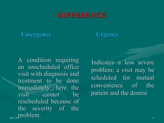
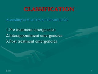
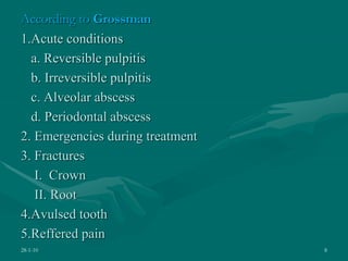
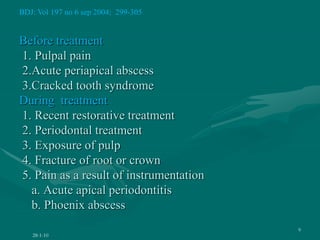
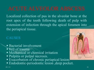
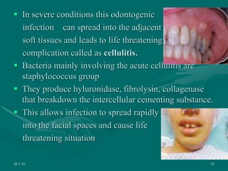
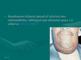
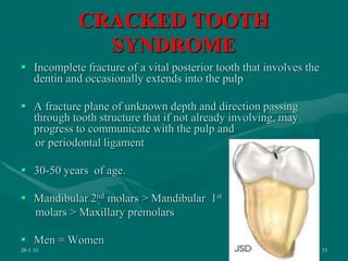
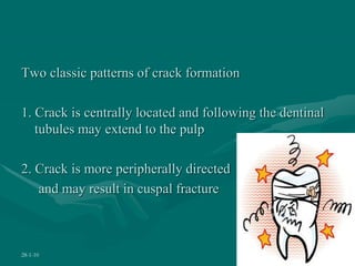
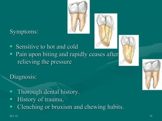
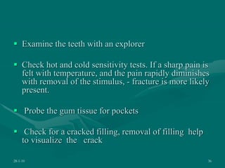
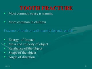
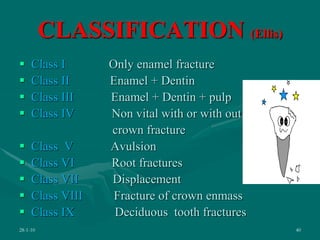
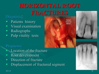
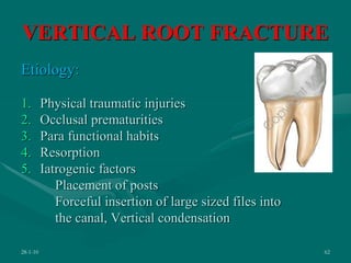
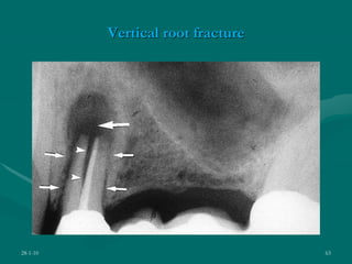
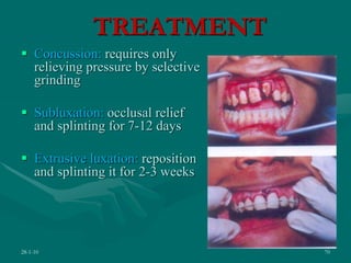
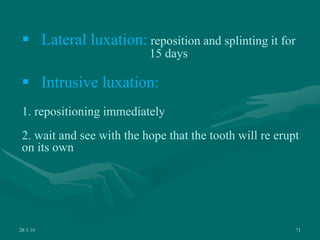
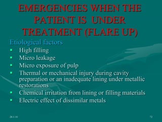
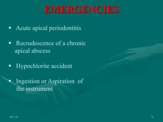
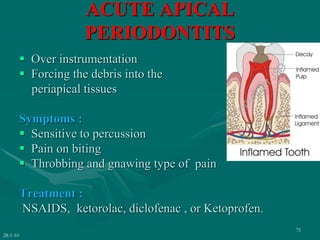
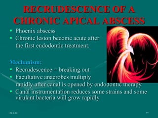
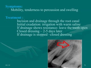
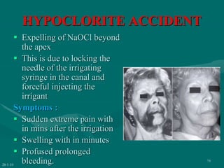
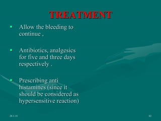
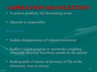
The document discusses various endodontic emergencies including their causes, symptoms, diagnosis and treatment. It defines an endodontic emergency as an unscheduled visit associated with pain or swelling from pulpoperiapical pathoses requiring immediate treatment. Common endodontic emergencies include acute pulpitis, acute apical abscess, cracked tooth syndrome, tooth fractures, and tooth avulsion. The document provides details on diagnosing and managing each emergency through methods like pulpectomy, apical trephination, incision and drainage, splinting or reimplantation depending on the specific emergency.
2.  Introduction
 Definition
 Difference between emergency and urgency
 Diagnosis and management
 Classification
 Acute pulpitis
 Acute alveolar abscess
 Esthetic emergencies
 Emergencies during the treatment
 Post endodontic treatment emergencies
 Conclusion
 References
28-1-10 2
3. INTRODUCTION
Emergency: Any urgent condition perceived by the
patient as requiring immediate medical or surgical
evaluation or treatment.
A sudden, urgent, usually unforeseen occurrence
requiring immediate attention.
28-1-10 3
4. Dental emergency :
An acute condition affecting the teeth, such as
inflammation of the soft tissues surrounding teeth or
past treatment complications of dental surgery
Endodontic emergency:
Defined as an unscheduled visit associated with pain
or swelling from pulpoperiapical pathoses requiring
immediate diagnosis and treatment
28-1-10 4
5.  1/3rd of all dental emergencies are from endodontic
origin
 90% of emergencies with pain as symptom, the pain
is pulpal or periapical
 In every year a number of on working days and
school days are lost due to dental pain in the world.
28-1-10 5
6. DIFFERENCE
Emergency
A condition requiring
an unscheduled office
visit with diagnosis and
treatment to be done
immediately , here the
visit cannot be
rescheduled because of
the severity of the
problem.
Urgency
Indicates a less severe
problem; a visit may be
scheduled for mutual
convenience of the
patient and the dentist
28-1-10 6
7. CLASSIFICATION
According to WALTON & TORABINEJAD
1.Pre treatment emergencies
2.Interappointment emergencies
3.Post treatment emergencies
28-1-10 7
8. According to Grossman
1.Acute conditions
a. Reversible pulpitis
b. Irreversible pulpitis
c. Alveolar abscess
d. Periodontal abscess
2. Emergencies during treatment
3. Fractures
I. Crown
II. Root
4.Avulsed tooth
5.Reffered pain
28-1-10 8
9. Before treatment
1. Pulpal pain
2.Acute periapical abscess
3.Cracked tooth syndrome
During treatment
1. Recent restorative treatment
2. Periodontal treatment
3. Exposure of pulp
4. Fracture of root or crown
5. Pain as a result of instrumentation
a. Acute apical periodontitis
b. Phoenix abscess
28-1-10
9
BDJ: Vol 197 no 6 sep 2004; 299-305
10. 3 D’s of successful
management
Diagnosis
Definitive treatment
Drugs
11. Diagnostic sequence
 Obtain information about the patient’s medical and dental
histories.
 Ask pointed subjective questions about the patient’s pain:
history, location, severity, duration, character, eliciting
stimuli.
 Perform extraoral examination
 Perform intraoral examination
 Perform pulp testing procedures.
 Use palpation and percussion sensitivity tests to determine
periapical status
 Proper Interpretation of radiographs
 Rapid and accurate diagnosis are important in the treatment of
endodontic emergencies
28-1-10 11
12. • SIGN - An objective evidence of a disease, such
evidence as is perceptable to the examining
physycian.
• SYMPTOM – It is a subjective , not usually visible to
others, symptoms are the patients experiences about
the illness, disease, or injury.
28-1-10 12
13. Most of the times patient
comes with chief
Complaint of
 Pain,
 Swelling,
 Transient loss of function.
 Esthetic abnormalities.
28-1-10 13
14. ACUTE PULPITIS
Acute reversible pulpitis:
 It is a mild to moderate inflammatory condition of the
pulp caused by noxious stimuli in which the pulp is
capable of returning to the uninflammed state
following removal of the stimuli
28-1-10 14
15. SYMPTOMS
 Short duration pain
 Does not linger
 Non tender to percussion
 Might be difficult to localize
 Might give an exaggerated response to vitality tests
 No radiographic significance.
28-1-10 15
16. CAUSES
 Caries close to the pulp
 Premature contact
 Recurrent caries
 Occlusal trauma
 Thermal Shock
 Microleakage
 Galvanic shock
 Chemical irritation - sweet or sour food
28-1-10 16
17. Choice of Treatment (Grossman)
The best treatment is prevention,
Protective base under restorations.
Avoid marginal leakage.
Occlusal reduction.
Avoid excessive heat during cavity preperation
Palliative treatment
Zinc oxide eugenol interim restoration
If pain disappears – restoration
If not - Pulp extirpation.
28-1-10 17
18. ACUTE IRREVERSIBLE
PULPITIS
 It is the persistent inflammatory condition of the
pulp, caused by a noxious stimuli
 Abnormal sensation to cold
 Abnormal sensation to hot
28-1-10 18
19. 28-1-10 19
SYMPTOMS
 There is often a history of spontaneous bouts of pain which
may last from a few seconds up to several hours
 When hot or cold fluids are applied, the pain elicited will be
more significant; cold may relieve the pain
 Pain may radiate initially, but once the periodontal ligament has
become involved the patient will be able to locate the tooth
 The tooth becomes tender to percussion once inflammation has
spread to the periodontal ligament
 A widened periodontal ligament may be seen on the
radiographs in the later stages
20. Causes
 Bacterial involvement of pulp through caries.
 Other factors-chemical, thermal, mechanical, or
galvanic irritation
 Reversible pulpitis may deteriorate in to irreversible
pulptis
28-1-10 20
21. Treatment
 Anesthetize the affected tooth.
 Apply rubber dam.
 Prepare access cavity.
 Remove the pulp from the chamber
 Locate the root canal orifice.
 Extirpate the pulp by sequentially. Instrumenting
with reamer or files or broaches to within 1mm
short of apex
28-1-10 21
22.  Irrigate with sterile saline solution, NaOCl solution.
 Dry the root canal with sterile absorbent points.
 Insert a medicated cotton pellet moistened with an
obtudant.
 Place a temp filling .
 Relieve the occlusal trauma.
 Analgesic if required
28-1-10 22
23. 28-1-10 23
PULPOTOMY
Conditions where pulpectomy is not possible in
multirooted teeth
 Anesthetize the affected tooth.
 Apply rubber dam.
 Prepare access cavity.
 Remove the pulp from the pulp chamber with
spoon excavator or round bur
 Cotton pellet moistened with formocresol is
placed in the cavity and it is sealed with
ZnOE
24. 28-1-10 24
ACUTE ALVEOLOR ABSCESS
Localized collection of pus in the alveolar bone at the
root apex of the tooth following death of pulp with
extension of infection through the apical foramen into
the periapical tissue.
CAUSES
 Bacterial involvement
 H/0 of trauma
 Mechanical or chemical irritation
 Pulpitis or pulpal necrosis.
 Exacerbation of chronic periapical lesion.
 Endodontic-periodontic lesion ,deep pocket.
25.  In severe conditions this odontogenic
infection can spread into the adjacent
soft tissues and leads to life threatening
complication called as cellulitis.
 Bacteria mainly involving the acute cellulitis are
staphylococcus group
 They produce hyluronidase, fibrolysin, collagenase
that breakdown the intercellular cementing substance.
 This allows infection to spread rapidly
into the facial spaces and cause life
threatening situation
28-1-10 25
26. 28-1-10 26
• Simultanious bilateral spread of infection into
submandibular, sublingual and submental space s is
called as ludwigs angina.
27. PHLEGMON:
 A massive cellulitis that does not go to suppuration
proceeds towards fast inflammatory infiltration of
subcutaneous tissue.
 Skin is bluish in color because of
tissue cyanosis
 Streptococcus haemolyticus
28-1-10 27
28. SYMPTOMS
Local reactions:
 Tenderness of tooth
 Severe throbbing pain
 Swelling
 Sinus tract
SYSTEMIC REACTIONS:
 Elevated temperature
 GI disturbances
 Malaise
 nausea
 Dizziness
 Lack of sleep
28-1-10 28
29. DIAGONSIS
 Pain and presence of swelling
 Mobility of tooth
 Non responsiveness to pulp testing
 Sensitiveness to percussion
 Small or large or diffuse radiolucency in radiographs
28-1-10 29
30. TREATMENT
 Local anesthesia
 Isolation
 Access cavity
 If drainage does not occur, apical foramen is
enlarged to 25, 30 no size to obtain the drainage. –
apical trephination .(weine)
 If time permits, complete cleaning and shaping of
the canals .
 Irrigation with NaOCl.
 Closed dressing given with ZnOE/ Placement of
intracanal medication
 Adjustment of occlusion
 If systemic involvement is present prescribing the
antibiotics and analgesics.
28-1-10 30
Apical trephination
31. BRITISH DENTAL JOURNAL VOL 198 NO. 12 JUNE 25 2005
• Incisional drainage is the first principle in
management of acute dentoalveolar infection.
• Penicillin-resistant bacteria are often present in acute
dental infection.
• The presence of penicillin resistant bacteria does not
adversely affect the outcome of treatment even if
penicillin is prescribed.
• It is likely that antibiotic therapy is often prescribed
unnecessarily in treatment of acute dental infection
28-1-10 31
32. TRAUMATIC AND
EASTHETIC EMERGENCIES
Traumatic injury to a tooth can cause
 1 cracked crown
 2. fracture crown
 3. fracture root
It can be broadly classified as
 1. Crown fracture
 2. Root fracture
28-1-10 32
33. CRACKED TOOTH
SYNDROME
 Incomplete fracture of a vital posterior tooth that involves the
dentin and occasionally extends into the pulp
 A fracture plane of unknown depth and direction passing
through tooth structure that if not already involving, may
progress to communicate with the pulp and
or periodontal ligament
 30-50 years of age.
 Mandibular 2nd molars > Mandibular 1st
molars > Maxillary premolars
 Men = Women
28-1-10 33
34. Two classic patterns of crack formation
1. Crack is centrally located and following the dentinal
tubules may extend to the pulp
2. Crack is more peripherally directed
and may result in cuspal fracture
28-1-10 34
35. Symptoms:
 Sensitive to hot and cold
 Pain upon biting and rapidly ceases after
relieving the pressure
Diagnosis:
 Thorough dental history.
 History of trauma,
 Clenching or bruxism and chewing habits.
28-1-10 35
36.  Examine the teeth with an explorer
 Check hot and cold sensitivity tests. If a sharp pain is
felt with temperature, and the pain rapidly diminishes
with removal of the stimulus, - fracture is more likely
present.
 Probe the gum tissue for pockets
 Check for a cracked filling, removal of filling help
to visualize the crack
28-1-10 36
38. 28-1-10 38
ASSESSMENT
OF TOOTH
LARGE
CRACK
WITHOUT PULP
INVOLVEMENT
WITH PULP
INVOLVEMENT
SMALL
CRACK
RESTORE WITH
COMPOSITE
•STABILIZE THE TOOTH.
•OCCLUSAL ADJUSTMENTS,
•PERMANENT STABILIZATION,
•BONDED OR CAST
RESTORATION
STABILIZE THE TOOTH ,
EXTIRPATE THE PULP,
ENDODONTIC THERAPY,
CAST RESTORATION
VERTICAL FRACTURES WITH HOPELESS
PROGNOSIS SHOULD BE EXTRACTED
Treatment
Dental traumatology 2006
39. TOOTH FRACTURE
 Most common cause is trauma,
 More common in children
Fracture of tooth or teeth mainly depends on the
 Energy of Impact
 Mass and velocity of object
 Resilience of the object
 Shape of the object
 Angle of direction
28-1-10 39
40. CLASSIFICATION (Ellis)
 Class I Only enamel fracture
 Class II Enamel + Dentin
 Class III Enamel + Dentin + pulp
 Class IV Non vital with or with out
crown fracture
 Class V Avulsion
 Class VI Root fractures
 Class VII Displacement
 Class VIII Fracture of crown enmass
 Class IX Deciduous tooth fractures
28-1-10 40
41. WHO CLASSIFICATION
 873.60 – enamel fracture
 873.61 – crown fracture involving enamel and dentin
with out pulp exposure
 873.62 – crown fracture with pulp exposure
 873.63 – root fractures
 873.64 – crown and root fractures
 873.66 – luxation
 873.67 – intrusion or extrusion
 873.68 – avulsion
 873.69 – other injuries
28-1-10 41
43. HEITHERSAY AND MORILE
28-1-10 43
 Class 1: fracture line does not extend below the level
of the attached gingiva
 Class 2: fracture line extends below the level of the
attached gingiva, till level of alveolar crest.
 Class 3:below the level of alveolar crest
 Class 4: fracture line with in the coronal third of the
root, below the level of the alveolar crest
44. 28-1-10 44
In general tooth fractures can be grouped into 5
major categories
 Fractures with out pulp
exposures
 Fractures with pulp
exposures
 Root fractures
 Tooth avulsion
 Luxation of tooth
45. TOOTH FRACTURE WITHOUT
PULP INVOLVEMENT
28-1-10 45
Ellis class I, II,
WHO classification 873.60, 61
 They are ranging from chipping of
enamel to deep dentinal fractures
Treatment :
 Only enamel fracture: Composite restoration
Dentin involvement :
 Immediate : applying hard setting calcium hydroxide
bonded resin restoration.base
46. FRACTURE OF CROWN WITH
PULP EXPOSURE
Vital pulp exposures
 Apex closed – root canal treatment
 Open apex –pulpotomy, partial
pulpotomy, or apexogensis,
Necrotic pulp exposures
 Open apex – apexification
 Closed apex- root canal treatment
28-1-10 46
47. TOOTH AVULSION
• Avulsed tooth is a dental and
emotional problem
• Main cause is trauma
• Ellis class V
28-1-10 47
48. Reimplantation
Time Treatment Success rate
½ hr Immediate
reimplantation then
RCT
80 %
< 2 hrs Reimplantation
followed by
immediate RCT
40 – 60 %
> 2 hrs First endodontic
treatment later
reimplantation
20 – 40 %
> 12 hrs Better to discard the
tooth.
-
28-1-10 48
49. MANAGEMENT (WEINE)
Part I (emergency treatment at the site of injury)
 Cleaning the tooth under the running water
 Reinsertion of tooth into the socket and
attain firm pressure .
 If reinsertion not possible place the tooth
in the transport media
28-1-10 49
50. Part II (emergency treatment at the dental office ):
 Place the tooth in the saline
 Health history, examination of area, radiographs
 Wiping the gross debris from the root surface with
wet sponge
 Irrigate the socket with saline
 Reimplant the tooth
 Check the position with radiographs
 Splint with arch wire and composite
28-1-10 50
51. Part III (completion of endodontic treatment)
 Closed apex - One week after replantation RCT is
adviced
 Open apex – watched with out pulp extirpation
 Stabilization period – 1 week
 Post operative instructions
 Soft diet
 Antibiotics and analgesics for 1-4 days
Use of emdogain is controversial
JCDA January 2000, Vol. 66, No. 1
28-1-10 51
52. TRANSPORT MEDIA
 Saline
 Distilled water
 Milk
 Oral vestibule, own saliva
 HBSS (Save A Tooth Solution)
 Coconut water
 Viaspan
28-1-10 52
54. Prognosis:
 Time elapsed between
the injury and
reimplantation
 Fracture of alveolus
 Blood clot in socket
 Direct mud at site
Follow up
 1w, 1m, 3m, 6m, 12m,
and annually for 5yrs
28-1-10 54
55. Fate of Reimplanted Tooth
 Tooth may maintain vitality
 Periodontally sound tooth may result
 Possible external resorption
 Ankylosis
 If no endodontic treatment is done –internal
resorption
28-1-10 55
56. ROOT FRACTURE
 Ellis class VI
 873.63
Can be divided into
 Vertical &
 Horizontal
1. Coronal
2. Middle
3. Apical third
28-1-10 56
57. On the basis of level of root fracture in
relation to various horizontal plane of
periodontium
• Class 1:fracture line nearing the
gingival attachment.
• Class 2: fracture line extends below
the level of the attached gingiva but
not below the alveolar crest
• Class 3: fracture line with in the
coronal one third of the root, but
below the level of alveolar crest
• Class 4: when fracture line extends
below the level of the alveolar crest,
but with in middle third of the root
• Class 5: when fracture line is in the
apical third of the root
28-1-10 57
58. HORIZONTAL ROOT
FRACTURES
Diagnosis:
 Patients history
 Visual examination
 Radiographs
 Pulp vitality tests
Prognosis:
 Location of the fracture
 Root development
 Direction of fracture
 Displacement of fractured segment
28-1-10 58
59. 28-1-10 59
Sequelae to root fractures (Andreasen)
 Healing with calcified tissues.
 Healing with interproximal connective tissue.
 Healing with interproximal bone.
 Healing with granulation tissue.
60. Treatment
Non surgical
 No mobilty – no treatment
 Mobile coronal fragment – repositioning
stabilization (4-6weeks)
 Nonseparated segments: Root canal therapy for both the
segments and stabilization with interradicular splints
 Separated segments: RCT for coronal segment only (apical
segment with vital tissue) . (cohen& burns)
28-1-10 60
61. 28-1-10 61
Surgical treatment (weine)
Apical 1/3rd fractures: rct of coronal segment only, apical
segment is left, it may heal, resorb in most instance does
not cause any problem.
Middle 1/3rd fractures: placement of cr-co pin joining
coronal and apical segment.
62. VERTICAL ROOT FRACTURE
Etiology:
1. Physical traumatic injuries
2. Occlusal prematurities
3. Para functional habits
4. Resorption
5. Iatrogenic factors
Placement of posts
Forceful insertion of large sized files into
the canal, Vertical condensation
28-1-10 62
65. 28-1-10 65
Periodontal examination:
Narrow deep isolated pocket – probe movement from
side to side is restricted
Radiographic examination:
1.Halo like bone loss (J shaped lesion )
2.Isolated bone loss
3.Radiolucent space between the long axis of the material
and canal wall
66. TREATMENT
28-1-10 66
Extraction :
 Single rooted teeth with vertical root fractures
 Multirooted teeth with multiple root fractures
Hemisection:
 Multirooted teeth with fracture confined to one root
only
 Multirooted teeth with fracture confined to furcation
only
67. LUXATION
 Abnormal mobility of the tooth with in the socket
with or without displacement as a result of trauma or
injury
Five types of luxation injuries (Andreasen)
 Concussion
 Subluxation
 Extrusive luxation
 Lateral luxation
 Intrusive luxation
28-1-10 67
68. 28-1-10 68
 Concussion : injury to supporting structures without
abnormal loosing or displacement
Subluxation: injury with abnormal
loosing
Extrusive luxation: partial displacment
of the tooth out of its socket
Lateral luxation: eccentric displacement
with fracture of alveolar socket
Intrusive luxation: displacement of
tooth deeper into the alveolar socket
Intrusion
70. TREATMENT
 Concussion: requires only
relieving pressure by selective
grinding
 Subluxation: occlusal relief
and splinting for 7-12 days
 Extrusive luxation: reposition
and splinting it for 2-3 weeks
28-1-10 70
71. 28-1-10 71
 Lateral luxation: reposition and splinting it for
15 days
 Intrusive luxation:
1. repositioning immediately
2. wait and see with the hope that the tooth will re erupt
on its own
72. EMERGENCIES WHEN THE
PATIENT IS UNDER
TREATMENT (FLARE UP)
Etiological factors
 High filling
 Micro leakage
 Micro exposure of pulp
 Thermal or mechanical injury during cavity
preparation or an inadequate lining under metallic
restorations
 Chemical irritation from lining or filling materials
 Electric effect of dissimilar metals
28-1-10 72
73. 28-1-10
73
 Periodontal treatment
 Exposure of pulp
 Fracture of root or crown
FACTORS (WALTON):
Irritants with in the pulp
system
Iatrogenic factors
Host factors
General systemic factors
74. EMERGENCIES
28-1-10 74
 Acute apical periodontitis
 Recrudescence of a chronic
apical abscess
 Hypochlorite accident
 Ingestion or Aspiration of
the instrument
75. ACUTE APICAL
PERIODONTITS
 Over instrumentation
 Forcing the debris into the
periapical tissues
Symptoms :
 Sensitive to percussion
 Pain on biting
 Throbbing and gnawing type of pain
Treatment :
NSAIDS, ketorolac, diclofenac , or Ketoprofen.
28-1-10
75
76. JCDA March 2003 vol 69 no 3. ( sackett)
1. Grade A – NSAIDS – preoperatively.
2. Grade B – Antibiotics – not recommended.
3. – NSAIDS used as a solution.
4. Grade C - Corticosteroids have a weak
5. evidence in pain management.
28-1-10 76
77. RECRUDESCENCE OF A
CHRONIC APICAL ABSCESS
 Phoenix abscess
 Chronic lesion become acute after
the first endodontic treatment.
Mechanism:
 Recrudescence = breaking out
 Facultative anaerobes multiply
rapidly after canal is opened by endodontic therapy
 Canal instrumentation reduces some strains and some
virulant bacteria will grow rapidly
28-1-10 77
78. 28-1-10 78
Symptoms:
Mobility, tenderness to percussion and swelling
Treatment :
Incision and drainage through the root canal
Initial exudation: irrigation with warm saline
If drainage shows resistance- leave the tooth open
Closed dressing – 2-5 days later
If drainage is stopped –closed dressing
79. HYPOCLORITE ACCIDENT
 Expelling of NaOCl beyond
the apex
 This is due to locking the
needle of the irrigating
syringe in the canal and
forceful injecting the
irrigant
Symptoms :
 Sudden extreme pain with
in mins after the irrigation
 Swelling with in minutes
 Profused prolonged
bleeding.
28-1-10
79
82. TREATMENT
 Allow the bleeding to
continue ,
 Antibiotics, analgesics
for five and three days
respectively .
 Prescribing anti
histamines (since it
should be considered as
hypersensitive reaction)
28-1-10 82
83. ASPIRATION OR INGESTION
 A serious problem, life threatening event.
 Operator is responsible
Recognition:
 Sudden disappearance of slipped instrument
 Sudden violent gagging or spasmodic coughing ,
wheezing, decrease breathing sounds by the patient .
 Radiographic evidence of presence of file in the
alimentary tract or airway
28-1-10
83
84. TREATMENT
Radiographic examination
• To know nature and size of the foreign body.
• Antero-posterior chest radiograph.
• Lateral chest radiograph.
• Lateral neck radiograph
• Supine abdominal radiograph.
28-1-10 84
85. The draw back with these radiographs is limitation for
radio opaque objects, in this scenario the following
are considered.
• Gastroscopy.
• Bronchoscopy.
• Computed tommography.
• Monitoring of physical signs.
• Test of stool for occult blood.
28-1-10 85
86.  If the instrument is in pharyngeal pouch and
oesophagus try to locate and remove with blunt long
tweezer
 Instrument is in lungs – surgical intervention.
 Bronchus – causing problem – remove it surgically .
 If it is in the alimentary tract monitor
radiographically and physical signs.
 Advice the patient to take fibre diet so that it provides
better movement.
 Usually it passes in faeces, if not remove surgically.
Prevention of these accidents is done by the universal
use of rubber dam and using a dental floss tied to the
instrument.
28-1-10 86
International Endodontic Journal, 41, 617–622, 2008
87. 28-1-10 87
International Endodontic Journal, 41, 617–622, 2008
At the time of ingestion
Lower part of stomach.
3 Hrs after , in small
intestine
2nd day, in the caecum
of large intestine.
3rd day no evidence
of instrument.
88. PREVENTION OF FLARE UPS
 Proper diagnosis
 Determination of correct working length
 Radiographs
 Apex locaters
 Complete extirpation of pulp
 Proper Irrigation
 Aviod filling too close to radiographic apex
28-1-10 88
89. 28-1-10 89
 Perform apical trephination only if necessary
 Reduce the tooth from occlusion if apex is severely
violated by over instrumentation
 Placement of intracanal medicament
 Prescription of mild analgesics and antibiotics whenever
condition warrants it
91. MANAGEMENT
 Occlusal correction and removal of high points in
the restoration
 Prescription of analgesics and, if the pain is more
severe and infection is present, antibiotics
 An attempt at removal of the root filling and
repreparation of the root canal
 Periradicular surgery
28-1-10 91
92. REFERENCES
1. ENDODONTIC PRACTICE – GROSSMAN
2. ENDODONTICS – WEINE
3. PRINCIPLES AND PRACTICE OF ENDODONTICS – WALTON
4. PATHWAYS OF PULP – STEPHEN COHEN AND BURNS
5. ENDODONTICS- INGLE AND BACKLAND
JOURNALS
• BDJ – vol 197 No 6, Sept 2004 p 299 – 305
• The crack tooth syndrome, JCDA Sept 2002, vol 168,No 8,
• Preoperative pain and medications used in emergency patients with
irrevercible acute pulpitis and acute apical periodontitis a prospective
comparitive study. Journal of orofacial pain.Volume 21, Number 4, 2007
• Emergency management of acute apical periodontitis, JCDA, Mar 2003
69, 169
28-1-10p 92
93. • Midtreatment flareups in endodontics, A dialemma. Dr Neeta shetty.
• Journal of orofacial pain 2007 vol 21, nov 4
• Pain associated with root canal treatment. 2009 feb 4 journal of orofacial
pain.
• Microflora in teeth associated with apical periodontis, IEJ 2009
• A double blind comparison of a supplimenal intraligamentary fantanyl
mepivacaine injection with 1;200000 epinephrine for irreversible pulpitis,
journal of pain and symptom management.
• Clinical management of avulsed permanent incisor, JCDA Jan 200 vol 66
No 1
• Analysis of 154 cases of teeth with cracks ,Dental traumatology 2006
• An outcome audit of the treatment of acute dentoalveolar infection , impact
of penicillin resistance.BDJ vol 198 no 12 jun 25 2005.
• A life threatening event from poorly managed dental pain. BDJ vol, 202 No
4Feb 24 2007
28-1-10 93
94. • Emergency management of acute apical periodontitis in permanent
dentition. review, March 2003 vol 69 no 3 JCDA
• A systematic review of the diagnostic classification of traumatic dental
injuries , Dental traumatology, 2006
• Gingival and bone necrosis caused by accidental NaOCl injection instead
of anaesthetic solution. IEJ Vol 41 2008
• Accidental injection with NaOCl; A case report. IEJ Vol 42 2009
• Palatal mucosal necrosis because of accidental NaOCl injection instead of
anaesthertic solution. IEJ Vol 39 2008
• Accidental swallowing of an endodontic file.IEJ Vol 41 2008
28-1-10 94