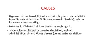
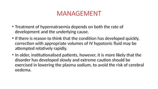
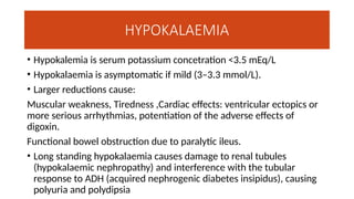
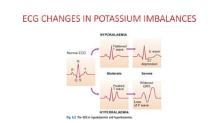
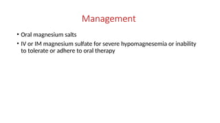
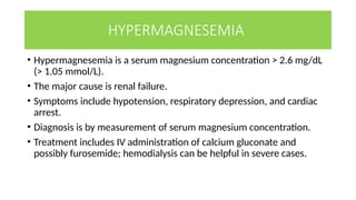
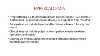
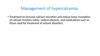
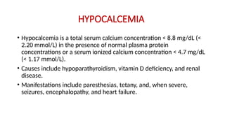
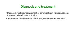
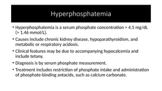
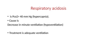
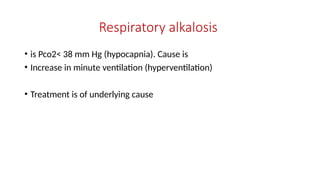
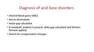
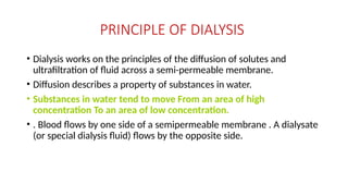
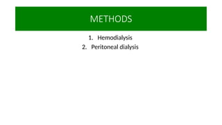
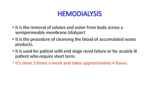
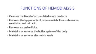
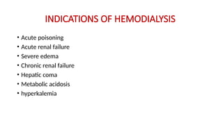
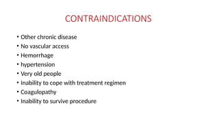
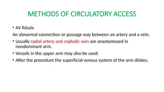
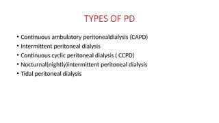
ELECTROLYTE IMBALANCES
AND DIALYSIS
BY SSONKO JOSEPH
Outline
• Imbalances in serum sodium levels
• Imbalances in serum potassium levels
• Imbalances in serum calcium levels
• Imbalances in serum magnesium levels
• Acid base imbalances
• Dialysis
Hemodialysis and peritoneal dialysis
HYPONATREMIA
• This is when the plasma sodium levels fall below 135mmol/L.
• It’s the most common electrolyte imbalance.
• Develops gradually and relatively asymptomatic
• More rapid changes in plasma osmolality and plasma sodium may be
associated with:
Anorexia, Nausea, Vomiting, Confusion, Lethargy, Seizures, Coma.
CAUSES OF HYPONATREMIA
• The causes of hyponatraemia are best organised according to any
associated change in ECF volume status.
Hypovolaemic (sodium deficit with a relatively smaller water deficit):
Renal Na loss (diuretics), GI Na loss (vomiting, diarrhoea).
Euvolaemic (water retention alone, i.e. ‘dilutional’): Primary polydipsia,
SIADH.
Hypervolaemic (sodium retention with relatively greater water
retention): Heart failure, cirrhosis and chronic kidney disease (without
water restriction
INVESTIGATIONS
• Plasma and urine electrolytes and osmolality are usually the only tests
required to classify the hyponatraemia.
MANAGEMENT
• The treatment for hyponatraemia is critically dependent on the rate
of development and severity, and on the underlying cause.
• If hyponatraemia has developed rapidly (over hours to days), and
there are signs of cerebral oedema (patient is obtunded or
convulsing), sodium levels should be rapidly restored to normal by
infusion of hypertonic (3%) sodium chloride solutions
• Rapid correction of hyponatraemia that has developed slowly (over
weeks to months) may lead to ‘central pontine myelinolysis’
Rx
• Specific treatment should be directed at the underlying cause.
• For hypovolaemic patients, this will involve controlling sodium loss
and giving IV saline if clinically warranted.
• Patients with dilutional hyponatraemia usually respond to fluid
restriction in the range of 600–1000 mL/day and removal of the
precipitating stimulus (e.g. a drug causing SIADH).
• Demeclocycline or oral urea supplements may be of use in SIADH
HYPERNATREMIA
• Plasma sodium levels >148mmol/L
• Reflects an inadequacy of the kidney in concentrating the urine in the
face of restricted water intake.
• Patients with hypernatraemia generally have reduced cerebral
function and cerebral dehydration.
• This triggers thirst and drinking, and if adequate water is obtained, is
self-limiting.
• If adequate water is not obtained, dizziness, confusion, weakness and
ultimately coma and death can result
CAUSES
• Hypovolemic (sodium deficit with a relatively greater water deficit):
Renal Na losses (diuretics), GI Na losses (colonic diarrhea), skin Na
losses (excessive sweating)
• Euvolaemic: Diabetes insipidus (central or nephrogenic.
• Hypervolaemic ;Enteral or parenteral nutrition, oral salt
administration, chronic kidney disease (during water restriction).
MANAGEMENT
• Treatment of hypernatraemia depends on both the rate of
development and the underlying cause.
• If there is reason to think that the condition has developed quickly,
correction with appropriate volumes of IV hypotonic fluid may be
attempted relatively rapidly.
• In older, institutionalised patients, however, it is more likely that the
disorder has developed slowly and extreme caution should be
exercised in lowering the plasma sodium, to avoid the risk of cerebral
oedema.
HYPOKALAEMIA
• Hypokalemia is serum potassium concetration <3.5 mEq/L
• Hypokalaemia is asymptomatic if mild (3–3.3 mmol/L).
• Larger reductions cause:
Muscular weakness, Tiredness ,Cardiac effects: ventricular ectopics or
more serious arrhythmias, potentiation of the adverse effects of
digoxin.
Functional bowel obstruction due to paralytic ileus.
• Long standing hypokalaemia causes damage to renal tubules
(hypokalaemic nephropathy) and interference with the tubular
response to ADH (acquired nephrogenic diabetes insipidus), causing
polyuria and polydipsia
ECG CHANGES IN POTASSIUM IMBALANCES
CAUSES
• Redistribution into cells: Alkalosis, insulin excess, β2-agonists.
Reduced potassium intake: Dietary, IV therapy. Excessive losses: These
may be renal or GI.
• Renal losses may be caused by the following factors: ● With
hypertension: primary/secondary hyperaldosteronism, Cushing’s
syndrome, corticosteroids, ectopic ACTH. ● With normal to low BP:
diuretics, renal tubular acidosis, post-obstructive diuresis, recovery
after acute tubular necrosis, inherited tubular disorders.
• GI losses may be caused by: ● Vomiting. ● Diarrhoea. ● Bowel
obstruction. ● Laxative abuse
INVESTIGATIONS
• Measurement of plasma electrolytes, bicarbonate, urine potassium
and sometimes calcium and magnesium is usually sufficient to
establish the diagnosis.
• Plasma renin activity and aldosterone levels identify patients with
primary hyperaldosteronism and other mineralocorticoid excess when
renin is suppressed; in other causes of hypokalaemia, renin is
elevated
MANAGEMENT
• Oral potassium supplements
• IV potassium supplements for severe hypokalemia or ongoing
potassium loses.
• If the problem is redistribution of potassium into cells, reversal of the
underlying cause (e.g. correction of alkalosis) may restore plasma
potassium without supplements
HYPERKALEMIA
• This is a serum potassium concentration >5.5mmol/L
• Hyperkalaemia typically presents with progressive muscular
weakness, but sometimes there are no symptoms until cardiac arrest
occurs (caused by the marked slowing of action potential conduction
in the presence of potassium levels >7 mmol/L).
CAUSES
• Redistribution out of cells: Acidosis, insulin deficiency, β-blockers,
severe hyperglycaemia.
• Increased intake: Exogenous (diet, IV therapy); endogenous
(haemolysis, rhabdomyolysis).
• Renal potassium retention: Renal failure (acute and chronic); tubular
secretory failure – drugs (NSAIDs, ACE inhibitors, spironolactone),
tubulo-interstitial disease, Addison’s disease.
• Spurious result: in vitro haemolysis
INVESTIGATION
• Plasma electrolyte, creatinine and bicarbonate results, together with
consideration of the clinical scenario, will usually provide the
explanation for hyperkalaemia.
• Addison’s disease should be excluded, unless there is an obvious
alternative diagnosis.
MANAGEMENT
HYPOMAGNESEMIA
• Hypomagnesemia is serum magnesium concentration < 1.8 mg/dL (<
0.70 mmol/L)
• Causes include inadequate magnesium intake and absorption or
increased excretion due to hypercalcemia or medications such as
furosemide
SIGNS AND SYMPTOMS
• Some patients are asymptomatic.
• Clinical features are often due to accompanying hypokalemia and
hypocalcemia.
• They include anorexia, nausea, vomiting, lethargy, weakness,
personality change, tetany (eg, positive Trousseau or Chvostek sign or
spontaneous carpopedal spasm, hyperreflexia), tremor, and muscle
fasciculations.
Management
• Oral magnesium salts
• IV or IM magnesium sulfate for severe hypomagnesemia or inability
to tolerate or adhere to oral therapy
HYPERMAGNESEMIA
• Hypermagnesemia is a serum magnesium concentration > 2.6 mg/dL
(> 1.05 mmol/L).
• The major cause is renal failure.
• Symptoms include hypotension, respiratory depression, and cardiac
arrest.
• Diagnosis is by measurement of serum magnesium concentration.
• Treatment includes IV administration of calcium gluconate and
possibly furosemide; hemodialysis can be helpful in severe cases.
HYPERCALCEMIA
• Hypercalcemia is a total serum calcium concentration > 10.4 mg/dL (>
2.60 mmol/L) or ionized serum calcium > 5.2 mg/dL (> 1.30 mmol/L).
• Principal causes include hyperparathyroidism, vitamin D toxicity, and
cancer.
• Clinical features include polyuria, constipation, muscle weakness,
confusion, and coma.
• Diagnosis is by measuring serum ionized calcium and parathyroid
hormone concentrations.
Management of hypercalcemia
• Treatment to increase calcium excretion and reduce bone resorption
of calcium involves saline, sodium diuresis, and medications such as
those used for treatment of seizure disorders.
HYPOCALCEMIA
• Hypocalcemia is a total serum calcium concentration < 8.8 mg/dL (<
2.20 mmol/L) in the presence of normal plasma protein
concentrations or a serum ionized calcium concentration < 4.7 mg/dL
(< 1.17 mmol/L).
• Causes include hypoparathyroidism, vitamin D deficiency, and renal
disease.
• Manifestations include paresthesias, tetany, and, when severe,
seizures, encephalopathy, and heart failure.
Diagnosis and treatment
• Diagnosis involves measurement of serum calcium with adjustment
for serum albumin concentration.
• Treatment is administration of calcium, sometimes with vitamin D.
Hyperphosphatemia
• Hyperphosphatemia is a serum phosphate concentration > 4.5 mg/dL
(> 1.46 mmol/L).
• Causes include chronic kidney disease, hypoparathyroidism, and
metabolic or respiratory acidosis.
• Clinical features may be due to accompanying hypocalcemia and
include tetany.
• Diagnosis is by serum phosphate measurement.
• Treatment includes restriction of phosphate intake and administration
of phosphate-binding antacids, such as calcium carbonate.
Hypophosphatemia
• Hypophosphatemia is a serum phosphate concentration < 2.5 mg/dL
(0.81 mmol/L).
• Causes include alcohol use disorder, burns, starvation, and diuretic
use.
• Clinical features include muscle weakness, respiratory failure, and
heart failure; seizures and coma can occur.
• Diagnosis is by serum phosphate concentration.
• Treatment consists of phosphate supplementation.
ACID BASE IMBALANCES
By SSONKO JOSEPH
INTRODUCTION
• Acid-base disorders are pathologic changes in carbon dioxide partial
pressure (Pco2) or serum bicarbonate (HCO3−) that typically produce
abnormal arterial pH values.
• Acidemia is serum pH < 7.35.
• Alkalemia is serum pH > 7.45.
• Acidosis refers to physiologic processes that cause acid accumulation
or alkali loss.
• Alkalosis refers to physiologic processes that cause alkali
accumulation or acid loss.
Metabolic acidosis
• is serum HCO3−< 24 mEq/L (< 24 mmol/L).
• Causes are;
①Increased acid production
②Acid ingestion
③Decreased renal acid excretion
④Gastrointestinal or renal HCO3− loss
• Treatment is;
1. Cause treated
2. Sodium bicarbonate (NaHCO3) primarily for severe acidemia, give with caution.
Metabolic alkalosis
• is serum HCO3−> 28 mEq/L (> 28 mmol/L).
• Causes are;
Acid loss
HCO3− retention
• Management;
I. Cause treated
II. IV 0.9% saline solution for chloride-responsive metabolic alkalosis
Respiratory acidosis
• is Pco2> 40 mm Hg (hypercapnia).
• Cause is
Decrease in minute ventilation (hypoventilation)
• Treatment is adequate ventilation
Respiratory alkalosis
• is Pco2< 38 mm Hg (hypocapnia). Cause is
• Increase in minute ventilation (hyperventilation)
• Treatment is of underlying cause
Diagnosis of acid base disorders
• Arterial blood gases (ABG)
• Serum electrolytes
• Anion gap calculated
• If metabolic acidosis is present, delta gap calculated and Winters
formula applied
• Search for compensatory changes
DIALYSIS
Presented by SSONKO JOSEPH
INTRODUCTION
• This is a process for removing waste and excess water from the blood,
and is used primarily as artificial replacement for lost kidney function
in people with renal failure.
• Dialysis is an artificial process used to remove water and waste
substances from the blood when the kidneys fail to function properly.
• It generally works through osmosis and filtration of fluid across a
semipermeable membrane with the use of a dialysate.
• Dialysis is the procedure used to correct fluid and electrolyte
imbalances and to remove waste products in renal failure.
PURPOSE
• To maintain fluid, electrolyte and acid base balance
• To remove endogenous and exogenous toxins
INDICATIONS
The need for dialysis may be acute or chronic.
1. Acute dialysis is indicated
A. when there is a high and rising level of serum potassium, fluid
overload, or impending pulmonary edema, increasing acidosis,
pericarditis, and severe confusion.
B. to remove certain medications or other toxins from the blood.
2. Chronic or maintenance dialysis is indicated in chronic renal failure,
known as end-stage renal disease (ESRD
PRINCIPLE OF DIALYSIS
• Dialysis works on the principles of the diffusion of solutes and
ultrafiltration of fluid across a semi-permeable membrane.
• Diffusion describes a property of substances in water.
• Substances in water tend to move From an area of high
concentration To an area of low concentration.
• . Blood flows by one side of a semipermeable membrane . A dialysate
(or special dialysis fluid) flows by the opposite side.
METHODS
1. Hemodialysis
2. Peritoneal dialysis
HEMODIALYSIS
• It is the removal of solutes and water from body across a
semipermeable membrane (dialyzer)
• It is the procedure of cleansing the blood of accumulated waste
products.
• It is used for patient with end stage renal failure or for acutely ill
patient who require short term.
• It's done 3 times a week and takes approximately 4 hours.
FUNCTIONS OF HEMODIALYSIS
• Cleanses the blood of accumulated waste products
• Removes the by-products of protein metabolism such as urea,
creatinine, and uric acid.
• Removes excessive fluids.
• Maintains or restores the buffer system of the body
• Maintains or restores electrolyte levels
INDICATIONS OF HEMODIALYSIS
• Acute poisoning
• Acute renal failure
• Severe edema
• Chronic renal failure
• Hepatic coma
• Metabolic acidosis
• hyperkalemia
CONTRAINDICATIONS
• Other chronic disease
• No vascular access
• Hemorrhage
• hypertension
• Very old people
• Inability to cope with treatment regimen
• Coagulopathy
• Inability to survive procedure
METHODS OF CIRCULATORY ACCESS
• AV fistula
An abnormal connection or passage way between an artery and a vein.
• Usually radial artery and cephalic vein are anastomosed in
nondominant arm.
• Vessels in the upper arm may also be used.
• After the procedure the superficial venous system of the arm dilates.
• By means of two large bore needles inserted into the dialated venous
system, blood may be obtained and passess through the dialyzer.
• The arterial end is used for the arterial flow and the distal end is used
for the reinfusion of dialysed blood.
• Healing of AVF requires at least 6 to 8 weeks; a central vein catheter is
used.
• If a patient isn't a candidate for AV fistula, an AV graft is considered
CENTRAL VENOUS CATHETER
• The third type of vascular access is a venous catheter.
• A venous catheter is a plastic tube which is inserted into a large vein
usually in the neck.
PROCEDURE
• Patient access is prepared and cannulated
• Heparin is administered
• Heparin and red blood flows through semipermeable dialysis in one
direction and dialysis solution surrounds the membrane and flows in
the opposite direction.
• Dialysis solution consist of highly purified water to which sodium,
potassium calcium, magnesium chloride, and dextrose have been
added, bicarbonate is added to achieve the the proper pH balance.
PROCEDURE...
• Through the process of diffusion solute in the form of electrolytes,
metabolic waste products acid base balance components can be
removed or added to the blood.
• Excess water is removed from the blood (ultrafiltration).
• The blood is then returned to the body through patient access.
COMPLICATIONS
• Infection
• Catheter clotting
• Central vein thrombosis
• Stenosis or thrombosis.
• Ischemia of the hand
• Aneurysm
PERITONEAL DIALYSIS
By SSONKO JOSEPH
Introduction
• Peritoneal dialysis uses the peritoneum as a natural permeable
membrane through which water and solutes can equilibrate.
Compared to hemodialysis, peritoneal dialysis is;
• Less physiologically stressful
• Does not require vascular access
• Can be done at home
• Allows patients much greater flexibility
• However, peritoneal dialysis requires much more patient involvement
than in-center hemodialysis.
COMPLICATIONS
The most important and common complications of peritoneal dialysis
are;
• Peritonitis
• Catheter tunnel exit-site infection
TYPES OF PD
• Continuous ambulatory peritonealdialysis (CAPD)
• Intermittent peritoneal dialysis
• Continuous cyclic peritoneal dialysis ( CCPD)
• Nocturnal(nightly)intermittent peritoneal dialysis
• Tidal peritoneal dialysis
References
• Davidson's principles and practices of medicine 24th edition
• MSD manual for professionals last updated in March 2023

Electrolyte imbalances and dialysis.pptx

  • 1.
  • 2.
    Outline • Imbalances inserum sodium levels • Imbalances in serum potassium levels • Imbalances in serum calcium levels • Imbalances in serum magnesium levels • Acid base imbalances • Dialysis Hemodialysis and peritoneal dialysis
  • 3.
    HYPONATREMIA • This iswhen the plasma sodium levels fall below 135mmol/L. • It’s the most common electrolyte imbalance. • Develops gradually and relatively asymptomatic • More rapid changes in plasma osmolality and plasma sodium may be associated with: Anorexia, Nausea, Vomiting, Confusion, Lethargy, Seizures, Coma.
  • 4.
    CAUSES OF HYPONATREMIA •The causes of hyponatraemia are best organised according to any associated change in ECF volume status. Hypovolaemic (sodium deficit with a relatively smaller water deficit): Renal Na loss (diuretics), GI Na loss (vomiting, diarrhoea). Euvolaemic (water retention alone, i.e. ‘dilutional’): Primary polydipsia, SIADH. Hypervolaemic (sodium retention with relatively greater water retention): Heart failure, cirrhosis and chronic kidney disease (without water restriction
  • 5.
    INVESTIGATIONS • Plasma andurine electrolytes and osmolality are usually the only tests required to classify the hyponatraemia.
  • 6.
    MANAGEMENT • The treatmentfor hyponatraemia is critically dependent on the rate of development and severity, and on the underlying cause. • If hyponatraemia has developed rapidly (over hours to days), and there are signs of cerebral oedema (patient is obtunded or convulsing), sodium levels should be rapidly restored to normal by infusion of hypertonic (3%) sodium chloride solutions • Rapid correction of hyponatraemia that has developed slowly (over weeks to months) may lead to ‘central pontine myelinolysis’
  • 7.
    Rx • Specific treatmentshould be directed at the underlying cause. • For hypovolaemic patients, this will involve controlling sodium loss and giving IV saline if clinically warranted. • Patients with dilutional hyponatraemia usually respond to fluid restriction in the range of 600–1000 mL/day and removal of the precipitating stimulus (e.g. a drug causing SIADH). • Demeclocycline or oral urea supplements may be of use in SIADH
  • 8.
    HYPERNATREMIA • Plasma sodiumlevels >148mmol/L • Reflects an inadequacy of the kidney in concentrating the urine in the face of restricted water intake. • Patients with hypernatraemia generally have reduced cerebral function and cerebral dehydration. • This triggers thirst and drinking, and if adequate water is obtained, is self-limiting. • If adequate water is not obtained, dizziness, confusion, weakness and ultimately coma and death can result
  • 9.
    CAUSES • Hypovolemic (sodiumdeficit with a relatively greater water deficit): Renal Na losses (diuretics), GI Na losses (colonic diarrhea), skin Na losses (excessive sweating) • Euvolaemic: Diabetes insipidus (central or nephrogenic. • Hypervolaemic ;Enteral or parenteral nutrition, oral salt administration, chronic kidney disease (during water restriction).
  • 10.
    MANAGEMENT • Treatment ofhypernatraemia depends on both the rate of development and the underlying cause. • If there is reason to think that the condition has developed quickly, correction with appropriate volumes of IV hypotonic fluid may be attempted relatively rapidly. • In older, institutionalised patients, however, it is more likely that the disorder has developed slowly and extreme caution should be exercised in lowering the plasma sodium, to avoid the risk of cerebral oedema.
  • 11.
    HYPOKALAEMIA • Hypokalemia isserum potassium concetration <3.5 mEq/L • Hypokalaemia is asymptomatic if mild (3–3.3 mmol/L). • Larger reductions cause: Muscular weakness, Tiredness ,Cardiac effects: ventricular ectopics or more serious arrhythmias, potentiation of the adverse effects of digoxin. Functional bowel obstruction due to paralytic ileus. • Long standing hypokalaemia causes damage to renal tubules (hypokalaemic nephropathy) and interference with the tubular response to ADH (acquired nephrogenic diabetes insipidus), causing polyuria and polydipsia
  • 12.
    ECG CHANGES INPOTASSIUM IMBALANCES
  • 13.
    CAUSES • Redistribution intocells: Alkalosis, insulin excess, β2-agonists. Reduced potassium intake: Dietary, IV therapy. Excessive losses: These may be renal or GI. • Renal losses may be caused by the following factors: ● With hypertension: primary/secondary hyperaldosteronism, Cushing’s syndrome, corticosteroids, ectopic ACTH. ● With normal to low BP: diuretics, renal tubular acidosis, post-obstructive diuresis, recovery after acute tubular necrosis, inherited tubular disorders. • GI losses may be caused by: ● Vomiting. ● Diarrhoea. ● Bowel obstruction. ● Laxative abuse
  • 14.
    INVESTIGATIONS • Measurement ofplasma electrolytes, bicarbonate, urine potassium and sometimes calcium and magnesium is usually sufficient to establish the diagnosis. • Plasma renin activity and aldosterone levels identify patients with primary hyperaldosteronism and other mineralocorticoid excess when renin is suppressed; in other causes of hypokalaemia, renin is elevated
  • 15.
    MANAGEMENT • Oral potassiumsupplements • IV potassium supplements for severe hypokalemia or ongoing potassium loses. • If the problem is redistribution of potassium into cells, reversal of the underlying cause (e.g. correction of alkalosis) may restore plasma potassium without supplements
  • 16.
    HYPERKALEMIA • This isa serum potassium concentration >5.5mmol/L • Hyperkalaemia typically presents with progressive muscular weakness, but sometimes there are no symptoms until cardiac arrest occurs (caused by the marked slowing of action potential conduction in the presence of potassium levels >7 mmol/L).
  • 17.
    CAUSES • Redistribution outof cells: Acidosis, insulin deficiency, β-blockers, severe hyperglycaemia. • Increased intake: Exogenous (diet, IV therapy); endogenous (haemolysis, rhabdomyolysis). • Renal potassium retention: Renal failure (acute and chronic); tubular secretory failure – drugs (NSAIDs, ACE inhibitors, spironolactone), tubulo-interstitial disease, Addison’s disease. • Spurious result: in vitro haemolysis
  • 18.
    INVESTIGATION • Plasma electrolyte,creatinine and bicarbonate results, together with consideration of the clinical scenario, will usually provide the explanation for hyperkalaemia. • Addison’s disease should be excluded, unless there is an obvious alternative diagnosis.
  • 19.
  • 20.
    HYPOMAGNESEMIA • Hypomagnesemia isserum magnesium concentration < 1.8 mg/dL (< 0.70 mmol/L) • Causes include inadequate magnesium intake and absorption or increased excretion due to hypercalcemia or medications such as furosemide
  • 21.
    SIGNS AND SYMPTOMS •Some patients are asymptomatic. • Clinical features are often due to accompanying hypokalemia and hypocalcemia. • They include anorexia, nausea, vomiting, lethargy, weakness, personality change, tetany (eg, positive Trousseau or Chvostek sign or spontaneous carpopedal spasm, hyperreflexia), tremor, and muscle fasciculations.
  • 22.
    Management • Oral magnesiumsalts • IV or IM magnesium sulfate for severe hypomagnesemia or inability to tolerate or adhere to oral therapy
  • 23.
    HYPERMAGNESEMIA • Hypermagnesemia isa serum magnesium concentration > 2.6 mg/dL (> 1.05 mmol/L). • The major cause is renal failure. • Symptoms include hypotension, respiratory depression, and cardiac arrest. • Diagnosis is by measurement of serum magnesium concentration. • Treatment includes IV administration of calcium gluconate and possibly furosemide; hemodialysis can be helpful in severe cases.
  • 24.
    HYPERCALCEMIA • Hypercalcemia isa total serum calcium concentration > 10.4 mg/dL (> 2.60 mmol/L) or ionized serum calcium > 5.2 mg/dL (> 1.30 mmol/L). • Principal causes include hyperparathyroidism, vitamin D toxicity, and cancer. • Clinical features include polyuria, constipation, muscle weakness, confusion, and coma. • Diagnosis is by measuring serum ionized calcium and parathyroid hormone concentrations.
  • 25.
    Management of hypercalcemia •Treatment to increase calcium excretion and reduce bone resorption of calcium involves saline, sodium diuresis, and medications such as those used for treatment of seizure disorders.
  • 26.
    HYPOCALCEMIA • Hypocalcemia isa total serum calcium concentration < 8.8 mg/dL (< 2.20 mmol/L) in the presence of normal plasma protein concentrations or a serum ionized calcium concentration < 4.7 mg/dL (< 1.17 mmol/L). • Causes include hypoparathyroidism, vitamin D deficiency, and renal disease. • Manifestations include paresthesias, tetany, and, when severe, seizures, encephalopathy, and heart failure.
  • 27.
    Diagnosis and treatment •Diagnosis involves measurement of serum calcium with adjustment for serum albumin concentration. • Treatment is administration of calcium, sometimes with vitamin D.
  • 28.
    Hyperphosphatemia • Hyperphosphatemia isa serum phosphate concentration > 4.5 mg/dL (> 1.46 mmol/L). • Causes include chronic kidney disease, hypoparathyroidism, and metabolic or respiratory acidosis. • Clinical features may be due to accompanying hypocalcemia and include tetany. • Diagnosis is by serum phosphate measurement. • Treatment includes restriction of phosphate intake and administration of phosphate-binding antacids, such as calcium carbonate.
  • 29.
    Hypophosphatemia • Hypophosphatemia isa serum phosphate concentration < 2.5 mg/dL (0.81 mmol/L). • Causes include alcohol use disorder, burns, starvation, and diuretic use. • Clinical features include muscle weakness, respiratory failure, and heart failure; seizures and coma can occur. • Diagnosis is by serum phosphate concentration. • Treatment consists of phosphate supplementation.
  • 30.
  • 31.
    INTRODUCTION • Acid-base disordersare pathologic changes in carbon dioxide partial pressure (Pco2) or serum bicarbonate (HCO3−) that typically produce abnormal arterial pH values. • Acidemia is serum pH < 7.35. • Alkalemia is serum pH > 7.45. • Acidosis refers to physiologic processes that cause acid accumulation or alkali loss. • Alkalosis refers to physiologic processes that cause alkali accumulation or acid loss.
  • 32.
    Metabolic acidosis • isserum HCO3−< 24 mEq/L (< 24 mmol/L). • Causes are; ①Increased acid production ②Acid ingestion ③Decreased renal acid excretion ④Gastrointestinal or renal HCO3− loss • Treatment is; 1. Cause treated 2. Sodium bicarbonate (NaHCO3) primarily for severe acidemia, give with caution.
  • 33.
    Metabolic alkalosis • isserum HCO3−> 28 mEq/L (> 28 mmol/L). • Causes are; Acid loss HCO3− retention • Management; I. Cause treated II. IV 0.9% saline solution for chloride-responsive metabolic alkalosis
  • 34.
    Respiratory acidosis • isPco2> 40 mm Hg (hypercapnia). • Cause is Decrease in minute ventilation (hypoventilation) • Treatment is adequate ventilation
  • 35.
    Respiratory alkalosis • isPco2< 38 mm Hg (hypocapnia). Cause is • Increase in minute ventilation (hyperventilation) • Treatment is of underlying cause
  • 36.
    Diagnosis of acidbase disorders • Arterial blood gases (ABG) • Serum electrolytes • Anion gap calculated • If metabolic acidosis is present, delta gap calculated and Winters formula applied • Search for compensatory changes
  • 37.
  • 38.
    INTRODUCTION • This isa process for removing waste and excess water from the blood, and is used primarily as artificial replacement for lost kidney function in people with renal failure.
  • 39.
    • Dialysis isan artificial process used to remove water and waste substances from the blood when the kidneys fail to function properly. • It generally works through osmosis and filtration of fluid across a semipermeable membrane with the use of a dialysate. • Dialysis is the procedure used to correct fluid and electrolyte imbalances and to remove waste products in renal failure.
  • 40.
    PURPOSE • To maintainfluid, electrolyte and acid base balance • To remove endogenous and exogenous toxins
  • 41.
    INDICATIONS The need fordialysis may be acute or chronic. 1. Acute dialysis is indicated A. when there is a high and rising level of serum potassium, fluid overload, or impending pulmonary edema, increasing acidosis, pericarditis, and severe confusion. B. to remove certain medications or other toxins from the blood. 2. Chronic or maintenance dialysis is indicated in chronic renal failure, known as end-stage renal disease (ESRD
  • 42.
    PRINCIPLE OF DIALYSIS •Dialysis works on the principles of the diffusion of solutes and ultrafiltration of fluid across a semi-permeable membrane. • Diffusion describes a property of substances in water. • Substances in water tend to move From an area of high concentration To an area of low concentration. • . Blood flows by one side of a semipermeable membrane . A dialysate (or special dialysis fluid) flows by the opposite side.
  • 43.
  • 44.
    HEMODIALYSIS • It isthe removal of solutes and water from body across a semipermeable membrane (dialyzer) • It is the procedure of cleansing the blood of accumulated waste products. • It is used for patient with end stage renal failure or for acutely ill patient who require short term. • It's done 3 times a week and takes approximately 4 hours.
  • 45.
    FUNCTIONS OF HEMODIALYSIS •Cleanses the blood of accumulated waste products • Removes the by-products of protein metabolism such as urea, creatinine, and uric acid. • Removes excessive fluids. • Maintains or restores the buffer system of the body • Maintains or restores electrolyte levels
  • 46.
    INDICATIONS OF HEMODIALYSIS •Acute poisoning • Acute renal failure • Severe edema • Chronic renal failure • Hepatic coma • Metabolic acidosis • hyperkalemia
  • 47.
    CONTRAINDICATIONS • Other chronicdisease • No vascular access • Hemorrhage • hypertension • Very old people • Inability to cope with treatment regimen • Coagulopathy • Inability to survive procedure
  • 48.
    METHODS OF CIRCULATORYACCESS • AV fistula An abnormal connection or passage way between an artery and a vein. • Usually radial artery and cephalic vein are anastomosed in nondominant arm. • Vessels in the upper arm may also be used. • After the procedure the superficial venous system of the arm dilates.
  • 49.
    • By meansof two large bore needles inserted into the dialated venous system, blood may be obtained and passess through the dialyzer. • The arterial end is used for the arterial flow and the distal end is used for the reinfusion of dialysed blood. • Healing of AVF requires at least 6 to 8 weeks; a central vein catheter is used. • If a patient isn't a candidate for AV fistula, an AV graft is considered
  • 50.
    CENTRAL VENOUS CATHETER •The third type of vascular access is a venous catheter. • A venous catheter is a plastic tube which is inserted into a large vein usually in the neck.
  • 51.
    PROCEDURE • Patient accessis prepared and cannulated • Heparin is administered • Heparin and red blood flows through semipermeable dialysis in one direction and dialysis solution surrounds the membrane and flows in the opposite direction. • Dialysis solution consist of highly purified water to which sodium, potassium calcium, magnesium chloride, and dextrose have been added, bicarbonate is added to achieve the the proper pH balance.
  • 52.
    PROCEDURE... • Through theprocess of diffusion solute in the form of electrolytes, metabolic waste products acid base balance components can be removed or added to the blood. • Excess water is removed from the blood (ultrafiltration). • The blood is then returned to the body through patient access.
  • 53.
    COMPLICATIONS • Infection • Catheterclotting • Central vein thrombosis • Stenosis or thrombosis. • Ischemia of the hand • Aneurysm
  • 54.
  • 55.
    Introduction • Peritoneal dialysisuses the peritoneum as a natural permeable membrane through which water and solutes can equilibrate.
  • 56.
    Compared to hemodialysis,peritoneal dialysis is; • Less physiologically stressful • Does not require vascular access • Can be done at home • Allows patients much greater flexibility • However, peritoneal dialysis requires much more patient involvement than in-center hemodialysis.
  • 57.
    COMPLICATIONS The most importantand common complications of peritoneal dialysis are; • Peritonitis • Catheter tunnel exit-site infection
  • 58.
    TYPES OF PD •Continuous ambulatory peritonealdialysis (CAPD) • Intermittent peritoneal dialysis • Continuous cyclic peritoneal dialysis ( CCPD) • Nocturnal(nightly)intermittent peritoneal dialysis • Tidal peritoneal dialysis
  • 59.
    References • Davidson's principlesand practices of medicine 24th edition • MSD manual for professionals last updated in March 2023