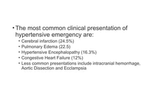
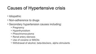
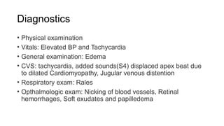
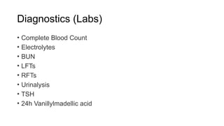
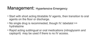
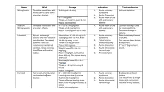
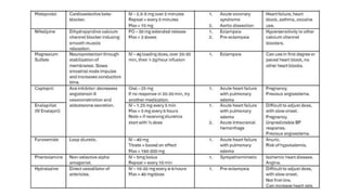
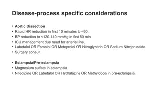
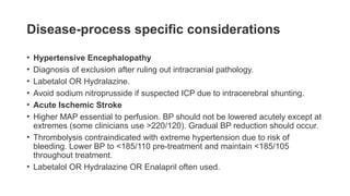
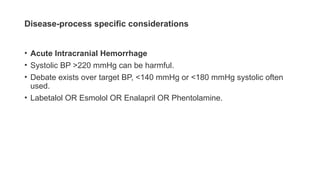
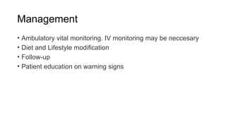
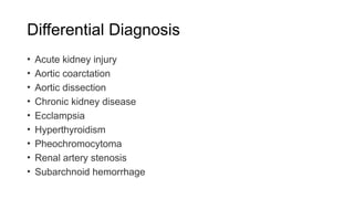
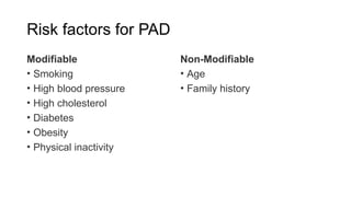
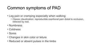
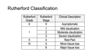
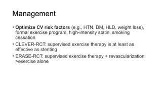
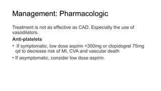
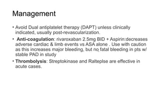
HYPERTENSIVE CRISES AND
PERIPHERAL ARTERIAL
DISEASES
MUTYABA MICHAEL SEBITOSI
Outline
• Definitions
• Hypertensive emergency
• Severe asymptomatic Hypertension
• Causes of hypertensive crisis
• Diagnostics
• Management
• Special Considerations
• Differential diagnosis
• Complications
Definitions
• Hypertensive Crisis: sudden severe elevation of blood
pressure above 180/120mmHg.
• Severe Hypertension: Term used broadly to define a very high
blood pressure (>180/120 mmHg)
• Hypertensive Emergency: Severe elevation in blood pressure
above 180/120 mmHg accompanied by acute life-threatening
end-organ damage.
• Severe Assymptomatic Hypertension: Severe elevation in
blood pressure above 180/120mmHg in absence of symptoms
or evidence of Acute end-organ damage.
• Malignant Hypertension: A subtype of hypertensive
emergency characterized by severely elevated blood pressure
accompanied by end-organ damage. Commonly papilledema.
• Accelerated Hypertension: Refers to subtype of hypertensive
emergency characterized by a recent significant increase over
baseline blood pressure with end-organ damage.
• Hypertensive Urgency: Other name for severe asymptomatic
hypertension
• Note: There is potential for end-organ damage even when the
BP is below the 180/110-120mmHg.
Severe asymptomatic HTN
• BP ≥180/120 without evidence of end-organ damage (may have
mild headache)
• Assess adherence to prior treatment before aggressively up-
titrating regimen to avoid overcorrection of BPs & hypotension
• Assess cause before treatment; commonly due to pain, anxiety,
urine retention, meds (e.g. steroids), OSA, nausea, withdrawal,
etc.
Hypertensive emergency
• Refers to a BP ≥180/120 mmHg with evidence of acute end-
organ damage.
• Rate of rise is more often more important than the actual BP.
This explains why patients with a prior diagnosis of
hypertension tolerate higher levels of BP without symptoms
compared to those presenting with hypertension for the first
time.
• There is a characteristic fibrinoid necrosis of the arterioles and
small arteries leading to the end organ damage.
End-organ damage
• Neurological: HTN encephalopathy (severe HA, seizure, AMS),
PRES, TIA, CVA (SAH, ICH)
• Occular: Retinopathy, Papilledema, Hemorrhage
• Respiratory: Pulmonary edema
• Cardiovascular: MI, Angina, Aortic dissection
• Hematological: Microangiopathic Hemolytic Anemia
• Renal: Acute Renal Failure, Hematuria, Proteinuria
•The most common clinical presentation of
hypertensive emergency are:
• Cerebral infarction (24.5%)
• Pulmonary Edema (22.5)
• Hypertensive Encephalopathy (16.3%)
• Congestive Heart Failure (12%)
• Less common presentations include intracranial hemorrhage,
Aortic Dissection and Ecclampsia
Causes of Hypertensive crisis
• Idiopathic
• Non-adherence to drugs
• Secondary hypertension causes including:
• Pregnancy
• Hyperthyroidism
• Pheochromocytoma
• Renal artery stenosis
• Use of cocaine or MAOIs
• Withdrawal of alcohol, beta-blockers, alpha stimulants
Diagnostics
• Physical examination
• Vitals: Elevated BP and Tachycardia
• General examination: Edema
• CVS: tachycardia, added sounds(S4) displaced apex beat due
to dilated Cardiomyopathy, Jugular venous distention
• Respiratory exam: Rales
• Opthalmologic exam: Nicking of blood vessels, Retinal
hemorrhages, Soft exudates and papilledema
Diagnostics (Labs)
• Complete Blood Count
• Electrolytes
• BUN
• LFTs
• RFTs
• Urinalysis
• TSH
• 24h Vanillylmadellic acid
Diagnostics: Imaging
• Chest radiograph: Cardial enlargement, pulmonary edema other
structures
• Transesophageal electrocardiography
• Renal angiography
• Electrocardiogram
Management: Severe Asymptomatic Hypertension
• Floor vs outpatient Management (with close follow up)
• ↓BP no more than 25-30% over hrs-days; then to goal as
outpatient.
• There is no evidence to support rapid lowering of BP in this case.
• If hospitalized for non-cardiac reason, worse outcomes with
intensifying anti-HTN immediately on admission
• Oral medication is preferred. IV or high-dose meds are avoided
due to increased risk for AKI, stroke and MI due to
hypoperfusion
• PO: start captopril, labetalol >> hydralazine (unpredict., reflex
tachy) & convert to long-acting before discharge OR start long-
acting agents
Management: Hypertensive Emergency
• Floor vs ICU
• Manage in the ICU if patient needs arterial line, anti hypertensive gtt
or if severe end organ damage.
• Continuous cardiac monitoring
• Frequent assessment of neurological status
• Monitoring fluid intake and output
• Excessive BP reduction will cause hypoperfusion. Thus:
• Reduce by max 25% within 1st hour and no lower than
160/100mmHg within 2-6 hours. Reduce to baseline over 24-48
hours.
Management: Hypertensive Emergency
• Start with short acting titratable IV agents, then transition to oral
agents on the floor or discharge.
• No single drug is recommeded, though IV: labetalol >>
hydralazine
• Rapid acting sublingual or oral medications (nitroglycerin and
captopril) may be used if there is no IV access.
Disease-process specific considerations
• Acute LV Failure with Pulmonary Edema
• Nitroglycerin OR Sodium Nitroprusside.
• Furosemide.
• Avoid hydralazine (increased cardiac work) and beta-blockers (decreased cardiac
contractility).
• Acute Coronary Syndromes
• Nitroglycerin OR Labetalol OR Esmolol.
• Avoid hydralazine (increased cardiac work).
Disease-process specific considerations
• Aortic Dissection
• Rapid HR reduction in first 10 minutes to <60.
• BP reduction to <120-140 mmHg in first 60 min
• ICU management due need for arterial line.
• Labetalol OR Esmolol OR Metoprolol OR Nitroglycerin OR Sodium Nitroprusside.
• Surgery consult
• Eclampsia/Pre-eclampsia
• Magnesium sulfate in eclampsia.
• Nifedipine OR Labetalol OR Hydralazine OR Methyldopa in pre-eclampsia.
Disease-process specific considerations
• Hypertensive Encephalopathy
• Diagnosis of exclusion after ruling out intracranial pathology.
• Labetalol OR Hydralazine.
• Avoid sodium nitroprusside if suspected ICP due to intracerebral shunting.
• Acute Ischemic Stroke
• Higher MAP essential to perfusion. BP should not be lowered acutely except at
extremes (some clinicians use >220/120). Gradual BP reduction should occur.
• Thrombolysis contraindicated with extreme hypertension due to risk of
bleeding. Lower BP to <185/110 pre-treatment and maintain <185/105
throughout treatment.
• Labetalol OR Hydralazine OR Enalapril often used.
Disease-process specific considerations
• Acute Intracranial Hemorrhage
• Systolic BP >220 mmHg can be harmful.
• Debate exists over target BP, <140 mmHg or <180 mmHg systolic often
used.
• Labetalol OR Esmolol OR Enalapril OR Phentolamine.
Management
• Ambulatory vital monitoring. IV monitoring may be neccesary
• Diet and Lifestyle modification
• Follow-up
• Patient education on warning signs
Differential Diagnosis
• Acute kidney injury
• Aortic coarctation
• Aortic dissection
• Chronic kidney disease
• Ecclampsia
• Hyperthyroidism
• Pheochromocytoma
• Renal artery stenosis
• Subarchnoid hemorrhage
Complications
• Renal Failure
• Vision loss
• Myocardial Infarction
• Stroke
Questions....?
Peripheral Arterial
Disease
Outline
• Definition
• Etiology
• Risk factors
• Presentation
• Diagnosis
• Management
• Acute Limb Ischemia
Definition
• PAD refers to tissue damage in the arms and legs arising from
a mismatch between blood supply relative to tissue metabolism
due to narrowing of the arteries.
• Peripheral artery disease is usually a sign of extensive
arteriosclerosis.
• The signature feature of PAD is claudication. Which refers to a
pain in the limb, normally the calves during walking. The papin
is normally relieved by rest.
Etiology
• Atherosclerosis.
• Swelling or irritation of blood vessels
• Injury/Trauma to the limbs
• Changes in muscles or ligaments
• Radiation exposure
Risk factors for PAD
Modifiable
• Smoking
• High blood pressure
• High cholesterol
• Diabetes
• Obesity
• Physical inactivity
Non-Modifiable
• Age
• Family history
Common symptoms of PAD
• Leg pain or cramping especially when walking:
• Classic claudication: reproducible exertional pain distal to occlusion,
relieved by rest.
• Numbness
• Coldness
• Sores
• Changes in skin color or teture
• Reduced or absent pulses in the limbs
Rutherford Classification
Diagnosis of PAD
• History
• Physical examination
• Checking local pulse
• Local examination of skin texture, temperature, hair or ulcers
• Labs
• Doppler Ultrasound
• MRA
• CTA
• Exercise testing: if high suspicion for PAD & normal resting ABIs
Doppler US findings
Management
• Optimize CV risk factors (e.g., HTN, DM, HLD, weight loss),
formal exercise program, high-intensity statin, smoking
cessation
• CLEVER-RCT: supervised exercise therapy is at least as
effective as stenting
• ERASE-RCT: supervised exercise therapy + revascularization
>exercise alone
Management
Ischemic ulcers
• Proper wound care (proper fitting breathable shoes,
Moisturising creams, Hygiene)
• Revascularization for appropriate healing depending on ABI
Management: Pharmacologic
Treatment is not as effective as CAD. Especially the use of
vasodilators.
Anti-platelets
• If symptomatic, low dose aspirin <300mg or clopidogrel 75mg
qd to decrease risk of MI, CVA and vascular death
• If asymptomatic, consider low dose aspirin.
Management
• Avoid Dual antiplatelet therapy (DAPT) unless clinically
indicated, usually post-revascularization.
• Anti-coagulation: rivaroxaban 2.5mg BID + Aspirin:decreases
adverse cardiac & limb events vs ASA alone . Use with caution
as this increases major bleeding, but no fatal bleeding in pts w/
stable PAD in study
• Thrombolysis: Streptokinase and Ralteplse are effective in
acute cases.
Management
• Cilostazol: Start 100mg BID. PDE3 inhibitor with vasodilatory
and anti-platelet effects. Adjunct for symptom relief refractory
to exercise therapy/smoking cessation. Use in combination with
ASA or anti platelet
• Only AHA/ACC recommended drug to increase exercise
capacity Contraindicated in HF. Ie helps relieve symptoms of
claudication.
• Angiogenic growth factors like VEGF and bFGF are being
studied. They are also been shown in reduce claudication.
Management
• Revascularisation:
• Endovascular repair (Percutaneous Transluminal Angioplasty
(PTA) vs stent vs Athrectomy) if threatened limb and/or severe
symptoms refractory to medical management. Assess benefit of
revascularization vs amputation.
Complications of PAD
• Tissue damage:
• Ulcers
• Gangrene
• Amputation secondary to critical limb ischemia
• Myocardial infarction
• Stroke
Acute Limb Ischemia
• Sudden decrease in limb perfusion threatening viability. It is the
most severe pattern of PAD. It develops over hours to days.
• It is a surgical emergency that requires immediate consultation
with vascular surgery
• Viable: no immediate threat of tissue loss; audible arterial
Doppler signal, intact motor/sensory
• Threatened: salvage requires prompt intervention; no audible
arterial Doppler signal, motor or sensory deficits
Etiologies
• Embolic (e.g., AF, endocarditis, proximal lesion)
• Thrombosis (e.g., atherosclerosis, APS, HITT)
• Trauma
Precipitating factors
• Dehydration
• Hypotension
• Abnormal posture (i.e. kneeling)
• Malignancy
• Hyperviscosity
• Hypercoagulability
Presentation
(6Ps)
• Pain
• Poikilothermia
• Pallor
• Pulselessness
• Paresthesia (unable to sense light touch)
• Paralysis
Diagnosis
• Pulse with Doppler ultrasonography
• Angiography
Treatment
• Urgent Vascular Surgery consult
• Anti-coagulation ± thrombolytic therapy
• Endovascular repair
Complications
• After treatment, monitor for:
• Reperfusion acidosis
• Hyperkalemia
• Myoglobinemia (AKI)
• Compartment syndrome
• Monitor for other complications like Stroke, MI etc
• Harrison’s Principles of Internal Medicine
• Up-to date
• Medscape
The end
• Questions???

Hypertensive emergency.pptxbbbbbbbbbbbbbbb

  • 1.
    HYPERTENSIVE CRISES AND PERIPHERALARTERIAL DISEASES MUTYABA MICHAEL SEBITOSI
  • 2.
    Outline • Definitions • Hypertensiveemergency • Severe asymptomatic Hypertension • Causes of hypertensive crisis • Diagnostics • Management • Special Considerations • Differential diagnosis • Complications
  • 3.
    Definitions • Hypertensive Crisis:sudden severe elevation of blood pressure above 180/120mmHg. • Severe Hypertension: Term used broadly to define a very high blood pressure (>180/120 mmHg) • Hypertensive Emergency: Severe elevation in blood pressure above 180/120 mmHg accompanied by acute life-threatening end-organ damage. • Severe Assymptomatic Hypertension: Severe elevation in blood pressure above 180/120mmHg in absence of symptoms or evidence of Acute end-organ damage.
  • 4.
    • Malignant Hypertension:A subtype of hypertensive emergency characterized by severely elevated blood pressure accompanied by end-organ damage. Commonly papilledema. • Accelerated Hypertension: Refers to subtype of hypertensive emergency characterized by a recent significant increase over baseline blood pressure with end-organ damage. • Hypertensive Urgency: Other name for severe asymptomatic hypertension
  • 5.
    • Note: Thereis potential for end-organ damage even when the BP is below the 180/110-120mmHg.
  • 6.
    Severe asymptomatic HTN •BP ≥180/120 without evidence of end-organ damage (may have mild headache) • Assess adherence to prior treatment before aggressively up- titrating regimen to avoid overcorrection of BPs & hypotension • Assess cause before treatment; commonly due to pain, anxiety, urine retention, meds (e.g. steroids), OSA, nausea, withdrawal, etc.
  • 7.
    Hypertensive emergency • Refersto a BP ≥180/120 mmHg with evidence of acute end- organ damage. • Rate of rise is more often more important than the actual BP. This explains why patients with a prior diagnosis of hypertension tolerate higher levels of BP without symptoms compared to those presenting with hypertension for the first time. • There is a characteristic fibrinoid necrosis of the arterioles and small arteries leading to the end organ damage.
  • 8.
    End-organ damage • Neurological:HTN encephalopathy (severe HA, seizure, AMS), PRES, TIA, CVA (SAH, ICH) • Occular: Retinopathy, Papilledema, Hemorrhage • Respiratory: Pulmonary edema • Cardiovascular: MI, Angina, Aortic dissection • Hematological: Microangiopathic Hemolytic Anemia • Renal: Acute Renal Failure, Hematuria, Proteinuria
  • 9.
    •The most commonclinical presentation of hypertensive emergency are: • Cerebral infarction (24.5%) • Pulmonary Edema (22.5) • Hypertensive Encephalopathy (16.3%) • Congestive Heart Failure (12%) • Less common presentations include intracranial hemorrhage, Aortic Dissection and Ecclampsia
  • 10.
    Causes of Hypertensivecrisis • Idiopathic • Non-adherence to drugs • Secondary hypertension causes including: • Pregnancy • Hyperthyroidism • Pheochromocytoma • Renal artery stenosis • Use of cocaine or MAOIs • Withdrawal of alcohol, beta-blockers, alpha stimulants
  • 11.
    Diagnostics • Physical examination •Vitals: Elevated BP and Tachycardia • General examination: Edema • CVS: tachycardia, added sounds(S4) displaced apex beat due to dilated Cardiomyopathy, Jugular venous distention • Respiratory exam: Rales • Opthalmologic exam: Nicking of blood vessels, Retinal hemorrhages, Soft exudates and papilledema
  • 12.
    Diagnostics (Labs) • CompleteBlood Count • Electrolytes • BUN • LFTs • RFTs • Urinalysis • TSH • 24h Vanillylmadellic acid
  • 13.
    Diagnostics: Imaging • Chestradiograph: Cardial enlargement, pulmonary edema other structures • Transesophageal electrocardiography • Renal angiography • Electrocardiogram
  • 14.
    Management: Severe AsymptomaticHypertension • Floor vs outpatient Management (with close follow up) • ↓BP no more than 25-30% over hrs-days; then to goal as outpatient. • There is no evidence to support rapid lowering of BP in this case. • If hospitalized for non-cardiac reason, worse outcomes with intensifying anti-HTN immediately on admission • Oral medication is preferred. IV or high-dose meds are avoided due to increased risk for AKI, stroke and MI due to hypoperfusion
  • 15.
    • PO: startcaptopril, labetalol >> hydralazine (unpredict., reflex tachy) & convert to long-acting before discharge OR start long- acting agents
  • 16.
    Management: Hypertensive Emergency •Floor vs ICU • Manage in the ICU if patient needs arterial line, anti hypertensive gtt or if severe end organ damage. • Continuous cardiac monitoring • Frequent assessment of neurological status • Monitoring fluid intake and output • Excessive BP reduction will cause hypoperfusion. Thus: • Reduce by max 25% within 1st hour and no lower than 160/100mmHg within 2-6 hours. Reduce to baseline over 24-48 hours.
  • 17.
    Management: Hypertensive Emergency •Start with short acting titratable IV agents, then transition to oral agents on the floor or discharge. • No single drug is recommeded, though IV: labetalol >> hydralazine • Rapid acting sublingual or oral medications (nitroglycerin and captopril) may be used if there is no IV access.
  • 20.
    Disease-process specific considerations •Acute LV Failure with Pulmonary Edema • Nitroglycerin OR Sodium Nitroprusside. • Furosemide. • Avoid hydralazine (increased cardiac work) and beta-blockers (decreased cardiac contractility). • Acute Coronary Syndromes • Nitroglycerin OR Labetalol OR Esmolol. • Avoid hydralazine (increased cardiac work).
  • 21.
    Disease-process specific considerations •Aortic Dissection • Rapid HR reduction in first 10 minutes to <60. • BP reduction to <120-140 mmHg in first 60 min • ICU management due need for arterial line. • Labetalol OR Esmolol OR Metoprolol OR Nitroglycerin OR Sodium Nitroprusside. • Surgery consult • Eclampsia/Pre-eclampsia • Magnesium sulfate in eclampsia. • Nifedipine OR Labetalol OR Hydralazine OR Methyldopa in pre-eclampsia.
  • 22.
    Disease-process specific considerations •Hypertensive Encephalopathy • Diagnosis of exclusion after ruling out intracranial pathology. • Labetalol OR Hydralazine. • Avoid sodium nitroprusside if suspected ICP due to intracerebral shunting. • Acute Ischemic Stroke • Higher MAP essential to perfusion. BP should not be lowered acutely except at extremes (some clinicians use >220/120). Gradual BP reduction should occur. • Thrombolysis contraindicated with extreme hypertension due to risk of bleeding. Lower BP to <185/110 pre-treatment and maintain <185/105 throughout treatment. • Labetalol OR Hydralazine OR Enalapril often used.
  • 23.
    Disease-process specific considerations •Acute Intracranial Hemorrhage • Systolic BP >220 mmHg can be harmful. • Debate exists over target BP, <140 mmHg or <180 mmHg systolic often used. • Labetalol OR Esmolol OR Enalapril OR Phentolamine.
  • 24.
    Management • Ambulatory vitalmonitoring. IV monitoring may be neccesary • Diet and Lifestyle modification • Follow-up • Patient education on warning signs
  • 25.
    Differential Diagnosis • Acutekidney injury • Aortic coarctation • Aortic dissection • Chronic kidney disease • Ecclampsia • Hyperthyroidism • Pheochromocytoma • Renal artery stenosis • Subarchnoid hemorrhage
  • 26.
    Complications • Renal Failure •Vision loss • Myocardial Infarction • Stroke
  • 27.
  • 28.
  • 29.
    Outline • Definition • Etiology •Risk factors • Presentation • Diagnosis • Management • Acute Limb Ischemia
  • 30.
    Definition • PAD refersto tissue damage in the arms and legs arising from a mismatch between blood supply relative to tissue metabolism due to narrowing of the arteries. • Peripheral artery disease is usually a sign of extensive arteriosclerosis. • The signature feature of PAD is claudication. Which refers to a pain in the limb, normally the calves during walking. The papin is normally relieved by rest.
  • 31.
    Etiology • Atherosclerosis. • Swellingor irritation of blood vessels • Injury/Trauma to the limbs • Changes in muscles or ligaments • Radiation exposure
  • 32.
    Risk factors forPAD Modifiable • Smoking • High blood pressure • High cholesterol • Diabetes • Obesity • Physical inactivity Non-Modifiable • Age • Family history
  • 33.
    Common symptoms ofPAD • Leg pain or cramping especially when walking: • Classic claudication: reproducible exertional pain distal to occlusion, relieved by rest. • Numbness • Coldness • Sores • Changes in skin color or teture • Reduced or absent pulses in the limbs
  • 34.
  • 35.
    Diagnosis of PAD •History • Physical examination • Checking local pulse • Local examination of skin texture, temperature, hair or ulcers • Labs • Doppler Ultrasound • MRA • CTA • Exercise testing: if high suspicion for PAD & normal resting ABIs
  • 36.
  • 37.
    Management • Optimize CVrisk factors (e.g., HTN, DM, HLD, weight loss), formal exercise program, high-intensity statin, smoking cessation • CLEVER-RCT: supervised exercise therapy is at least as effective as stenting • ERASE-RCT: supervised exercise therapy + revascularization >exercise alone
  • 38.
    Management Ischemic ulcers • Properwound care (proper fitting breathable shoes, Moisturising creams, Hygiene) • Revascularization for appropriate healing depending on ABI
  • 39.
    Management: Pharmacologic Treatment isnot as effective as CAD. Especially the use of vasodilators. Anti-platelets • If symptomatic, low dose aspirin <300mg or clopidogrel 75mg qd to decrease risk of MI, CVA and vascular death • If asymptomatic, consider low dose aspirin.
  • 40.
    Management • Avoid Dualantiplatelet therapy (DAPT) unless clinically indicated, usually post-revascularization. • Anti-coagulation: rivaroxaban 2.5mg BID + Aspirin:decreases adverse cardiac & limb events vs ASA alone . Use with caution as this increases major bleeding, but no fatal bleeding in pts w/ stable PAD in study • Thrombolysis: Streptokinase and Ralteplse are effective in acute cases.
  • 41.
    Management • Cilostazol: Start100mg BID. PDE3 inhibitor with vasodilatory and anti-platelet effects. Adjunct for symptom relief refractory to exercise therapy/smoking cessation. Use in combination with ASA or anti platelet • Only AHA/ACC recommended drug to increase exercise capacity Contraindicated in HF. Ie helps relieve symptoms of claudication. • Angiogenic growth factors like VEGF and bFGF are being studied. They are also been shown in reduce claudication.
  • 42.
    Management • Revascularisation: • Endovascularrepair (Percutaneous Transluminal Angioplasty (PTA) vs stent vs Athrectomy) if threatened limb and/or severe symptoms refractory to medical management. Assess benefit of revascularization vs amputation.
  • 43.
    Complications of PAD •Tissue damage: • Ulcers • Gangrene • Amputation secondary to critical limb ischemia • Myocardial infarction • Stroke
  • 44.
    Acute Limb Ischemia •Sudden decrease in limb perfusion threatening viability. It is the most severe pattern of PAD. It develops over hours to days. • It is a surgical emergency that requires immediate consultation with vascular surgery • Viable: no immediate threat of tissue loss; audible arterial Doppler signal, intact motor/sensory • Threatened: salvage requires prompt intervention; no audible arterial Doppler signal, motor or sensory deficits
  • 45.
    Etiologies • Embolic (e.g.,AF, endocarditis, proximal lesion) • Thrombosis (e.g., atherosclerosis, APS, HITT) • Trauma
  • 46.
    Precipitating factors • Dehydration •Hypotension • Abnormal posture (i.e. kneeling) • Malignancy • Hyperviscosity • Hypercoagulability
  • 47.
    Presentation (6Ps) • Pain • Poikilothermia •Pallor • Pulselessness • Paresthesia (unable to sense light touch) • Paralysis
  • 48.
    Diagnosis • Pulse withDoppler ultrasonography • Angiography
  • 49.
    Treatment • Urgent VascularSurgery consult • Anti-coagulation ± thrombolytic therapy • Endovascular repair
  • 50.
    Complications • After treatment,monitor for: • Reperfusion acidosis • Hyperkalemia • Myoglobinemia (AKI) • Compartment syndrome • Monitor for other complications like Stroke, MI etc
  • 51.
    • Harrison’s Principlesof Internal Medicine • Up-to date • Medscape
  • 52.

Editor's Notes

  • #37 30-40 minutes supervised exercise program