Short Term Analysis of Clinical, Functional Radiological Outcome of Total Kne...iosrjce
IOSR Journal of Dental and Medical Sciences is one of the speciality Journal in Dental Science and Medical Science published by International Organization of Scientific Research (IOSR). The Journal publishes papers of the highest scientific merit and widest possible scope work in all areas related to medical and dental science. The Journal welcome review articles, leading medical and clinical research articles, technical notes, case reports and others.
Background:
The anterolateral ligament (ALL) is a true well-defined ligament in the knee first described in 1879 by Segond. After the work of Claes et al., several studies were conducted about biomechanics and its role in stability of the knee. The anatomical existence of the ALL has been studied by and various radiographic diagnostic modalities and in cadavers. It originates from lateral femoral epicondyle and is inserted between Gerdy’s tubercle and the fibular head. There has been controversy about the existence of ALL in pediatric patients. The aim of this work was to confirm the presence of ALL in pediatric patients by using MRI.
Materials and Methods:
We reviewed the knee MRI scans of 100 pediatric patients (ages between one and 12 yr) who had no knee injury or congenital deformity and had been evaluated by an expert radiologist.
Results:
The ALL was detected in 90% of the pediatric patients with the use of MRI.
Conclusions:
The main finding of this study was that ALL can be seen in pediatric patients using MRI. Despite numerous studies, additional research is needed to further define the role of the ALL in knee function.
Level of Evidence:
Level IV.
Arthroscopic Anterior Cruciate Ligament Reconstruction Using Four-Strand Hams...Apollo Hospitals
In this study, we analyzed the clinical outcomes at two years following reconstruction of the anterior cruciate ligament with use of a four-strand hamstring tendon autograft in patients who had presented with a symptomatic torn anterior cruciate ligament.
A review of the reverse total shoulder replacement surgery and it's clinical implications for both physical rehabilitation and functional anatomy.
Objectives:
Understand basic anatomy of the shoulder complex and its implications for shoulder replacement
Understand indications for shoulder replacement
Understand differences between standard and reverse total shoulder replacements
Understand precautions following rTSA
Understand important concepts in rehabilitation following rTSA
By replacing all or a portion of the meniscus with donor cartilage, the patient can regain the natural “shock absorber” in the knee and experience many additional years of activity, even in the presence of arthritis. Being "bone on bone" does not always mean that the joint needs to be artificially replaced, often the "bone on bone" is isolated to a portion of the knee joint and this can be repaired using meniscus transplant alone or in combination with any of the Biologic Knee Replacement procedures.
Correlation between acl injury and involvement of the anterolateral ligament ...Prof. Hesham N. Mustafa
Background:
Clinical testing has demonstrated the role of the anterolateral ligament (ALL) in controlling anterolateral laxity and knee instability at high angles of flexion. Few studies have discussed the association between an anterior cruciate ligament (ACL) injury and ALL injury, specifically after residual internal rotation and a post-ACL reconstruction positive pivot-shift that could be attributed to ALL injury. The goal of this study was to assess the correlation between ALL injury and ALL injury with concomitant ACL injury using MRI.
Material and Methods:
This was a retrospective study of 246 patients with unilateral ACL knee injuries from a database that was reexamined to identify whether ALL injuries occurred in association with ACL injuries. We excluded the postoperative reconstructed cases. The charts were reviewed on the basis of the presence or absence of diagnosed ACL injury with no regard for age or sex.
Results:
Of the 246 patients with ACL injury, there were 165 (67.1%) patients with complete tears, 55 (22.4%) with partial tears, and 26 (10.6%) with sprains. There were 176 (71.5%) patients with ALL and associated ACL injuries, whereas 70 (28.5%) did not have associated ACL injuries. There was a significant statistical relationship between ACL and ALL injuries (P<0.0001).
Conclusions:
There is high incidence of ALL tears associated with ACL injuries. Clinicians should be aware of this injury and consider the possibility of simultaneous ALL and ACL repair to prevent further knee instability.
Level of Evidence:
Level IV.
The treatment for sarcoma cancer is done only through the surgical methods in which the bone and soft-tissue of limb of the patient is saved from extremity tumour cases.
Crimson Publishers-Comparison of Minimal Invasive Subvastal Approach with Sta...crimsonpublishersOOIJ
Comparison of Minimal Invasive Subvastal Approach with Standard Medial Parapatellar Approach in Total Knee Replacement by Mohamed Nabil in Orthoplastic Surgery & Orthopedic Care International Journal
Open debridement and radiocapitellar replacement in primary and post-traumati...Alberto Mantovani
Background: Postmortem and clinical studies have shown an early and prevalent involvement of the radiohumeral
joint in primary and secondary arthritis of the elbow. The lateral resurfacing elbow (LRE) prosthesis
has recently been developed for the treatment of lateral elbow arthritis. However, few data have been
published on LRE results.
Materials and methods: A prospective multicenter study was designed to assess LRE preliminary results.
There were 20 patients (average age, 55 years). Preoperative diagnosis were primary osteoarthritis in 11
and post-traumatic osteoarthritis in 9. All patients underwent open debridement and LRE prosthesis.
Patients were evaluated preoperatively and postoperatively with the Mayo Elbow Performance Score
(MEPS), modified American Shoulder Elbow Surgeons (m-ASES) elbow assessment, and the Quick
Disabilities of the Arm, Shoulder and Hand (Quick-DASH). Mean follow-up was 22.6 months.
Results: At the last follow-up, the mean improvement of MEPS and m-ASES was 35 (P ¼ .001) and 34
(P ¼ .001) respectively; the average Quick DASH decreased by 29 (P ¼ .001). Average range of motion
was improved by 35 (P ¼.001). MEPI results were excellent in 12 patients, good in 2, and fair and poor in
3 each. Mild overstuffing was observed in 5 patients, and an implant malpositioning in 3. The implant
survival rate was 100%.
Conclusion: LRE showed promising results in this prospective investigation. Most patients had an
uneventful postoperative course and have shown a painless elbow joint, with satisfactory functional
recovery at short-term follow-up. Further studies with longer follow-up are warranted.
Comparison Results between Patients with Developmental Hip Dysplasia Treated ...CrimsonPublishersOPROJ
Comparison Results between Patients with Developmental Hip Dysplasia Treated with Either Salter or Pemberton Osteotomy by Dello Russo Bibiana* in Orthopedic Research Online Journal
The value of dynamic MRI in cervical spondylotic myelopathy: About 24 cases.T...Michel Triffaux
Dynamic magnetic resonance imaging (MRI) of the cervical spine is extremely useful in assessing
pathological changes at the spinal cord, vertebrae, discs, ligaments and facet joints. we attempted to
document the radiological changes that the cervical spine undergoes during dynamic maneuvers and the
effects of Dynamic MRI in management of cervical myelopathy, emphasizing on the changes in treatment
protocol effected by the new findings discovered. Our work is based on 24 consecutive patients with
cervical spondylotic myelopathy had cervical MR imaging in neutral position, in flexion and extension of
the cervical spine between January 2021 and December 2021. The result found the mean age was 57.9
years (range 26-85 years). Among these 24 patients, there were 11 males and 13 females. Total number of
levels of compression were 47 and the additional levels of involvement were 17. Additional levels of
compression were noted in 12 patients, among these 17 new levels, 7 were in the posterior and 10 in the
anterior. The most affected level was C5C6 with 16 cases. All additional levels of compression were noted
in extension; Reduction of the cervical canal was observed in 20 patients only in extension. In the bending
sequences we have noticed an increase of the canal diameter in 3 patients. The location of the
compression is in 15 cases anterior, 2 cases posterior and 5 cases are mixed anterior and posterior
Surgery was considered in 17 patients. Anterior procedures were 11 (ACDF/ corpectomy and fusion) and
Posterior surgeries were 6 (laminoplasty/laminectomy), and. The rest of the patients did not require
surgery and was conservatively treated. A change of the signal was found in 3 patients during the
acquisition in extension position a. Most studies have shown a reduction of the root canal with an increase
of the compression level, which was the case in our study. MRI is a useful tool for diagnosis of CM, it does
not give an exact idea as to which is the offending level in a multilevel compression that requires surgery.
Even the approach and procedure cannot be decided on a static examination and hence are subject to
significant interpractitioner the role of extension MRI in determining cervical compression levels. Thus,
dynamic cervical spine MRI should be an important investigation before we decide to write off surgical
treatment in patients with cervical myelopathy and cord signal changes without definitive compression on
static MRI. Flexion and extension MRI is an important tool for decision making and planning appropriate
management in cervical compressive myelopathy.
Study of current pattern of Cervical Spondylotic Myelopathy and to evaluate t...iosrjce
IOSR Journal of Dental and Medical Sciences is one of the speciality Journal in Dental Science and Medical Science published by International Organization of Scientific Research (IOSR). The Journal publishes papers of the highest scientific merit and widest possible scope work in all areas related to medical and dental science. The Journal welcome review articles, leading medical and clinical research articles, technical notes, case reports and others.
Periprosthetic fractures are the third most common reason for revision total hip arthroplasty. Surgical treatment of periprosthetic fractures belongs to the most difficult procedures due to the extensive surgery, elderly polymorbid patients and the high frequency of other complications. The aim of this study was to evaluate the results of operatively treated periprosthetic femoral fractures after total hip arthroplasty.
We evaluated 47 periprosthetic fractures in 40 patients (18 men and 22 women) operated on between January 2004 and December 2010. The mean follow-up period was 27 months (within a range of 12-45 months). For the clinical evaluation, we used modified Merle d'Aubigné scoring system.
In group of Vancouver A fractures, 3 patients were treated with a mean score of 15,7 points (good result). We recorded a mean score of 14,2 points (fair result) in 6 patients with Vancouver B1 fractures, 12,4 points (fair result) in 24 patients with Vancouver B2 fractures and 12,8 points (fair result) in 7 patients with Vancouver B3 fractures. In group of Vancouver C fractures, we found a mean score of 16,2 points (good result) in 7 patients.
Therapeutic algorithm based on the Vancouver classification system is, in our opinion, satisfactory. Accurate differentiation of B1 and B2 type of fractures is essential. Preoperative radiographic images may not be reliable and checking the stability of the prosthesis fixation during surgery should be performed.
A comparative study on the clinical and functional outcome of limb salvage su...NAAR Journal
The aim of this study was to analyze the survival, recurrence, complications as well as the quality of life (QOL) in tibial osteosarcoma (OSA) patients managed by limb salvage surgery (LSS), either by a prosthesis, resection or graft or by amputation. 106 tibial osteosarcoma patients were enrolled where 39 had custom-designed endoprosthetic arthroplasty (LSS1), 36 underwent resection and bone graft (LSS2) while only 31 underwent amputation. A Comparison was done based on post-operative survival rates, postoperative recurrence, and complications. The impact of the patient’s QOL was also evaluated.
Report Back from SGO 2024: What’s the Latest in Cervical Cancer?bkling
Are you curious about what’s new in cervical cancer research or unsure what the findings mean? Join Dr. Emily Ko, a gynecologic oncologist at Penn Medicine, to learn about the latest updates from the Society of Gynecologic Oncology (SGO) 2024 Annual Meeting on Women’s Cancer. Dr. Ko will discuss what the research presented at the conference means for you and answer your questions about the new developments.
TEST BANK for Operations Management, 14th Edition by William J. Stevenson, Ve...kevinkariuki227
TEST BANK for Operations Management, 14th Edition by William J. Stevenson, Verified Chapters 1 - 19, Complete Newest Version.pdf
The prostate is an exocrine gland of the male mammalian reproductive system
It is a walnut-sized gland that forms part of the male reproductive system and is located in front of the rectum and just below the urinary bladder
Function is to store and secrete a clear, slightly alkaline fluid that constitutes 10-30% of the volume of the seminal fluid that along with the spermatozoa, constitutes semen
A healthy human prostate measures (4cm-vertical, by 3cm-horizontal, 2cm ant-post ).
It surrounds the urethra just below the urinary bladder. It has anterior, median, posterior and two lateral lobes
It’s work is regulated by androgens which are responsible for male sex characteristics
Generalised disease of the prostate due to hormonal derangement which leads to non malignant enlargement of the gland (increase in the number of epithelial cells and stromal tissue)to cause compression of the urethra leading to symptoms (LUTS
Ozempic: Preoperative Management of Patients on GLP-1 Receptor Agonists Saeid Safari
Preoperative Management of Patients on GLP-1 Receptor Agonists like Ozempic and Semiglutide
ASA GUIDELINE
NYSORA Guideline
2 Case Reports of Gastric Ultrasound
NVBDCP.pptx Nation vector borne disease control programSapna Thakur
NVBDCP was launched in 2003-2004 . Vector-Borne Disease: Disease that results from an infection transmitted to humans and other animals by blood-feeding arthropods, such as mosquitoes, ticks, and fleas. Examples of vector-borne diseases include Dengue fever, West Nile Virus, Lyme disease, and malaria.
Knee anatomy and clinical tests 2024.pdfvimalpl1234
This includes all relevant anatomy and clinical tests compiled from standard textbooks, Campbell,netter etc..It is comprehensive and best suited for orthopaedicians and orthopaedic residents.
Couples presenting to the infertility clinic- Do they really have infertility...Sujoy Dasgupta
Dr Sujoy Dasgupta presented the study on "Couples presenting to the infertility clinic- Do they really have infertility? – The unexplored stories of non-consummation" in the 13th Congress of the Asia Pacific Initiative on Reproduction (ASPIRE 2024) at Manila on 24 May, 2024.
Title: Sense of Taste
Presenter: Dr. Faiza, Assistant Professor of Physiology
Qualifications:
MBBS (Best Graduate, AIMC Lahore)
FCPS Physiology
ICMT, CHPE, DHPE (STMU)
MPH (GC University, Faisalabad)
MBA (Virtual University of Pakistan)
Learning Objectives:
Describe the structure and function of taste buds.
Describe the relationship between the taste threshold and taste index of common substances.
Explain the chemical basis and signal transduction of taste perception for each type of primary taste sensation.
Recognize different abnormalities of taste perception and their causes.
Key Topics:
Significance of Taste Sensation:
Differentiation between pleasant and harmful food
Influence on behavior
Selection of food based on metabolic needs
Receptors of Taste:
Taste buds on the tongue
Influence of sense of smell, texture of food, and pain stimulation (e.g., by pepper)
Primary and Secondary Taste Sensations:
Primary taste sensations: Sweet, Sour, Salty, Bitter, Umami
Chemical basis and signal transduction mechanisms for each taste
Taste Threshold and Index:
Taste threshold values for Sweet (sucrose), Salty (NaCl), Sour (HCl), and Bitter (Quinine)
Taste index relationship: Inversely proportional to taste threshold
Taste Blindness:
Inability to taste certain substances, particularly thiourea compounds
Example: Phenylthiocarbamide
Structure and Function of Taste Buds:
Composition: Epithelial cells, Sustentacular/Supporting cells, Taste cells, Basal cells
Features: Taste pores, Taste hairs/microvilli, and Taste nerve fibers
Location of Taste Buds:
Found in papillae of the tongue (Fungiform, Circumvallate, Foliate)
Also present on the palate, tonsillar pillars, epiglottis, and proximal esophagus
Mechanism of Taste Stimulation:
Interaction of taste substances with receptors on microvilli
Signal transduction pathways for Umami, Sweet, Bitter, Sour, and Salty tastes
Taste Sensitivity and Adaptation:
Decrease in sensitivity with age
Rapid adaptation of taste sensation
Role of Saliva in Taste:
Dissolution of tastants to reach receptors
Washing away the stimulus
Taste Preferences and Aversions:
Mechanisms behind taste preference and aversion
Influence of receptors and neural pathways
Impact of Sensory Nerve Damage:
Degeneration of taste buds if the sensory nerve fiber is cut
Abnormalities of Taste Detection:
Conditions: Ageusia, Hypogeusia, Dysgeusia (parageusia)
Causes: Nerve damage, neurological disorders, infections, poor oral hygiene, adverse drug effects, deficiencies, aging, tobacco use, altered neurotransmitter levels
Neurotransmitters and Taste Threshold:
Effects of serotonin (5-HT) and norepinephrine (NE) on taste sensitivity
Supertasters:
25% of the population with heightened sensitivity to taste, especially bitterness
Increased number of fungiform papillae
micro teaching on communication m.sc nursing.pdfAnurag Sharma
Microteaching is a unique model of practice teaching. It is a viable instrument for the. desired change in the teaching behavior or the behavior potential which, in specified types of real. classroom situations, tends to facilitate the achievement of specified types of objectives.
- Video recording of this lecture in English language: https://youtu.be/lK81BzxMqdo
- Video recording of this lecture in Arabic language: https://youtu.be/Ve4P0COk9OI
- Link to download the book free: https://nephrotube.blogspot.com/p/nephrotube-nephrology-books.html
- Link to NephroTube website: www.NephroTube.com
- Link to NephroTube social media accounts: https://nephrotube.blogspot.com/p/join-nephrotube-on-social-media.html
These lecture slides, by Dr Sidra Arshad, offer a quick overview of physiological basis of a normal electrocardiogram.
Learning objectives:
1. Define an electrocardiogram (ECG) and electrocardiography
2. Describe how dipoles generated by the heart produce the waveforms of the ECG
3. Describe the components of a normal electrocardiogram of a typical bipolar leads (limb II)
4. Differentiate between intervals and segments
5. Enlist some common indications for obtaining an ECG
Study Resources:
1. Chapter 11, Guyton and Hall Textbook of Medical Physiology, 14th edition
2. Chapter 9, Human Physiology - From Cells to Systems, Lauralee Sherwood, 9th edition
3. Chapter 29, Ganong’s Review of Medical Physiology, 26th edition
4. Electrocardiogram, StatPearls - https://www.ncbi.nlm.nih.gov/books/NBK549803/
5. ECG in Medical Practice by ABM Abdullah, 4th edition
6. ECG Basics, http://www.nataliescasebook.com/tag/e-c-g-basics
Tom Selleck Health: A Comprehensive Look at the Iconic Actor’s Wellness Journeygreendigital
Tom Selleck, an enduring figure in Hollywood. has captivated audiences for decades with his rugged charm, iconic moustache. and memorable roles in television and film. From his breakout role as Thomas Magnum in Magnum P.I. to his current portrayal of Frank Reagan in Blue Bloods. Selleck's career has spanned over 50 years. But beyond his professional achievements. fans have often been curious about Tom Selleck Health. especially as he has aged in the public eye.
Follow us on: Pinterest
Introduction
Many have been interested in Tom Selleck health. not only because of his enduring presence on screen but also because of the challenges. and lifestyle choices he has faced and made over the years. This article delves into the various aspects of Tom Selleck health. exploring his fitness regimen, diet, mental health. and the challenges he has encountered as he ages. We'll look at how he maintains his well-being. the health issues he has faced, and his approach to ageing .
Early Life and Career
Childhood and Athletic Beginnings
Tom Selleck was born on January 29, 1945, in Detroit, Michigan, and grew up in Sherman Oaks, California. From an early age, he was involved in sports, particularly basketball. which played a significant role in his physical development. His athletic pursuits continued into college. where he attended the University of Southern California (USC) on a basketball scholarship. This early involvement in sports laid a strong foundation for his physical health and disciplined lifestyle.
Transition to Acting
Selleck's transition from an athlete to an actor came with its physical demands. His first significant role in "Magnum P.I." required him to perform various stunts and maintain a fit appearance. This role, which he played from 1980 to 1988. necessitated a rigorous fitness routine to meet the show's demands. setting the stage for his long-term commitment to health and wellness.
Fitness Regimen
Workout Routine
Tom Selleck health and fitness regimen has evolved. adapting to his changing roles and age. During his "Magnum, P.I." days. Selleck's workouts were intense and focused on building and maintaining muscle mass. His routine included weightlifting, cardiovascular exercises. and specific training for the stunts he performed on the show.
Selleck adjusted his fitness routine as he aged to suit his body's needs. Today, his workouts focus on maintaining flexibility, strength, and cardiovascular health. He incorporates low-impact exercises such as swimming, walking, and light weightlifting. This balanced approach helps him stay fit without putting undue strain on his joints and muscles.
Importance of Flexibility and Mobility
In recent years, Selleck has emphasized the importance of flexibility and mobility in his fitness regimen. Understanding the natural decline in muscle mass and joint flexibility with age. he includes stretching and yoga in his routine. These practices help prevent injuries, improve posture, and maintain mobilit
Tom Selleck Health: A Comprehensive Look at the Iconic Actor’s Wellness Journey
eidelman2016.pdf
1. Intermediate to Long-Term Results of Femoral Neck
Lengthening (Morscher Osteotomy)
Mark Eidelman, MD,* Doron Keshet, MD,w Scott Nelson, MD,z and Noam Bor, MDy
Background: Femoral neck lengthening and transfer of the
greater trochanter were introduced by Morscher as treatment
for patients with coxa breva and overgrowth of the greater
trochanter. In this study we evaluated intermediate and long-
term results of this operation.
Methods: We reviewed clinical and radiographic results of 18
patients (20 hips) who were treated by Morscher osteotomy. Ten
patients had Perthes disease, 4 had developmental dysplasia of
the hip, and 4 had avascular necrosis of the hip. The median age
at surgery was 16 years [interquartile range (IQR): 14 to 17.5].
The median follow-up period was 7 years (IQR: 4.5 to 10). All
patients were evaluated clinically by means of Harris Hip Score
(HHS) before and after surgery.
Results: Preoperative clinical examination revealed that all pa-
tients had a limp and a positive Trendelenburg test. Median
HHS was 72.5 (IQR: 69 to 83). Postoperatively, the Trende-
lenburg test was negative in 14 hips and positive in 6 hips.
Postoperative median HHS was 94.5 (IQR: 89 to 96). Radio-
graphic examination showed progression of osteoarthritis in 3
patients. One operation failed and was converted to total ar-
throplasty (total hip replacement) after 4 years. Two hips re-
quired total hip replacement 10 years after the operation. Leg
length discrepancy was reduced in 17/20 hips. Overall patient
satisfaction level was good-excellent in 12 patients, fair in 4, and
bad in 2 patients. Postoperative complications included blade
migration (1 patient (and wire breakage (2 patients).
Conclusions: Morscher osteotomy can be effective for the
treatment of patients with short femoral neck and overgrowth of
the greater trochanter with a positive Trendelenburg test and
mild leg length discrepancy. A congruent nonarthritic hip joint
is a prerequisite for the success of the operation.
Level of Evidence: Level IV.
Key Words: femoral neck lengthening, Morscher osteotomy,
coxa breva
(J Pediatr Orthop 2016;00:000–000)
Coxa brevis (short femoral neck) and overgrowth of
the greater trochanter are deformities of the proximal
femur believed to be related to an ischemic impairment of
the upper femoral epiphysis. Several pathologies can
cause this deformity: Perthes disease, developmental
dysplasia of the hip (DDH), postinfectious state, post-
traumatic state and idiopathic avascular necrosis
(AVN).1,2 Regardless of the underlying cause, the ische-
mic necrosis of the proximal femoral epiphysis leads to
morphologic changes of the femoral head and femoral
neck. These changes include diminished longitudinal
growth of the femoral neck and a relative greater tro-
chanteric “overgrowth.” Subsequently, these morpho-
logic changes have direct influence on hip biomechanics
and clinical presentation.
The short femoral neck reduces the lever arm of the
hip abductor muscles and the high greater trochanter
reduces the tension of the abductor muscles, contributing
to abductor muscle insufficiency and subsequent
Trendelenburg sign and gait. Other common clinical
findings are mild leg length discrepancy (LLD), reduced
hip range of motion, impingement of the overgrown
greater trochanter in abduction, and anterior impinge-
ment in forward flexion.1–3
The purpose of surgical treatment for coxa brevis
and overgrowth of the greater trochanter is restoration of
the normal anatomy of the proximal femur and restora-
tion of limb length, thereby improving hip biomechanics,
gait, abductor force, and hip range of motion. In 1980,
Morscher developed the femoral neck lengthening os-
teotomy (FNLO) for correction of this complex de-
formity. The principle of the surgical technique is
lateralization of the femoral shaft along a double os-
teotomy, thereby lengthening the neck and the limb si-
multaneously due to the obliquity of the osteotomy.4 The
purpose of this study was to evaluate the intermediate to
long-term results of the operation,by assessing Harris Hip
Score (HHS), LLD and satisfaction at long-term follow-
up compared with preoperative assessment, and to de-
termine what proportion of hips have gone on to ar-
throplasty at long-term follow-up. To the best of our
knowledge this study contains the largest group of pa-
tients with the longest follow-up.
METHODS
The study is a retrospective case series. All data
were extracted in 3 medical centers by 3 orthopaedic
From the *Pediatric Orthopedics Unit; wRambam Health Care Center,
Haifa; yPediatric Orthopedics Unit, Ha-Emek Hospital, Afula,
Israel; and zOrthopaedic Surgery, Loma Linda University, Loma
Linda, CA.
No funding received for this study from any organization.
None of the authors received financial support for this study.
The authors declare no conflicts of interest.
Reprints: Doron Keshet, MD, Rakefet 14, Atlit, Israel. E-mail: doron.
keshet@gmail.com.
Copyright r 2016 Wolters Kluwer Health, Inc. All rights reserved.
ORIGINAL ARTICLE
J Pediatr Orthop Volume 00, Number 00, ’’ 2016 www.pedorthopaedics.com | 1
Copyright r 2016 Wolters Kluwer Health, Inc. Unauthorized reproduction of this article is prohibited.
2. surgeons, using the same criteria. We used both electronic
and paper charts, x-rays were assessed on digital software
and on hard copy radiographs. Between the years 1990
and 2013, we operated on 18 patients (20 hips). The me-
dian age at surgery was 16 years [interquartile range
(IQR): 14 to 17.5]. The median follow-up period was 7
years (IQR: 4.5 to 10). Three patients had prior pelvic
osteotomies to address acetabular pathologies. All pa-
tients that had FNLO were included in the study.
Clinical evaluation included measurement of LLD,
Trendelenburg sign and hip range of motion. Level of
function, pain and restriction of daily activity were
documented and expressed via the HHS. This was eval-
uated at the last examination before the surgery and at
the latest follow up after surgery.5,6
Radiologic assessment included plain pelvic radio-
graphs and long limb x-rays, taken preoperatively and
during follow-up.
Surgical Technique
The patient lies in the supine position on a radio-
lucent operating table, with a 20-degree tilt toward the
contralateral side. The key point of the procedure is ac-
curate placement of the first K-wire. This wire should
create a neck-shaft angle of 130 degrees to assure the
correct final neck-shaft angle (Fig. 1A). It should also be
positioned in the correct anteversion, parallel to the ref-
erence wire (Fig. 1B).
Additional proximal wires are inserted at the level
of the greater trochanter and at the level of the proximal
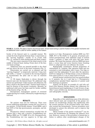
FIGURE 1. Placement of the first K-wire. This wire should create a neck-shaft angle of 130 degrees to assure the correct final neck-
shaft angle (A). It should also be positioned in the correct anteversion, parallel to the reference wire (B). Additional proximal wires
are inserted, at the level of the greater trochanter and at the level of the proximal border of femoral neck in a parallel manner, the
“moving fragment” is marked in white background and black stripes (C).
FIGURE 2. Additional wires are inserted parallel to the chisel
and just above the level of the lesser trochanter.
FIGURE 3. Using multiple drills, a “tunnel” is created in the
moving fragment to accommodate for the plate and to ease its
insertion.
Eidelman et al J Pediatr Orthop Volume 00, Number 00, ’’ 2016
2 | www.pedorthopaedics.com Copyright r 2016 Wolters Kluwer Health, Inc. All rights reserved.
Copyright r 2016 Wolters Kluwer Health, Inc. Unauthorized reproduction of this article is prohibited.
3. border of the femoral neck in a parallel manner. We
coined the bone fragment between these 2 osteotomies as
the “moving fragment,” usually 15 to 25 mm wide
(Fig. 1C, marked in white background and black stripes).
The next step is insertion of the chisel exactly in the
middle of femoral head, halting 10 mm short of the fem-
oral head cartilage.
Additional wires are inserted parallel to the chisel
and just above the level of the lesser trochanter (Fig. 2).
Osteotomies are carried out as planned and the
“moving fragment” is temporarily removed. Using mul-
tiple drills, a “tunnel” is created in the moving fragment
to accommodate the plate and to ease its insertion
(Fig. 3).
A 130 degree blade-plate is driven through the
femoral neck into the femoral head. No additional valg-
ization was performed. Under traction and abduction, the
plate is fixed to the femoral shaft. Tension band wiring is
used for fixation of the greater trochanter. Recently, an
additional solid screw has been used for further stability
of the fixation (Figs. 4A, B).
Postoperative management consists of restricted
weight-bearing for 6 weeks, followed by weight-bearing as
tolerated.
RESULTS
No patients were lost for follow-up. There were
several underlying pathologies causing the coxa brevis in
our patients: 10 had Perthes disease, 4 had DDH, and 4
had AVN of the hip (posttraumatic, postseptic, and idi-
opathic).
Preoperative clinical examination revealed that all
patients had a limp and a positive Trendelenburg test.
Median HHS was 72.5 (IQR: 69 to 83). Postoperatively,
the Trendelenburg test was negative in 14 hips and
positive in 6 hips. Postoperative median HHS was 94.5
(IQR: 89.5 to 96) (Table 1). In general, all patients pre-
sented postoperatively with unlimited range of motion
except 2 patients (3 hips) with prior hip joint incon-
gruence. We found that patients with low HHS had more
pain, a worse limp and worse function hence breaking
down to subgroups does not influence the results.
Radiographic examination showed progression of
osteoarthritis in 3 patients. One operation failed and was
converted to total arthroplasty after 4 years. Two hips re-
quired total hip replacement 10 years after the operation.
LLD was reduced in 17/20 hips (mean 1.3 cm of reduction,
from mean LLD of 2.1 cm to mean LLD of 0.8cm).
Overall patient satisfaction level was good-excellent
in 12 patients, fair in 4 and bad in 2. Postoperative
complications included blade migration (1 patient (and
wire breakage (2 patients).
Figures 5A–D show an example of a girl who suf-
fered from bilateral Perthes disease who underwent bi-
lateral femoral neck lengthening at 13 and 14 years of age.
After 7 years of follow-up, she demonstrated good clinical
and radiographic outcome.
DISCUSSION
Regardless of the underlying cause of the deformity,
short femoral neck and overgrowth of the greater tro-
chanter are morphologic changes that significantly alter
the normal anatomy of the proximal femur and, hence,
alter normal hip biomechanics. Various procedures were
developed to correct this deformity and to improve hip
biomechanics.7–10
Distal transfer of the greater trochanter8,9 can im-
prove hip biomechanics by changing the abductor lever
arm. However, success of this procedure is limited due to
failure to address other morphological changes of the
FIGURE 4. A and B, The plate is fixed to the femoral shaft. Tension band wiring is used for fixation of the greater trochanter and
an additional screw is used for further stability of the fixation.
J Pediatr Orthop Volume 00, Number 00, ’’ 2016 Femoral Neck Lengthening
Copyright r 2016 Wolters Kluwer Health, Inc. All rights reserved. www.pedorthopaedics.com | 3
Copyright r 2016 Wolters Kluwer Health, Inc. Unauthorized reproduction of this article is prohibited.
4. TABLE 1. Patient and Hip Characteristics
Patient
No./Sex Etiology
Age at
Surgery
Follow-
up (y) Side
Trendelenburg
Preoperative
Trendelenburg
Postoperative
LLD
Preoperative
LLD
Postoperative
Harris Hip Score
Preoperative
Harris Hip Score
Postoperative Remarks
1/F Perthes 16 4 R + + 2 0 69 76 THR
2/M Perthes 17 24 R + 2 0.5 83 96
3/M Perthes 20 10 L + + 2 0 70 71 THR
4/M Perthes 20 10 R + + 0 1.5 74 70 THR
5/M Perthes 16 19 R + 2.5 1 76 93 Wire
breakage
6/M Perthes 14 14 L + + 2.5 0 70 90
7/F Perthes 13 11 L + 3 1.5 89 97
8/F DDH 20 8 L + 3 1 79 96
9/M Septic hip 17 8 R + 4 2 60 89 Wire
breakage
10/M Perthes 14 4 R + 3 1 76 93
11/F DDH 13 7 R + 3 0.5 71 96 Blade
migration
12/F DDH 13 4 L + 2 0.5 88 96
13/M Perthes 15 7 R + 2.5 1 69 99
14/M Perthes 16 5 R + 2.3 1 67 97
15/M Posttraumatic
AVN
17 10 L + 3 0 69 88
16/F Idiopathic
AVN
16 3 L + 2.5 1 83 99
17/F Perthes 13 7 R + 2 0.5 89 96
18/F Perthes 14 6 L + 0.5 1.5 89 96
19/M Septic hip 25 4 L + + 0 0 60 88
20/F DDH 18 3 L + 1.9 1.5 61 91
AVN indicates avascular necrosis; DDH, developmental dysplasia of the hip; F, female; L, left; LLD, leg length discrepancy; M, male; R, right; THR, total hip replacement.
Eidelman
et
al
J
Pediatr
Orthop
Volume
00,
Number
00,
’’
2016
4
|
www.pedorthopaedics.com
Copyright
r
2016
Wolters
Kluwer
Health,
Inc.
All
rights
reserved.
Copyright
r
2016
Wolters
Kluwer
Health,
Inc.
Unauthorized
reproduction
of
this
article
is
prohibited.
5. femoral head.8 Valgus intertrochanteric osteotomy11,12
can improve neck-shaft angle and can theoretically im-
prove abductor mechanism, but it does not change rela-
tions between overriding trochanter and the femoral head
nor the relation between the femoral head and the ace-
tabulum. Recently developed relative neck lengthening8,13
may address pathologies of the femoral head and improve
abductor mechanism by distal transfer of the greater
trochanter. Inspection of the acetabulum is an essential
part of this procedure that allows treatment of acetabular
pathology. However, this is a technically demanding
procedure with need of surgical hip dislocation, devel-
opment of an extended retinacular flap with the potential
risk of AVN of the femoral head.
In 1980 Morscher developed the FNLO to address
correction of neck length, LLD, and abductor tension.
Preliminary results were published in 1988, describing 15
patients who underwent the surgery, confirming good
outcome with regard to biomechanics and function.4
In 1990, Morscher published a series of 37 patients
who underwent FNLO, with a mean follow-up period of
8 years (range, 1 to 16 y), and concluded that it improves
leg length, abductor force and gait, provided that there
are no or minimal arthritic changes in the hip joint.14
All patients presented in this study, when evaluated
before surgery, had abnormal gait, positive Trendelen-
burg sign, LLD, pain and fatigue during activity. On
postoperative follow-up, LLD was reduced in all patients,
with mean 1.3 cm of reduction, from mean LLD of 2.1 cm
to mean LLD of 0.8 cm, similar to the results presented by
Morscher in his work.14 Most of the patients had negative
Trendelenburg sign, improved gait pattern and reported
better function, reduced fatigue, and less hip discomfort.
Data were validated by an objective criteria measured by
the HHS (preoperatively and at the latest follow-up) and
demonstrated improvement in all parameters with a me-
dian increase of 17 points (IQR: 8.2 to 27.2).
There were several complications in our series:
hardware failure in 3, wire breakage in 2, and blade mi-
gration requiring revision surgery in 1. There were no
cases of nonunion or infection.
During follow-up, 3 patients presented clinical and ra-
diologic signs of osteoarthritis. One patient demonstrated
rapid deterioration to end-stage osteoarthritis, requiring total
FIGURE 5. A–D, Bilateral femoral neck lengthening in a 13-year-old girl. Seven-year follow-up radiographs demonstrates good
outcome.
J Pediatr Orthop Volume 00, Number 00, ’’ 2016 Femoral Neck Lengthening
Copyright r 2016 Wolters Kluwer Health, Inc. All rights reserved. www.pedorthopaedics.com | 5
Copyright r 2016 Wolters Kluwer Health, Inc. Unauthorized reproduction of this article is prohibited.
6. hip replacement within 4 years. Another patient who un-
derwent bilateral femoral neck lengthening had slow pro-
gression to end-stage osteoarthritis, requiring bilateral total
arthroplasty after 10 years of follow-up. It should be noted
that all 3 hips showed radiographic signs of incongruity and
osteoarthritis before femoral neck lengthening, emphasizing
the importance of having a correct indication for this oper-
ation. The limitation of this study is that despite using the
same surgical technique, the procedure was performed in 3
different medical centers.
CONCLUSIONS
We believe that the Morsher’s operation requires
meticulous preoperative planning and execution. A pre-
requisite for a favorable outcome is congruent hip joints
with no radiographic signs of osteoarthritis.
This long-term follow-up study demonstrates that
Morsher’s operation is a safe and predictable procedure
for restoration of normal hip anatomy and biomechanics
in adolescents and young adults with coxa brevis and
trochanteric overgrowth, combined with mild LLD.
REFERENCES
1. Stevens PM, Coleman SS. Coxa breva: its pathogenesis and a
rationale for its management. J Pediatr Orthop. 1985;5:515–521.
2. Iwersen LJ, Kalen V, Eberle C. Relative trochanteric overgrowth
after ischemic necrosis in congenital dislocation of the hip. J Pediatr
Orthop. 1989;9:381–385.
3. Schneidmueller D, Carstens C, Thomsen M. Surgical treatment of
overgrowth of the greater trochanter in children and adolescents.
J Pediatr Orthop. 2006;26:486–490.
4. Buess P, Morscher E. Osteotomy to lengthen the femur neck with
distal adjustment of the trochanter major in coxa vara after hip
dislocation. Orthopade. 1988;17:485–490.
5. Harris WH. Etiology of osteoarthritis of the hip. Clin Orthop.
1986;213:20–33.
6. d’Entremont AG, Cooper AP, Johari A, et al. What clinimetric
evidence exists for using hip-specific patient-reported outcome
measures in pediatric hip impingement? Clin Orthop Relat Res. 2015;
473:1361–1367.
7. Anderson LA, Erickson JA, Severson EP, et al. Sequelae of Perthes
disease: treatment with surgical hip dislocation and relative femoral
neck lengthening. J Pediatr Orthop. 2010;30:758–766.
8. Garrido IM, Moltó FJ, Lluch DB. Distal transfer of the greater
trochanter in acquired coxa vara: clinical and radiographic results.
J Pediatr Orthop B. 2003;12:38–43.
9. Givon U, Schindler A, Ganel A, et al. Distal transfer of the greater
trochanter revisited: long-term follow-up of nine hips. J Pediatr
Orthop. 1995;15:346–348.
10. Standard SC. Treatment of coxa brevis. Orthop Clin North Am.
2011;42:373–387.
11. Notzli HP, Chou LB, Ganz R. Open-reduction and intertrochan-
teric osteotomy for osteonecrosis and extrusion of the femoral head
in adolescents. J Pediatr Orthop. 1995;15:16–20.
12. Santore RF. Intertrochanteric osteotomy for osteonecrosis. Semin
Arthroplasty. 1991;2:208–213.
13. Ganz R, Huff TW, Leunig M. Extended retinacular soft-tissue flap
for intra-articular hip surgery: surgical technique, indications, and
results of application. Instr Course Lect. 2009;58:241–255.
14. Hasler CC, Morscher EW. Femoral neck lengthening osteotomy
after growth disturbance of the proximal femur. J Pediatr Orthop B.
1999;8:271–275.
Eidelman et al J Pediatr Orthop Volume 00, Number 00, ’’ 2016
6 | www.pedorthopaedics.com Copyright r 2016 Wolters Kluwer Health, Inc. All rights reserved.
Copyright r 2016 Wolters Kluwer Health, Inc. Unauthorized reproduction of this article is prohibited.