Stem cells Used to Develop Mini Human Brain & Stem Cells for Spinal Cord Inju...Ankita-rastogi
Stem cell derived from human umbilical cord or bone marrow improves mobility with spinal cord injuries providing the first physical evidence that the therapeutic use of these cells can help restore motor skills lost from acute spinal cord tissue damage. For more information visit: http://www.cryobanksindia.com/moms-corner/case-studies/
Electrophysiology of Human Native Receptors in Neurological and Mental DisordersInsideScientific
Dr. Agenor Limon presents research integrating functional metrics with large anatomical, transcriptomic, and proteomic datasets to evaluate the relationship between synaptic E/I ratio and behavioral abnormalities across postmortem intervals and brain banks.
Alterations in synaptic function have been found in transcriptomic, genetic, and proteomic studies of neurological and mental disorders. Clinical and preclinical studies suggest that synaptic dysfunction and behavioral abnormalities in disorders like Alzheimer’s disease and schizophrenia may be mechanistically linked to the emergence of imbalances between excitatory (E) and inhibitory (I) receptors. However, until recently, the electrophysiological E/I synaptic ratio had only been measured in animal models.
Using pioneering methods developed in the lab including reactivation and microtransplantation of synaptic receptors from frozen human brains, Dr. Agenor Limon’s research team has obtained electrophysiological metrics of global synaptic E/I ratios in cortical brain regions of subjects that were affected by Alzheimer’s Disease and synaptic measurements in schizophrenia.
In this webinar, Dr. Agenor Limon will present recent research integrating functional metrics with large anatomical, transcriptomic, and proteomic datasets to evaluate the relationship between synaptic E/I ratio and behavioral abnormalities across postmortem intervals and brain banks.
Key Topics Include:
- Understand the global synaptic excitation to inhibition ratio between excitatory and inhibitory synaptic receptors determined from reactivated frozen human brain tissue
- Understand the relationship between electrophysiological metrics of receptor function and multi-omic data in neurodegenerative disorders
- Understand the role of deviations of the excitation to inhibition ratio with clinical presentation in Alzheimer’s disease
The Brain as a Whole: Executive Neurons and Sustaining Homeostatic GliaInsideScientific
Carl Petersen and Alexei Verkhratsky share their research on homeostatic neuroglia and imaging of neuronal network function. This webinar is brought to you by APS’ new journal, Function, and part of their Physiology in Focus learning series.
During this exclusive live webinar, Carl Petersen and Alexei Verkhratsky discuss astrocyte-mediated homeostatic control of the central nervous system, and how optical and 2-photon microscopy can be used for functional neuroimaging.
Imaging Neuronal Function
Carl Petersen, PhD
Highly dynamic and spatially distributed neuronal circuits in the brain control mammalian behavior. Through technological advances, optical measurements of neuronal function can now be carried out in behaving mice at multiple scales. Wide-field imaging allows the dynamic interactions between different brain areas to be studied as sensory information is processed and transformed into behavioral output. Within a brain region, two-photon microscopy can be used to image the neuronal network activity with cellular resolution allowing different types of projection neurons to be distinguished. Together optical methods provide versatile tools for causal mechanistic understanding of neuronal network function in mice.
Astrocytes: indispensable neuronal supporters in sickness and in health
Alexei Verkhratsky, MD, PhD, DSc
The nervous system is composed of two arms: the executive neurons and the homeostatic neuroglia. The neurons require energy, support, and protection, all of which is provided by the neuroglia. Astrocytes, the principal homeostatic cells of the brain and spinal cord, are tightly integrated into the neural networks and act within the context of the neural tissue. As astrocytes control the homeostasis of the central nervous system at all levels of organization, from the molecular to the whole organ level, we can begin to define and understand brain vulnerabilities to aging and diseases.
Monitoring neural activities by optical imagingMd Kafiul Islam
Monitoring neural activities by optical imaging along with the use of genetic modification provides better spatio-temporal resolution to study single neural firing and hence very useful in understanding the neural process and dynamics. This is just a glimpse of few articles reported their outcome of such imaging.
Functional Ultrasound (fUS) Imaging in the Brain of Awake Behaving MiceInsideScientific
To watch the webinar, visit:
https://insidescientific.com/webinar/functional-ultrasound-imaging-brain-awake-behaving-mice-neurotar-iconeus
Functional ultrasound (fUS) imaging is a new kid on the block in neuroimaging. It combines high spaciotemporal resolution with deep tissue penetration, which enables non-invasive whole-brain imaging in mice.
This exciting new technology complements and extends classical imaging modalities: it enables more straightforward, unobstructed and non-invasive functional measurements in mouse models of CNS diseases. Sensitive to changes in cerebral blood volume, fUS imaging is used to characterize brain networks with functional connectivity analysis and to measure the responses to sensory stimuli and pharmacological challenges.
fUS imaging performed in the brain of awake mice removes the biases and artifacts associated with the use of general anesthesia, which is no longer a “necessary evil” of translational imaging. Besides that: fUS imaging in awake mice allows integrating functional imaging with behavioral readouts starting from open field locomotion tracking to maze navigation and sociability studies.
In this webinar, you will learn:
– Functional ultrasound (fUS) imaging methodology
– How translational fUS neuroimaging helps to advance basic neuroscience research and preclinical drug discovery
– The main advantages and limitations of using functional ultrasound compared to other techniques such as BOLD fMRI
– The benefits of imaging in awake, head-restrained but otherwise freely moving mice
– Imaging functional activation, connectivity and pharmacologically-induced changes in awake and behaving mice
– How to combine fUS imaging with behavioral observation
Studying Epilepsy in Awake Head-Fixed Mice Using Microscopy, Electrophysiolog...InsideScientific
Epilepsy research employs sophisticated research methods such as fluorescence optical imaging and optogenetics, as well as novel electrophysiological techniques, to address unresolved questions about seizure generation and propagation on the cellular and circuitry levels. Since epilepsy research is most relevant when performed in non-anesthetized mice, it requires specialized tools that ensure stable head fixation during high-precision imaging and recordings.
In this webinar, Dr. Anthony Umpierre (Prof. LongJun Wu group, Mayo Clinic, USA) and Prof. Rob Wykes (UCL, UK) present their research on microglial calcium signaling and epileptic networks carried out in awake head-fixed mice. In addition to sharing exciting new findings, the presenters address the challenges of working with awake mice.
Key topics will include…
- Mesoscopic investigations of seizure dynamics and propagation using widefield calcium imaging
- Generating full-bandwidth electrophysiological recordings enabled by graphene micro-transistors to detect spreading depolarizations and seizures
- On-demand optogenetic induction of spreading depolarizations to investigate pharmacological suppression in the awake brain
- The impact of acute versus chronic window preparations on microglial calcium activity
- The use of genetically encoded calcium indicators to study calcium dynamics in microglia
- The effects of bi-directional shifts in neuronal activity caused by kainate-triggered status epilepticus and isoflurane anesthesia on microglial calcium
Making Optical and Electrophysiological Measurements in the Brain of Head-Fix...InsideScientific
A growing number of researchers are moving from reduced preparations such as dissociated cultured neurons or brain slices, to experimentation in live animals - in vivo - using advanced methods such as two-photon microscopy or combined optogenetics and patch-clamp recordings. In order to immobilize the animal during these challenging applications general anesthesia is often administered; however, the use of anesthetics greatly distorts brain function.
Is there a better way?
In this exclusive webinar sponsored by Neurotar Ltd, leading experts in the technology will discuss methodology, best-practices and show attendees how to immobilize the rodent’s head without restraining its body using the Mobile HomeCage™. The result is a controlled research environment for studying brain function in awake, freely-moving subjects with no stress to the animal. Discussion around how this technique can be applied to the study of neuronal plasticity, neurodegeneration, addiction, brain trauma and other pathophysiological conditions in longitudinal experiments will be included. Furthermore, presenters will demonstrate how this methodology is best combined with microscopy and electrophysiology techniques – all in vivo.
Endocytosis and Endosome Trafficking: Roles in Coronavirus Uptake and Cell Si...InsideScientific
To learn more and watch the webinar, visit:
https://insidescientific.com/webinar/endocytosis-endosome-trafficking-coronavirus-uptake-cell-signaling-aps
Endocytosis and Endosome Trafficking: Roles in Coronavirus Uptake and Cell Signaling
Click to share on Twitter (Opens in new window)Click to share on LinkedIn (Opens in new window)Click to share on Facebook (Opens in new window)Click to share on Email (Opens in new window)
ON DEMAND
Ole Petersen, Roop Mallik and Erwin Neher share late-breaking research looking at endocytosis and calcium signaling in the context of SARS-CoV-2, organelle transport and calcium imaging. This webinar is brought to you by APS’ new journal, Function, and part of their Physiology in Focus learning series.
REGISTER
During this exclusive live webinar, Ole Petersen, Roop Mallik and Erwin Neher discuss how the COVID-19 virus uses receptor-mediated endocytosis to gain entry into host cells, how motor proteins guide endosomes and phagosomes from the cell surface to lysosomes, and how intracellular calcium buffering can be used to modulate cell signaling and calcium imaging.
Endocytic Uptake of SARS-CoV-2: The Critical Roles of pH, Ca2+ and NAADP
Ole Petersen, CBE, FRS
Very recent work shows that SARS-CoV-2 enters our cells through receptor-mediated endocytosis, dependent on an endosomal bafilomycin-sensitive proton pump as well as two-pore channels (TPCs). Physiological intracellular Ca2+ signals, mediated by the messenger nicotinic acid adenine dinucleotide phosphate (NAADP), depend on the very same proton pump and TPCs. Two hitherto completely separate research fields, namely molecular virology and cellular Ca2+ signaling physiology are now coming together, creating exciting new research opportunities.
Trafficking of Endosomes and Phagosomes: Geometry, Force and Cholesterol
Roop Mallik, PhD
Uptake of material from the external world by endocytosis/phagocytosis supplies nutrients to cells, and is also critical for cell signaling. The journey of endosomes/phagosomes begins at the cell periphery and ends at lysosomes near the cell center. I will discuss how the balance of forces generated by antagonistic motor proteins guides this journey, and how lipids are emerging as a master-controller of this balance.
Calcium Buffering in Endo- and Exocytosis Studies
Erwin Neher, FRS
Researchers use calcium-chelators (buffers) to manipulate levels of free intracellular calcium concentration ([Ca2+]i) and to shape Calcium signals. Unlike pH buffers, which are used to strictly control pH levels, calcium buffers inside a living cell may not influence the steady-state level of [Ca2+]i at all, but rather slow-down [Ca2+]i-changes induced either endogenously or by the experimenter. Such properties and their consequences on Ca2+-imaging will be discussed.
Cerebral Open Flow Microperfusion (cOFM) for in vivo Cerebral Fluid Sampling ...InsideScientific
Cerebral open flow microperfusion (cOFM) is a minimally invasive, in vivo sampling technology that allows continuous long-term sampling of cerebral fluid in living animals. The decisive advantage of cOFM is that the cOFM probe is membrane–free and comprises macroscopic openings which offer the possibility for a multitude of applications without restriction regarding size, lipophilicity or protein binding effects of the collected substances. The cOFM probe is designed to elicit minimal tissue reactions and allows for reconstitution of the blood-brain barrier (BBB). Thus, cOFM can sample cerebral fluids in living and freely moving animals with intact BBB.
During this webinar, Dr. Joanna Hummer introduces cOFM and presents how cOFM is used as an in vivo sampling technology in neuroscience for drug development.
Dr. Florie Le Prieult, presents data her team collected using cOFM during a pharmacokinetic studies of therapeutic antibodies. Her study includes head-to-head comparison of cOFM and microdialysis.
Sensorimotor Network Development During Early Postnatal Life in the Awake and...InsideScientific
In the last decades, electrophysiological and imaging-based approaches provided significant new insights into the mechanisms of neuronal development. Nevertheless, many important questions remain unanswered. How does the fine control of a motor output develop? How does sensorimotor integration in the early and subsequent phases of brain development shape behavior? How does sensorimotor development evolve in awake and sleeping states? What role do myoclonic twitches play in this process?
Answering these questions requires performing high-precision tests in the brain of non-anesthetized animals across sleep and wake during the early stages of their postnatal development. Such tests require head-fixation apparatus suitable for neonatal and juvenile rodents. The Mobile HomeCage combines a stable head-fixation with an air-lifted cage that closely resembles laboratory rodents’ natural habitat – an optimal platform for studying early postnatal brain development.
In this webinar, Dr. Cavaccini (Prof. Karayannis’s lab at the Brain Research Institute, University of Zurich) and Dr. Dooley (Prof. Blumberg’s lab at the University of Iowa), share their insights into the development of rodent sensorimotor neuronal circuits during early postnatal life. They elucidate the cortical and subcortical mechanisms involved in the development of sensorimotor circuitry during wakefulness (in a mouse model) and REM sleep (in a rat model).
Key Takeaways
Dr. Anna Cavaccini:
- Anatomical and functional changes occur at the striatal level before and after the onset of different sensory modalities
- Locomotor activity changes throughout early development, and it correlates with striatal function
- Sensory information coming from whiskers affects locomotion and striatal function before and after the onset of different sensory modalities
Dr. James Dooley:
- Myoclonic twitches in REM sleep continue to trigger cortical and thalamic activity beyond the early postnatal period
- Twitch-related thalamic activity is spatiotemporally refined by the third postnatal week
- Motor thalamus activity reflects an internal model of movement produced by twitches and is dependent on the cerebellar output
Single-Cell Electrophysiology and 2-Photon Imaging in Awake Mice with 2D-Loco...InsideScientific
In this webinar sponsored by Neurotar, experts present their research utilizing the Mobile HomeCage®, an experimental tool which ensures the stability required for high-precision neurophysiological techniques while allowing mice to navigate and explore their environment.
Case Study #1:
Dr. Sarah Stuart and Dr. Jon Palacios-Filardo of the University of Bristol present their studies combining analysis of goal-directed behavior with whole-cell recordings from the hippocampus of awake mice. The researchers share useful tips for the surgery protocol and for adjusting the head fixation angle in order to facilitate mouse motility and exploratory behavior.
Case Study #2:
Dr. Alexander Dityatev and Weilun Sun from the German Center for Neurodegenerative Diseases (DZNE) discuss 2-photon imaging of fluorescently labeled microglia in vivo in the context of neurodegenerative disease. They also present their recent data on the effects of different anesthetics on the microglial response to localized laser injury.
Case Study #3:
Dr. Norbert Hájos from the Hungarian Academy of Sciences presents his lab’s research into the amygdala’s role in reward-driven behavior. He shares the challenges of making single-unit recordings using silicon probes during mouse locomotion and subsequent morphological identification of active neurons in the amygdala.
Key topics covered during this webinar include:
- Requirements for stable single-cell recordings and 2-photon imaging in behaving mice
- Challenges of combining high-precision techniques with behavioral research
- Methodological considerations for improving exploratory behavior in head-fixed mice
- Quantitative analysis of microglial function using 2-photon microscopy in awake mice
- Recording neuronal activity in the amygdala of awake mice followed by morphological identification of recorded neurons
In Vitro Neuroscience Services-Creative Biolabscailynnjohnson
Creative Biolabs' dedicated team of neuroscience CRO scientists want the same thing as you do: to find a cure for the devastating diseases of the central nervous system.
https://neuros.creative-biolabs.com/
lecture 5 from a college level introduction to psychology course taught Fall 2011 by Brian J. Piper, Ph.D. (psy391@gmail.com) at Willamette University, includes Golgi, Cajal, parts of the neuron, action potentials, synapse, neurotransmitters, agonist, antagonist, parts of the nervous system
Nervous System -Autonomic Nervous System-Neurons -Ganglia - Nerves Copy.Home
The nervous system is the body's communication network, coordinating and regulating all bodily functions. Comprising the central nervous system (CNS) and peripheral nervous system (PNS), it consists of neurons, specialized cells transmitting electrical and chemical signals. The CNS, consisting of the brain and spinal cord, interprets and processes information. The PNS extends from the CNS, transmitting signals between the brain, spinal cord, and the rest of the body. Sensory neurons detect stimuli, while motor neurons control muscle movement. This intricate system enables sensory perception, voluntary and involuntary actions, and regulates bodily processes, ensuring homeostasis and facilitating responses to the environment.
The presentation focuses on one of the important aspects of Neurophysiology-- The sesnsorimotor integration for planning and execution of movement.
It highlights on the brain regions associated with motor functions, the crosstalk between association areas, hierarchical levels of movement execution and the diseases related to it.
PowerPoint giving a summary on research in stem cells (brief historical overview), and the explanatory component of the papers which changed the game of stem cell research Yamanka's Nuclear Reprogramming.
Powerpoint accompanying research paper on the Teratogenic effects of Fipronil (GABAergic antagonist) on the neurological development of zebrafish (as model of vertebrates).
Abstract:
Long‐term potentiation (LTP) and Long‐term depression (LTD) are the best understood mechanisms by which biological systems achieve the sculpting of neural pathways necessary for learning and memory. Foundational to the understanding of LTP and LTD is the model of Hebbian learning. Although research has demonstrated that neural networks can be developed and trained in silico to mimic the computational function of neural ensembles, the development of a truly realistic model remains elusive. We construct a simulation of Hebbian learning implementing the Hopfield network model. We then propose an experimental setup that will use the light‐directed electrical stimulation of neurons cultured on silicon wafers in order to gain information on the parameters of synaptic strength and firing frequencies for all neurons in the system simultaneously (determinants of LTP and LTD). This will allow the parallel running of neural networks in the in vivo and in silico setting, allowing statistical comparison between the biological model and computer simulation and additional refinement to create a more biologically relevant computational model.
Keywords: short and long-term potentiation (LTP), short and long-term depression (LTD), neural plasticity, Hebbian learning, Hopfield Network, parallel neural network training, light-directed electrical stimulation (LDES).
Paper Biology 280 S Minireview Advances In Cancer Detection And TherapeuticsJoshua Mendoza-Elias
Abstract:
Nanotechnology is a field that has made significant advances in the engineering of technologies that allow earlier detection and treatment of disease – specifically cancer. In the modern era, these technologies define the emerging field of nanomedicine and seek to redefine detection and treatment with the promise of more effective, sensitive and cost effective high throughput systems. Compared to conventional treatment options, these next generation cancer therapeutics also seek to overcome the invasiveness of surgery, chemotherapy, radiotherapy, immunotherapy, and hormonal therapy. These technologies include nanoshells, quantum-dots, ultraoxide particles, DNA microarrays, liposomes, dendrimers, and fullerines. Highlighted in this paper, microwires and microcantilevers (“biofinger”) show promise in becoming multiplex platforms (“Labs on a chip”) that detect a variety of biological markers at low concentration in real time. In addition, DNA based diagnostic computer constructs (DNAdc) that release therapeutic nucleic acid sequences in a gene expression specific manner will be highlighted as well. The potential for a wide range of clinical applications to disease, including cancer, makes a basic understanding of the field of nanomedicine important to the biomedical sciences. In addition, nanomedicine will have a huge impact in developing a repertoire of nano-based therapeutics for other diseases.
Keywords: “Biofinger”-lab on a chip, DNA diagnostic computer construct (DNAdc), DNA microarrays, nanoshells, quantum-dots, ultraoxide particles, liposomes, dendrimers, fullerines, single wall carbon nanotubes (SWNT).
Paper Biology 205 L The Teratogenic Effects Of Fipronil On Zebrafish Neurolog...Joshua Mendoza-Elias
Abstract:
Growing research has implicated Fipronil as a teratogen in the development of chordates. Fipronil is a pesticide that acts to disrupt and inhibit normal nerve activity. The result is excessive nerve stimulation caused by overstimulation of gamman-aminobuytric (GABA) gated chloride ion channels. Experiments using zebrafish (Danio rerio) indicate Fipronil is teratogenic at concentrations above 0.7 μM. In this study, the effect of increasing concentrations (0.7 μM, 1.1 μM, 2.3 μM) of Fipronil on neurological development were examined. Our results showed that within six hours of GABA-receptor expression (36 hours-high pec) there was neurological damage as indicated by an abnormal behavioral escape touch response (resembling the accordion class phenotype), abnormal morphology (ventrally curved long fin), and damage to neurological tissue (notochord damage concentrated in the dorsal area of the trunk-tail interface). Within 48 hours (long-pec) damage was more extreme as observed in behavior, long tail morphology, and damage to neurological tissue. Furthermore, distinct regions of necrotic tissue were visible, accompanied by aberrant circulation. This data demonstrates Fipronil teratogenicity increases with exposure and concentration and is correlated with increasing damage to the notochord during development. This vertebrate study may have implications for diseases such as fetal alcohol syndrome (FAS) and autism on the phenotypic impact of Fipronil on the development of GABAergic neural pathways.
Keywords: zebrafish, fipronil, teratogen, GABA channels, notochord, necrosis.
Presentation of our data and results from the first antibody staining of the new allele, evePJ4 (created by Dr. Amy Bejsovec\'s lab) and the wgNE2 allele.
Factory Supply Best Quality Pmk Oil CAS 28578–16–7 PMK Powder in Stockrebeccabio
Factory Supply Best Quality Pmk Oil CAS 28578–16–7 PMK Powder in Stock
Telegram: bmksupplier
signal: +85264872720
threema: TUD4A6YC
You can contact me on Telegram or Threema
Communicate promptly and reply
Free of customs clearance, Double Clearance 100% pass delivery to USA, Canada, Spain, Germany, Netherland, Poland, Italy, Sweden, UK, Czech Republic, Australia, Mexico, Russia, Ukraine, Kazakhstan.Door to door service
Hot Selling Organic intermediates
TEST BANK for Operations Management, 14th Edition by William J. Stevenson, Ve...kevinkariuki227
TEST BANK for Operations Management, 14th Edition by William J. Stevenson, Verified Chapters 1 - 19, Complete Newest Version.pdf
Ethanol (CH3CH2OH), or beverage alcohol, is a two-carbon alcohol
that is rapidly distributed in the body and brain. Ethanol alters many
neurochemical systems and has rewarding and addictive properties. It
is the oldest recreational drug and likely contributes to more morbidity,
mortality, and public health costs than all illicit drugs combined. The
5th edition of the Diagnostic and Statistical Manual of Mental Disorders
(DSM-5) integrates alcohol abuse and alcohol dependence into a single
disorder called alcohol use disorder (AUD), with mild, moderate,
and severe subclassifications (American Psychiatric Association, 2013).
In the DSM-5, all types of substance abuse and dependence have been
combined into a single substance use disorder (SUD) on a continuum
from mild to severe. A diagnosis of AUD requires that at least two of
the 11 DSM-5 behaviors be present within a 12-month period (mild
AUD: 2–3 criteria; moderate AUD: 4–5 criteria; severe AUD: 6–11 criteria).
The four main behavioral effects of AUD are impaired control over
drinking, negative social consequences, risky use, and altered physiological
effects (tolerance, withdrawal). This chapter presents an overview
of the prevalence and harmful consequences of AUD in the U.S.,
the systemic nature of the disease, neurocircuitry and stages of AUD,
comorbidities, fetal alcohol spectrum disorders, genetic risk factors, and
pharmacotherapies for AUD.
Couples presenting to the infertility clinic- Do they really have infertility...Sujoy Dasgupta
Dr Sujoy Dasgupta presented the study on "Couples presenting to the infertility clinic- Do they really have infertility? – The unexplored stories of non-consummation" in the 13th Congress of the Asia Pacific Initiative on Reproduction (ASPIRE 2024) at Manila on 24 May, 2024.
These simplified slides by Dr. Sidra Arshad present an overview of the non-respiratory functions of the respiratory tract.
Learning objectives:
1. Enlist the non-respiratory functions of the respiratory tract
2. Briefly explain how these functions are carried out
3. Discuss the significance of dead space
4. Differentiate between minute ventilation and alveolar ventilation
5. Describe the cough and sneeze reflexes
Study Resources:
1. Chapter 39, Guyton and Hall Textbook of Medical Physiology, 14th edition
2. Chapter 34, Ganong’s Review of Medical Physiology, 26th edition
3. Chapter 17, Human Physiology by Lauralee Sherwood, 9th edition
4. Non-respiratory functions of the lungs https://academic.oup.com/bjaed/article/13/3/98/278874
Pulmonary Thromboembolism - etilogy, types, medical- Surgical and nursing man...VarunMahajani
Disruption of blood supply to lung alveoli due to blockage of one or more pulmonary blood vessels is called as Pulmonary thromboembolism. In this presentation we will discuss its causes, types and its management in depth.
Tom Selleck Health: A Comprehensive Look at the Iconic Actor’s Wellness Journeygreendigital
Tom Selleck, an enduring figure in Hollywood. has captivated audiences for decades with his rugged charm, iconic moustache. and memorable roles in television and film. From his breakout role as Thomas Magnum in Magnum P.I. to his current portrayal of Frank Reagan in Blue Bloods. Selleck's career has spanned over 50 years. But beyond his professional achievements. fans have often been curious about Tom Selleck Health. especially as he has aged in the public eye.
Follow us on: Pinterest
Introduction
Many have been interested in Tom Selleck health. not only because of his enduring presence on screen but also because of the challenges. and lifestyle choices he has faced and made over the years. This article delves into the various aspects of Tom Selleck health. exploring his fitness regimen, diet, mental health. and the challenges he has encountered as he ages. We'll look at how he maintains his well-being. the health issues he has faced, and his approach to ageing .
Early Life and Career
Childhood and Athletic Beginnings
Tom Selleck was born on January 29, 1945, in Detroit, Michigan, and grew up in Sherman Oaks, California. From an early age, he was involved in sports, particularly basketball. which played a significant role in his physical development. His athletic pursuits continued into college. where he attended the University of Southern California (USC) on a basketball scholarship. This early involvement in sports laid a strong foundation for his physical health and disciplined lifestyle.
Transition to Acting
Selleck's transition from an athlete to an actor came with its physical demands. His first significant role in "Magnum P.I." required him to perform various stunts and maintain a fit appearance. This role, which he played from 1980 to 1988. necessitated a rigorous fitness routine to meet the show's demands. setting the stage for his long-term commitment to health and wellness.
Fitness Regimen
Workout Routine
Tom Selleck health and fitness regimen has evolved. adapting to his changing roles and age. During his "Magnum, P.I." days. Selleck's workouts were intense and focused on building and maintaining muscle mass. His routine included weightlifting, cardiovascular exercises. and specific training for the stunts he performed on the show.
Selleck adjusted his fitness routine as he aged to suit his body's needs. Today, his workouts focus on maintaining flexibility, strength, and cardiovascular health. He incorporates low-impact exercises such as swimming, walking, and light weightlifting. This balanced approach helps him stay fit without putting undue strain on his joints and muscles.
Importance of Flexibility and Mobility
In recent years, Selleck has emphasized the importance of flexibility and mobility in his fitness regimen. Understanding the natural decline in muscle mass and joint flexibility with age. he includes stretching and yoga in his routine. These practices help prevent injuries, improve posture, and maintain mobilit
These lecture slides, by Dr Sidra Arshad, offer a quick overview of physiological basis of a normal electrocardiogram.
Learning objectives:
1. Define an electrocardiogram (ECG) and electrocardiography
2. Describe how dipoles generated by the heart produce the waveforms of the ECG
3. Describe the components of a normal electrocardiogram of a typical bipolar leads (limb II)
4. Differentiate between intervals and segments
5. Enlist some common indications for obtaining an ECG
Study Resources:
1. Chapter 11, Guyton and Hall Textbook of Medical Physiology, 14th edition
2. Chapter 9, Human Physiology - From Cells to Systems, Lauralee Sherwood, 9th edition
3. Chapter 29, Ganong’s Review of Medical Physiology, 26th edition
4. Electrocardiogram, StatPearls - https://www.ncbi.nlm.nih.gov/books/NBK549803/
5. ECG in Medical Practice by ABM Abdullah, 4th edition
6. ECG Basics, http://www.nataliescasebook.com/tag/e-c-g-basics
Explore natural remedies for syphilis treatment in Singapore. Discover alternative therapies, herbal remedies, and lifestyle changes that may complement conventional treatments. Learn about holistic approaches to managing syphilis symptoms and supporting overall health.
Report Back from SGO 2024: What’s the Latest in Cervical Cancer?bkling
Are you curious about what’s new in cervical cancer research or unsure what the findings mean? Join Dr. Emily Ko, a gynecologic oncologist at Penn Medicine, to learn about the latest updates from the Society of Gynecologic Oncology (SGO) 2024 Annual Meeting on Women’s Cancer. Dr. Ko will discuss what the research presented at the conference means for you and answer your questions about the new developments.
Report Back from SGO 2024: What’s the Latest in Cervical Cancer?
EGR 183 Bow Tie Presentation
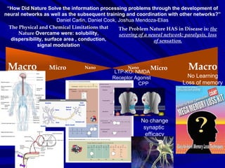
1. “ How Did Nature Solve the information processing problems through the development of neural networks as well as the subsequent training and coordination with other networks?” Daniel Carlin, Daniel Cook, Joshua Mendoza-Elias Macro Micro Nano Macro Micro The Physical and Chemical Limitations that Nature Overcame were: solubility, dispersibility, surface area , conduction, signal modulation The Problem Nature HAS in Disease is: the severing of a neural network: paralysis, loss of sensation. Nano LTP-KO: NMDA Receptor Agonist CPP No change synaptic efficacy No Learning Loss of memory
2. “ How Did Nature Solve the information processing problems through the development of neural networks as well as the subsequent training and coordination with other networks?” Daniel Carlin, Daniel Cook, Joshua Mendoza-Elias Macro Micro Nano Macro Micro The Physical and Chemical Limitations that Nature Overcame were: solubility, dispersibility, surface area , conduction, signal modulation The Problem Nature HAS in Disease is: the severing of a neural network: paralysis, loss of sensation. Nano
3.
4.
5. Component Design Nervous System by function CNS (encased in bone) PNS Brain Spinal Chord Somatic System Autonomic System Parasympathetic Enteric Sympathetic Movement Coordination Receive external stimuli 8 cervical 12 thoraic 5 lumbar 5 sacral 11 coccygeal Cerrvical Spinal: C1-C4 Brachial Plexus: C5-T1 Lateral Chord: C5-C6 Posterior Chord: C6-C8 Medial Chord: C7-T1 Rest Relaxation Digestion Flight/Fight
9. Material Selection: Neurons Sensory Neuron - Converts external stimuli to electrical signals - Chemoreceptors (e.g. olfactory signals) - Mechanoreceptors (e.g. joint position detection) Motor Neuron: - Stimulated by interneurons (small feedback loops or from ANS/PNS) - Activates effectors (glands, muscles, ...) Interneuron: - Data processing, stimulated by: - sensory neurons - other interneurons or both. - many unknown types remain A: Cortical pyramidal cell - primary excitatory neurons of cerebral cortex B: Purjinke cell of cerebellum. Transmit output of cerebral cortex C: Stellate cell - provides inhibitory input to cerebral cortex Example neurons from the brain: In Out Process Basic building blocks of nervous system are neurons. Hundreds of different types, many uncatalogued. Three main categories:
10. Material Selection: Glia in the CNS - Structural & metabolic support (feed neurons) - Transmitter reuptake: express transporters for neurotransmitters - Regulate ion concentrations (potassium) - Act as immune cells of the nervous system - Responsible for myelin sheathing of axons - Single oligodendrocyte myelinates 10-15 axons - Modulates axon conduction speed Astrocytes Microglial cells Oligodendrocyte Approx. 3:1 ratio of glial cells to neurons in the brain Modulate signal propagation and neurotransmitter uptake at the synaptic cleft Provide scaffold for neural development, help in injury recovery
11.
12. Materials Performance http://vadim.oversigma.com/MAS862/Project.html Number of neurons (adult)* 20,000,000,000 - 50,000,000,000 Number of neurons in cerebral cortex (adult)* about 20,000,000,000 (some sources have incorrect number 8,000,000) Number of synapses (adult) 1014 (2,000-5,000 per neuron) Weight Birth 0.3 kg, 1 y/o 1 kg, puberty 1.3 kg, adult 1.5 kg Power consumption (adult) 20-40 Watts (0.5-4 nW/neuron) Percentage of body 2% weight, 0.04-0.07% cells, 20-44% power consumption Genetic code influence 1 bit per 10,000-1,000,000 synapses Atrophy/death of neurons 50,000 per day (between ages 20 and 75) Sleep requirement (adult) average 7.5 hours/day or 31% Normal operating temperature 37 ± 2°C Maximum firing frequency of neuron 250-2,000 Hz (0.5-4 ms intervals) Signal propagation speed inside axon 90 m/s sheathed, <0.1 m/s unsheathed Processing of complex stimuli 0.5s or 100-1,000 firings
13.
14.
15.
16. “ How Did Nature Solve the information processing problems through the development of neural networks as well as the subsequent training and coordination with other networks?” Daniel Carlin, Daniel Cook, Joshua Mendoza-Elias Macro Micro Nano Macro Micro The Physical and Chemical Limitations that Nature Overcame were: solubility, dispersibility, surface area , conduction, signal modulation The Problem Nature HAS in Disease is: the severing of a neural network: paralysis, loss of sensation. Nano
17. Design Methodology: Define the function component to carry a load Material Selection Component Design Tentative component design Approximate stress analysis Tentative choice of material Assemble Materials Data Analysis of Materials Performance iterate Detailed Specifications and Design Choice of Production Methods Prototype Testing Establish Production Further Development iterate iterate iterate
29. Detailed Specifications & Design - Neurochemical Transport Cell membrane Small molecules Large molecules Active transport Targeting AA, peptide frags, Ions, H 2 O FA, aggregate complexes Clathrin COPI/II pH Signal Transduction ATP Kinase Endocytosis Secretion Processing Diffusion
30. Choice of Production Methods Cells arise from previous cells. Genome encoded functions: -juxtracrine & paracrine signaling Germline eventually develops tissues and cellular domains. Neural tissues develop from ectoderm Ideally, we would like to work with neural precursor cells that have undetermined cell fates. .
31.
32. “ How Did Nature Solve the information processing problems through the development of neural networks as well as the subsequent training and coordination with other networks?” Daniel Carlin, Daniel Cook, Joshua Mendoza-Elias Macro Micro Nano Macro Micro The Physical and Chemical Limitations that Nature Overcame were: solubility, dispersibility, surface area , conduction, signal modulation The Problem Nature HAS in Disease is: the severing of a neural network: paralysis, loss of sensation. Nano
34. Neurotransmitters Release Function Functions of Neurotransmitters Neurotransmitters are chemicals that are used to relay, amplify and modulate signals between a neuron and another cell. According to the prevailing beliefs of the 1960s, a chemical can be classified as a neurotransmitter if it meets the following conditions: (i.) It is synthesized endogenously, that is, within the presynaptic neuron; (ii.) It is available in sufficient quantity in the presynaptic neuron to exert an effect on the postsynaptic neuron; (iii.) Externally administered, it must mimic the endogenously-released substance; and (iv.) A biochemical mechanism for inactivation must be present. Functions of Vesicles A vesicle is a relatively small and enclosed compartment, separated from the cytosol by at least one lipid bilayer. If there is only one lipid bilayer, they are called unilamellar vesicles; otherwise they are called multilamellar. Vesicles store, transport, or digest cellular products and waste. Functions of Protein Pumps Ion protein pumps establish the electrochemical gradient required for the transmission of action potentials. Functions of Receptors Receptors are responsible for binding neurochemicals and causing propogation of the signal being transmitted by the synapse. Receptors are also responsible for the upregulation of Cellular Pathways that form the basis of “learning.” Functions of Cellular Pathways in LTP and LTD Once a receptor binds a neurochemical, many changes begin to occur within the neuron that determines if the connection will be reinforced or weakened. The timing, frequency, and strength of stimulus will determine which genetic programs are activated. In terms of learning, genes associated with long-term potentiation (LTP) “learning” or long-term depression “unlearning” will be activated.
35. Synapse Function - Protein Pumps/Channels Types of Proteins Establishing Ion Gradients: Ion transporters use energy (ATP) to establish electrochemical gradients. Ion Channels use diffusion down chemical gradients. Ion balance will “prime” the neuron and also determine parameters of time as firing cannot occur until the neuron is reset. Resetting results in a return in ion balance and neurotransmitter levels.
36. Synapse Function - Neurotransmitters Types of Neurotransmitters: Acetylcholine - voluntary movement of the muscles Norepinephrine - wakefulness or arousal Dopamine - voluntary movement and motivation, "wanting" Serotonin - memory, emotions, wakefulness, sleep and temperature regulation GABA (gamma aminobutyric acid) - inhibition of motor neurons Glycine - spinal reflexes and motor behaviour Neuromodulators - sensory transmission-especially pain *Neurotransmitters achieve neurotransmission by binding neuroreceptors.
37. Synapse Function - Vesicles Types of Macromolecules in Vesicles: V and T Snares: Proteins that facililtate vesicle fusion and release COPI/COPII/Clathrin: Protein responsible for targeting of vesicles. Neurotransmitters are loaded onto vesicles for release into the synaptic cleft.
38. Receptors are any membrane bound protein that binds a neurochemical and facilitates synaptic transmission. There is a large repertoire of receptors that bind neurochemicals in different ways and illicit different effects in the neuron. This is achieved by differences in: (i.) affinity for the neurochemical (ii.) specialized receptors that activate specific secondary mechanisms and/or cellular pathways. Synapse Function - Receptors A B C
39. Long-term potentiation (LTP) is the mechanism by which neural networks are “trained”. LTP: High fast peak rise in Ca+2 increase efficacy Activates LTP second messengers, phosphorylation pathways (short-term) increrase Ampa receptors genetic pathways e.g. thicker axons (long-term) *All proteins involved in LTP have not been fully characterized. Synapse: The Process of Learning - LTP and LTD
40. Repeated stimulation causes changes and activation of LTP causes reinforcement of learning. Synapse: The Process of Learning cont’d - LTP and LTD
41. Synapse: The Process of Learning cont’d - LTP and LTD Long-term depression (LTD) is The mechanism by which neurons are “detrained.” LTD: Slow rise peak in Ca+2 decrease synaptic efficacy Activated LTP secondary messengers (phosphatases) (short-term) decrease Ampa Receptors genetic pathways e.g. thinner axons (long-term). Similar short-term and long-term changes are observed as in LTP. *All of the proteins involved involved in LTD have not been fully characterized.
42. Timing is important for LTP and LTD. -Connections that are established by LTP are specific . -Connections may be associative in that “neurons that fire together, fire together.” Synapse: The Process of Learning cont’d - Spike Driven Plasticity
43. “ How Did Nature Solve the information processing problems through the development of neural networks as well as the subsequent training and coordination with other networks?” Daniel Carlin, Daniel Cook, Joshua Mendoza-Elias Macro Micro Nano Macro Micro The Physical and Chemical Limitations that Nature Overcame were: solubility, dispersibility, surface area , conduction, signal modulation The Problem Nature HAS in Disease is: the severing of a neural network: paralysis, loss of sensation. Nano LTP-KO: NMDA Receptor Agonist CPP No change synaptic efficacy No Learning Loss of memory
44.
45. NMDAr in LTP NMDAr (i.) Binds glutamate (ii.) Relieves Mg+2 block (iii.)Allows for characteristic rapid spike in Ca +2 levels that (iv.) cause LTP mechanisms to be activated.
56. “ How Did Nature Solve the information processing problems through the development of neural networks as well as the subsequent training and coordination with other networks?” Daniel Carlin, Daniel Cook, Joshua Mendoza-Elias Macro Micro Nano Macro Micro The Physical and Chemical Limitations that Nature Overcame were: solubility, dispersibility, surface area , conduction, signal modulation The Problem Nature HAS in Disease is: the severing of a neural network: paralysis, loss of sensation. Nano LTP-KO: NMDA Receptor Agonist CPP No change synaptic efficacy No Learning Loss of memory
58. Loss of function: NMDA Receptor Antagonist Knocking out LTP Agonist - def - An antagonist is a chemical agent that is able to prevent the normal functioning and of a neuroreceptor. CPP ( 3-(2-Carboxypiperazin-4-yl)propyl-1-phosphonic acid ) blocks the normal functioning of the NMDA receptor and by causing loss of function of the NMDA receptor. The NMDAr is an important part component in the mechanisms of LTP and LTD. (C.) Loss of LTP causes the inability of neurons to create synaptic pathways (attractors) that will store information. Meanwhile LTD will cause information degradation by “detraining” established synaptic pathways. The result is a loss of memory. (A.) Loss of LTD causes the inability to degrade information. (B.) Normal functioning of LTP and LTD in concert Nature Neuroscience . 5 (1): 48-52 (2002) Nature Neuroscience . 5 (1): 6-8. (2002)
59. Recovery of CPP Antagonist KO effect The antagonist effect of CPP can be compensated by increasing the amount of NMDA receptors, increasing the amount of AMPA receptors, or a combination of both. In addition, overtime receptors will be replaced and new ones that have not bound CPP will begin to compensate. The process of replacement can also be accelerated by using monoclonal antibodies against CPP to aid in elimination of CPP bound NMDA receptors from the body. Increase NMDAr expression Increase AMPAr expression Antibodies against CPP Time
68. Types of Neuron Inhibitory Neurons Input Neuron Output Neuron Neuron is voltage-clamped, presynaptic to all neurons in model Inhibitory neurons depress post-synaptic neurons Excitatory Neurons Average firing rates solved at each time step Learning rule determines change in synaptic strength inhibitory synapse excitatory synapse Key: 1 2 3 4 α = - 1 4
69. Synaptic strengths 1.0 1.0 t=t 0 t=t 0+1 + α In phase Out of phase + α Two neurons are firing full-speed: Strengths increase by factor of alpha 1.0 0.0 - β One neuron v i is firing but v j is not: Strengths decrease by factor of beta - β v i v j v i v j v i v j v i v j
70. Inhibitory Neurons t=t 0 t=t 0+1 Excitatory Inhibitory = ...+ w i,j v i +... = ...- w i,j v i +... An inhibitory neuron v i is firing, depressing the post-synaptic neuron Weighted v i is summed negatively into v j Weighted v i is summed positively into v j An excitatory neuron v i is firing, potentiating the post-synaptic neuron v i v j v j v i v i v j v i v j
71.
72.
73. Output Neuron Maxima & Minima Local max near minimum Local min near maximum Maximum at maximum time firing rate Input Neuron
74.
75. Biological parallel with In Silico Starvoytov et al. 2005 Light-drected stimulation of neurons on silicon wafers. J Neurophysiol 93 : 1090-1098.