easylearningwithned.blogspot.com-What is CELL Discovery Types structure and Functions of cell.pdf
•
0 likes•3 views

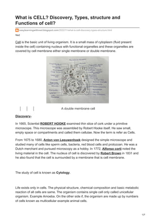
Cell is the basic unit of all living organisms. It was first observed by Robert Hooke in 1665 under a microscope where he saw compartments in cork and named them 'cells'. Key parts of the cell include the cell membrane, cytoplasm, organelles, and a nucleus. Cells can be either prokaryotic with no nucleus or eukaryotic with a nucleus surrounded by a membrane. Cells come in different shapes and sizes and perform specialized functions depending on their type.

Recommended
More Related Content
Similar to easylearningwithned.blogspot.com-What is CELL Discovery Types structure and Functions of cell.pdf
Similar to easylearningwithned.blogspot.com-What is CELL Discovery Types structure and Functions of cell.pdf (20)
More from Home
More from Home (11)
Recently uploaded
Recently uploaded (20)
easylearningwithned.blogspot.com-What is CELL Discovery Types structure and Functions of cell.pdf
- 1. 1/7 Ned What is CELL? Discovery, Types, structure and Functions of cell? easylearningwithned.blogspot.com/2022/11/what-is-cell-discovery-types-structure.html Cell is the basic unit of living organism. It is a small mass of cytoplasm (fluid present inside the cell) containing nucleus with functional organelles and these organelles are covered by cell membrane either single membrane or double membrane. A double membrane cell Discovery- In 1665, Scientist ROBERT HOOKE examined thin slice of cork under a primitive microscope. This microscope was assembled by Robert Hooke itself. He saw small, empty space or compartments and called them cellulae. Now the term is refer as Cells. From 1675 to 1680, Anton von Leeuwenhoek designed the simple microscope and studied many of cells like sperm cells, bacteria, red blood cells and protozoan. He was a Dutch merchant and pursued microscopy as a hobby. In 1772, Alfonso corti noted the living material in the cell. The nucleus of cell is discovered by Robert Brown in 1831 and he also found that the cell is surrounded by a membrane that is cell membrane. The study of cell is known as Cytology. Life exists only in cells. The physical structure, chemical composition and basic metabolic reaction of all cells are same. The organism contains single cell only called unicellular organism. Example Amoeba. On the other side if, the organism are made up by numbers of cells known as multicellular example animal cells.
- 2. 2/7 Unicellular organisms- The organisms have only single cell is known as unicellular. A single cell carries all their life functions, so that its life span is short. If cell get injured it cannot be recovered, it lead to death of the organism. Example of unicellular is Amoeba, Paramecium, Euglena, Yeast. Multicellular Organisms- the organisms have more than one cells is known as multicellular organisms. All cells divide their work, so that its life span is more than unicellular organisms. If cell get injured it did not lead to death of organism. Some dead cells can keep functioning. Example of multicellular organism is Human, Plants, Animal. Types of cells- Cells are basically two types. 1) Prokaryotic Cells 2) Eukaryotic cells Prokaryotic cells- are the most primitive cells, about 3.5 billion years ago. These are single membrane system and no membrane enveloping on the genetic material. Its membrane bears respiratory enzymes and may enfold to form mesosomas. Cell membrane control the movement of molecules from in to out of the cell. In prokaryotic cells the 70S ribosomes presents. It may be lie free in the cytoplasm or attached on the cell membrane. The general size of prokaryotic cells is 1 to 10micrometer and they have different types of shapes like Sphere, Commas, Rods, Helices. forms of bacteria
- 3. 3/7 1) Spheres- Cocci 2) Commas - Vibrio's 3) Rods - Bacilli 4) Helices - Spirillum Prokaryotic cells are occurred in Bacteria, Cyanobacteria, Mycoplasmas, Chlamydia's and Rickettsia's. Bacteria is observed under the microscope by stained it using gram stain. If bacteria retain the stain that known as Gram positive bacteria and if it loses the stain it known as gram negative bacteria. A Bacteria Cell Membrane:- Three types of layers present on gram negative bacteria. i) The first layer of bacteria is coated with a slime layer and capsule. This slime layer and capsule are formed of polysaccharides, but also contain polypeptide. This layer protects the cell from any virus attack. It did not allow to retain the stain. ii) Then the second layer is cell wall, it maintains its shape and structure of cell. It prevents bursting of cells. It consists with peptidoglycan cross linked by short- peptides. Small thread like Pili attached with cell wall. It helps bacteria to attached on surface. iii) The third layer is cell membrane, it maintains the movement of material in and out and bears respiratory enzymes. It also holds receptor molecules to detect chemical in the surroundings. Cytoplasm:-
- 4. 4/7 i) The cytoplasm contain only Ribosomes as a organelle. 70s Ribosomes are present in it. It may be present freely or in group. The ribosomes in group known as polysomes. ii) The cytoplasm also contain biosynthetic enzymes that is transfer RNA (tRNAs) and mRNAs. iii) It contain variety of organic and inorganic molecules like fat droplets, protein droplets. iv) Some gas vacuoles move freely in prokaryotic cells. The tail like Solid flagellum is originate from cytoplasm. Its one end is inside the cytoplasm and the other end is move freely outside the environment. It provide the motility to the organism. Nucleus:- The genetic material lie directly in the cytoplasm without any envelope and the shape of genetic material is circular and Helical. These cells have only one copy of chromosome. Eukaryotic cells- have double-membrane system. First membrane surrounds the cell and second membrane surround the nucleus. Its cell membrane lacks the respiratory enzymes. 80S ribosomes are present in eukaryotic cells. These ribosomes lie freely or attached to endoplasmic reticulum and nuclear envelope. Most of Eukaryotic are sexual organisms and they are great in size. The range of eukaryotic cell size is about 10 to 100micrometer and the Shape of eukaryotic cell is may be:- oval, spherical, disc-like, cuboidal, columnar, spindle like, irregular. The Eukaryotic cells are occurred in algae, fungi, plants and animals.
- 5. 5/7 Shapes of cells Animal cell is observed under electron microscope. Cell Membrane:- Animal cell contains number of organelles and covered with thin, flexible, cell membrane. This cell membrane maintains the shape of cell, protect from any virus attack and allow the movement of material inside or outside by exocytosis, pinocytosis and phagocytosis. Microvilli are present on cell membrane for the secretion or adsorption. Cytoplasm:-Inside the cell membrane, the cytoplasm contains all organelles like Endoplasmic reticulum Mitochondria Golgi complex Lysosomes
- 6. 6/7 Centrosome Microfilaments Intermediate fibers Microtubules Microbodies Ribosomes. These all organelles keep their functions. Nucleus:- The Nucleus present center of the cytoplasm, so that it called controller of cell. It covered with nuclear envelope known as second membrane and the fluidic part present inside the nuclear membrane is known as nucleoplasm. Nucleus contain chromosomes and nucleolus. This nucleolus is known as small nucleus, which synthesize and store the RNA. The shape of genetic material is linear and circular. Function of cell:- 1) Cell provide the shape of body. 2) It control the movement of material. 3) It maintain homeostasis. 4) It store the genetic information. 5) Some cells may die but still keep functioning. Example- Xylem vessels and horny cells. Some Important Questions
- 7. 7/7 Q1) Who introduced the term Cell? Ans- Scientist ROBERT HOOKE introduced the term cell. Q2) Who discovered the Nucleus? Ans- ROBERT BROWN discovered the nucleus. Q3) What is the control center of cell? Ans- Nucleus is the control center of the cell. Q4) Where is nucleolus found and what is its another name? Ans- Nucleolus found in nucleus and the other name of nucleolus is small nucleus. Who is the Father of Biology ? What is the unit of Living organism ?