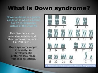
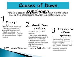
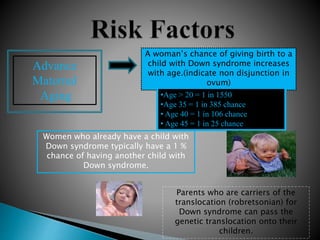
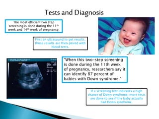
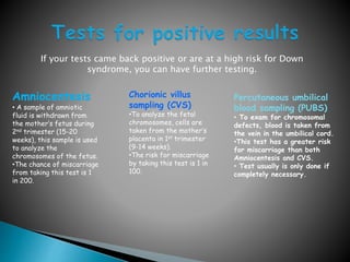
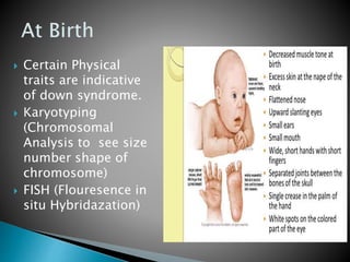
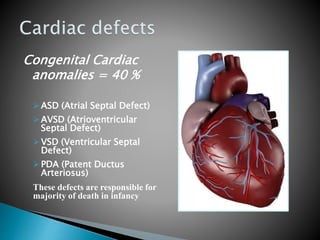
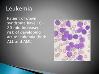
Down Syndrome
By: Aamir Sharif
What is Down syndrome?
Down syndrome is a genetic
condition in which a person
has 47 chromosomes
instead of the usual 46.
This disorder causes
mental retardation and
other problems, occurs in
1 in 700 births.
Down syndrome ranges
in severity, so
developmental
problems may range
from mild to serious.
 21 chromosome carries about 500 genes,
170 of them served as protein coding genes
and 5 are miRNA.
 Two chromosomes genes DYRK1A, (codes for
threonine kinase) and RCAN1 (Regulator of
calcineurin 1), which codes for a protein that
inhibits a cellular phosphatase enzyme
(calcineurin) are mainly involved in causing
down syndrome.
Causes of Down
syndromeThere are 3 possible abnormalities which all result in extra genetic
material from chromosome 21,which causes Down syndrome.
1Trisomy
21• About 95 percent of cases with
Down syndrome are caused by
Trisomy 21 (non disjunction).
• With Trisomy 21 there are 3
copies of chromosome 21 in every
cell, instead of the usual 2 copies
(total 47).
2 Mosaic Down
syndrome
• rare form (1 % ) of Down
syndrome
• With Mosaic Down syndrome
some cells have an extra copy of
chromosome 21, but not all.
• Mosaic Down syndrome is
caused by abnormal cell division
after fertilization (during
embryogenesis).
3 Translocatio
n Down
syndrome
• occurs when part of a chromosome
21 becomes attached onto another
chromosome, usually chromosome 14,
before or at conception.
• Children with Translocation have the
usual 2 copies of chromosome 21, but
have additional material from
chromosome 21 stuck to the
translocated chromosome.
MOST cases of Down syndrome are NOT inherited.
Individuals with Down syndrome have a widely recognized appearance.
Common physical signs include:
• flattened nose
• small ears
• small mouth
• upward slanting eyes
• wide, short hands with short fingers
• separated joints between the bones of the skull
• decreased muscle tone at birth
• protruding tongue
• small head
Advance
Maternal
Aging
A woman’s chance of giving birth to a
child with Down syndrome increases
with age.(indicate non disjunction in
ovum)
•Age > 20 = 1 in 1550
•Age 35 = 1 in 385 chance
• Age 40 = 1 in 106 chance
• Age 45 = 1 in 25 chance
Women who already have a child with
Down syndrome typically have a 1 %
chance of having another child with
Down syndrome.
Parents who are carriers of the
translocation (robretsonian) for
Down syndrome can pass the
genetic translocation onto their
children.
Tests and Diagnosis
The most efficient two step
screening is done during the 11th
week and 14th week of pregnancy.
First an ultrasound to get results,
those results are then paired with
blood tests.
“When this two-step screening
is done during the 11th week
of pregnancy, researchers say it
can identify 87 percent of
babies with Down syndrome.”
If a screening test indicates a high
chance of Down syndrome, more tests
are done to see if the baby actually
had Down syndrome.
If your tests came back positive or are at a high risk for Down
syndrome, you can have further testing.
Amniocentesis
• A sample of amniotic
fluid is withdrawn from
the mother’s fetus during
2nd trimester (15-20
weeks), this sample is used
to analyze the
chromosomes of the fetus.
•The chance of miscarriage
from taking this test is 1
in 200.
Chorionic villus
sampling (CVS)
•To analyze the fetal
chromosomes, cells are
taken from the mother’s
placenta in 1st trimester
(9-14 weeks).
•The risk for miscarriage
by taking this test is 1 in
100.
Percutaneous umbilical
blood sampling (PUBS)
• To exam for chromosomal
defects, blood is taken from
the vein in the umbilical cord.
•This test has a greater risk
for miscarriage than both
Amniocentesis and CVS.
• Test usually is only done if
completely necessary.
 Certain Physical
traits are indicative
of down syndrome.
 Karyotyping
(Chromosomal
Analysis to see size
number shape of
chromosome)
 FISH (Flouresence in
situ Hybridazation)
Congenital Cardiac
anomalies = 40 %
ASD (Atrial Septal Defect)
AVSD (Atrioventricular
Septal Defect)
VSD (Ventricular Septal
Defect)
PDA (Patent Ductus
Arteriosus)
These defects are responsible for
majority of death in infancy
Patient of down
syndrome have 10-
20 fold increased
risk of developing
acute leukemia (both
ALL and AML)
Alzheimer Disease
 Gene of Amyloid
Precursor Protein
(APP) located on
chromosome 21.
 Trisomy lead to
increase production
of APP that will lead
to Aß which in turn
result in Alzheimer.
 Infectious Diseases
Especially to lungs
and thyroid, due to
abnormal immune
response, that might
be due to abnormal T
cell functions.

Down syndrome

  • 1.
  • 2.
    What is Downsyndrome? Down syndrome is a genetic condition in which a person has 47 chromosomes instead of the usual 46. This disorder causes mental retardation and other problems, occurs in 1 in 700 births. Down syndrome ranges in severity, so developmental problems may range from mild to serious.
  • 3.
     21 chromosomecarries about 500 genes, 170 of them served as protein coding genes and 5 are miRNA.  Two chromosomes genes DYRK1A, (codes for threonine kinase) and RCAN1 (Regulator of calcineurin 1), which codes for a protein that inhibits a cellular phosphatase enzyme (calcineurin) are mainly involved in causing down syndrome.
  • 4.
    Causes of Down syndromeThereare 3 possible abnormalities which all result in extra genetic material from chromosome 21,which causes Down syndrome. 1Trisomy 21• About 95 percent of cases with Down syndrome are caused by Trisomy 21 (non disjunction). • With Trisomy 21 there are 3 copies of chromosome 21 in every cell, instead of the usual 2 copies (total 47). 2 Mosaic Down syndrome • rare form (1 % ) of Down syndrome • With Mosaic Down syndrome some cells have an extra copy of chromosome 21, but not all. • Mosaic Down syndrome is caused by abnormal cell division after fertilization (during embryogenesis). 3 Translocatio n Down syndrome • occurs when part of a chromosome 21 becomes attached onto another chromosome, usually chromosome 14, before or at conception. • Children with Translocation have the usual 2 copies of chromosome 21, but have additional material from chromosome 21 stuck to the translocated chromosome. MOST cases of Down syndrome are NOT inherited.
  • 5.
    Individuals with Downsyndrome have a widely recognized appearance. Common physical signs include: • flattened nose • small ears • small mouth • upward slanting eyes • wide, short hands with short fingers • separated joints between the bones of the skull • decreased muscle tone at birth • protruding tongue • small head
  • 6.
    Advance Maternal Aging A woman’s chanceof giving birth to a child with Down syndrome increases with age.(indicate non disjunction in ovum) •Age > 20 = 1 in 1550 •Age 35 = 1 in 385 chance • Age 40 = 1 in 106 chance • Age 45 = 1 in 25 chance Women who already have a child with Down syndrome typically have a 1 % chance of having another child with Down syndrome. Parents who are carriers of the translocation (robretsonian) for Down syndrome can pass the genetic translocation onto their children.
  • 7.
    Tests and Diagnosis Themost efficient two step screening is done during the 11th week and 14th week of pregnancy. First an ultrasound to get results, those results are then paired with blood tests. “When this two-step screening is done during the 11th week of pregnancy, researchers say it can identify 87 percent of babies with Down syndrome.” If a screening test indicates a high chance of Down syndrome, more tests are done to see if the baby actually had Down syndrome.
  • 8.
    If your testscame back positive or are at a high risk for Down syndrome, you can have further testing. Amniocentesis • A sample of amniotic fluid is withdrawn from the mother’s fetus during 2nd trimester (15-20 weeks), this sample is used to analyze the chromosomes of the fetus. •The chance of miscarriage from taking this test is 1 in 200. Chorionic villus sampling (CVS) •To analyze the fetal chromosomes, cells are taken from the mother’s placenta in 1st trimester (9-14 weeks). •The risk for miscarriage by taking this test is 1 in 100. Percutaneous umbilical blood sampling (PUBS) • To exam for chromosomal defects, blood is taken from the vein in the umbilical cord. •This test has a greater risk for miscarriage than both Amniocentesis and CVS. • Test usually is only done if completely necessary.
  • 9.
     Certain Physical traitsare indicative of down syndrome.  Karyotyping (Chromosomal Analysis to see size number shape of chromosome)  FISH (Flouresence in situ Hybridazation)
  • 10.
    Congenital Cardiac anomalies =40 % ASD (Atrial Septal Defect) AVSD (Atrioventricular Septal Defect) VSD (Ventricular Septal Defect) PDA (Patent Ductus Arteriosus) These defects are responsible for majority of death in infancy
  • 11.
    Patient of down syndromehave 10- 20 fold increased risk of developing acute leukemia (both ALL and AML)
  • 12.
    Alzheimer Disease  Geneof Amyloid Precursor Protein (APP) located on chromosome 21.  Trisomy lead to increase production of APP that will lead to Aß which in turn result in Alzheimer.  Infectious Diseases Especially to lungs and thyroid, due to abnormal immune response, that might be due to abnormal T cell functions.

Editor's Notes