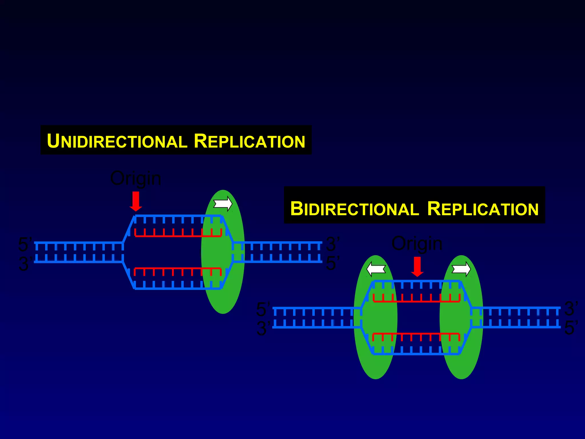
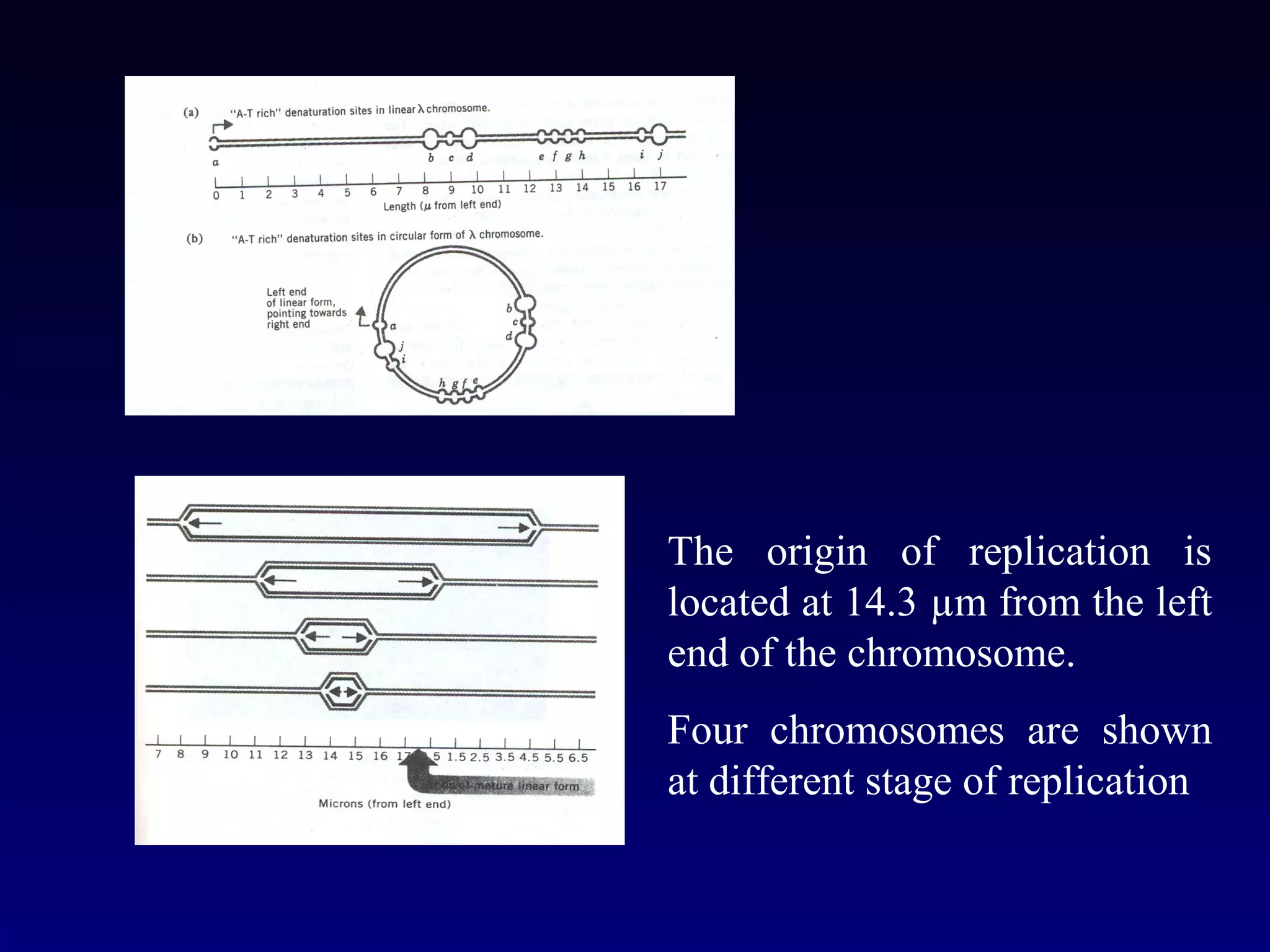
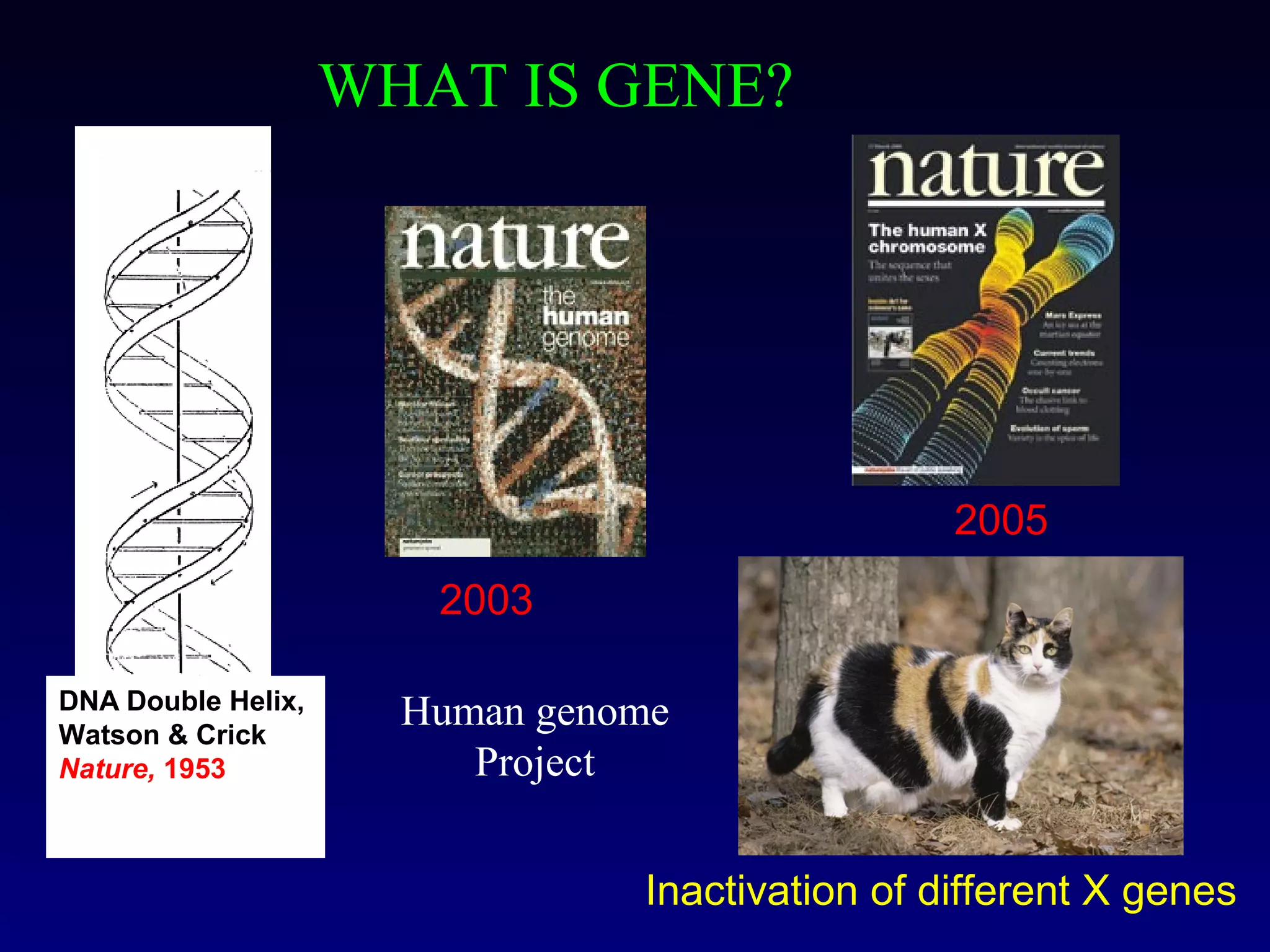
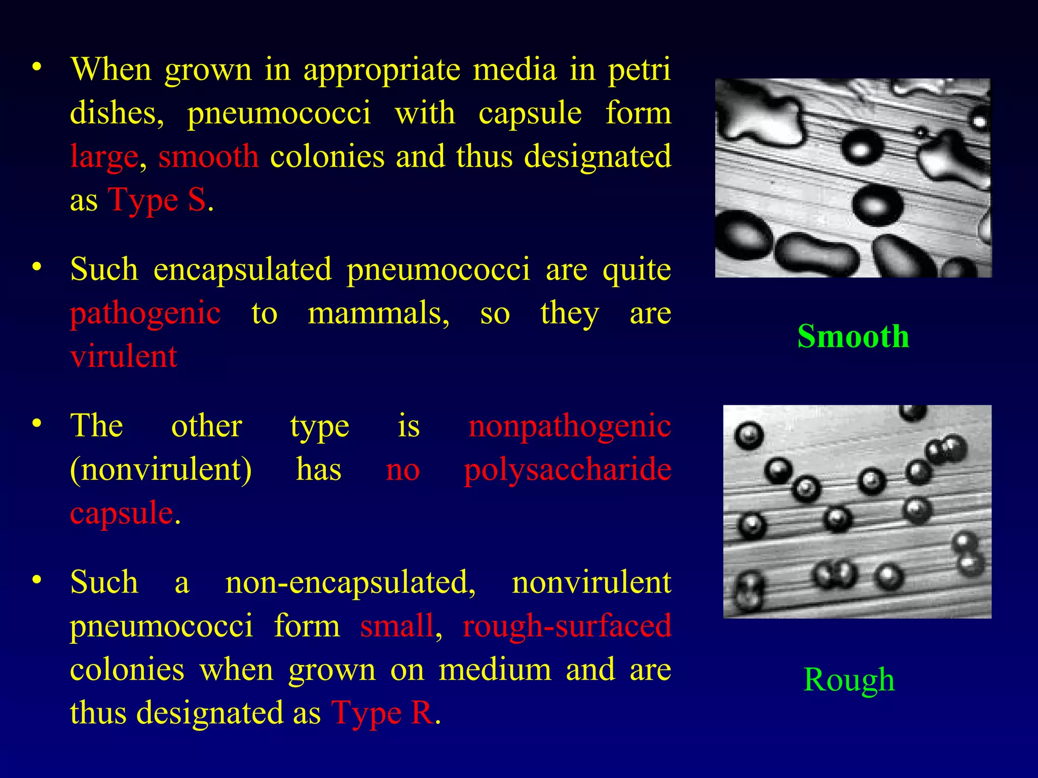
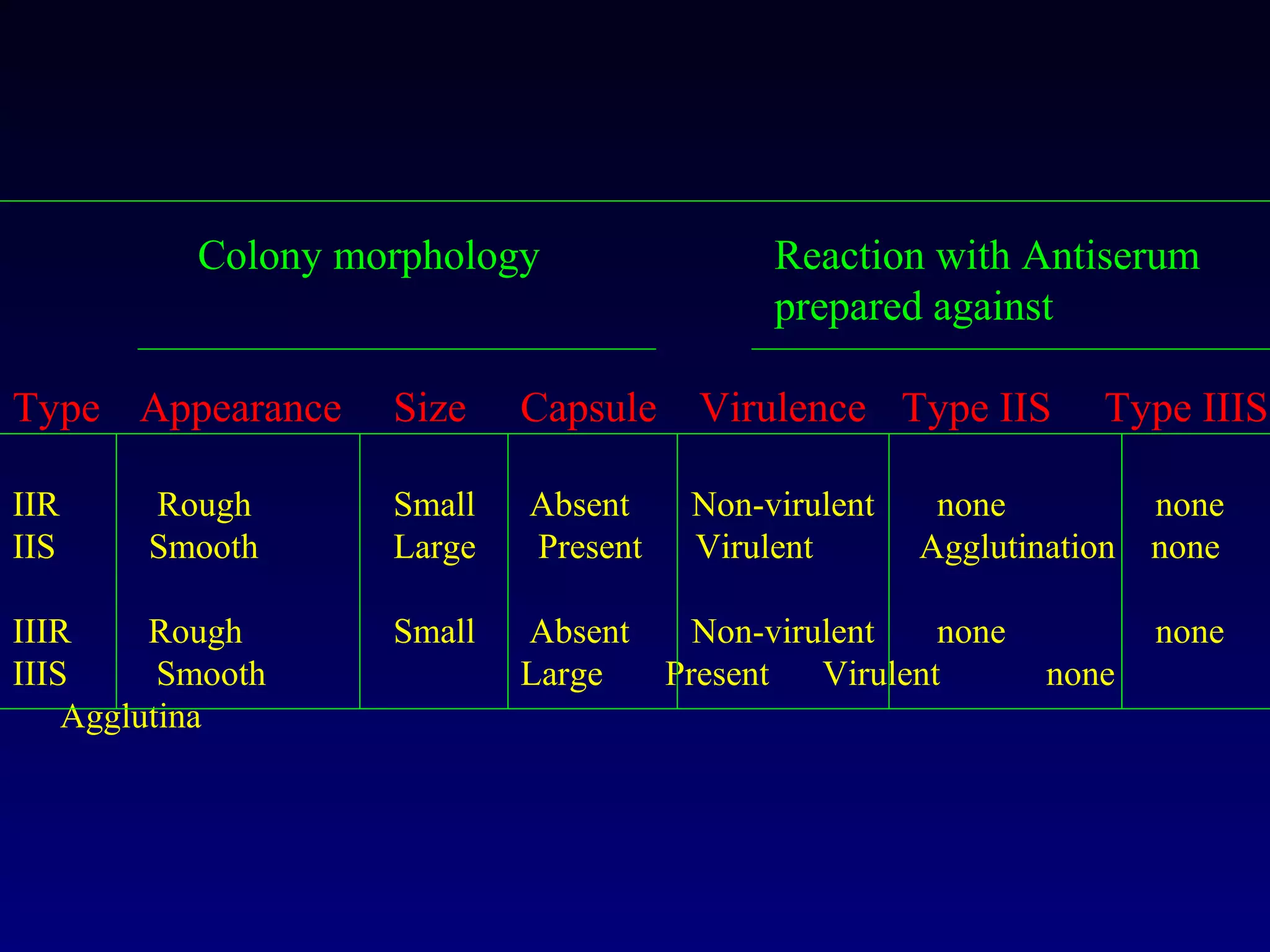
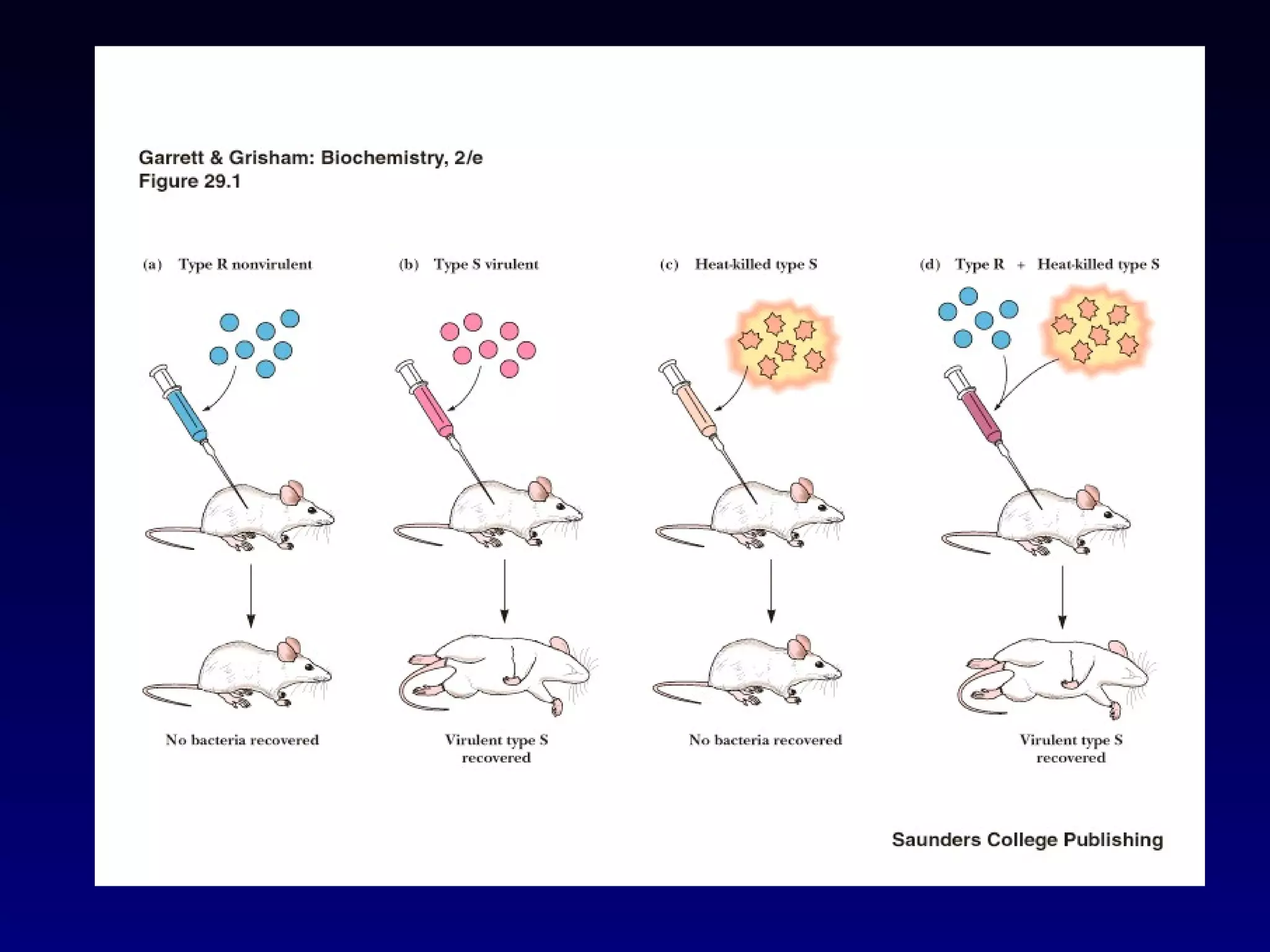
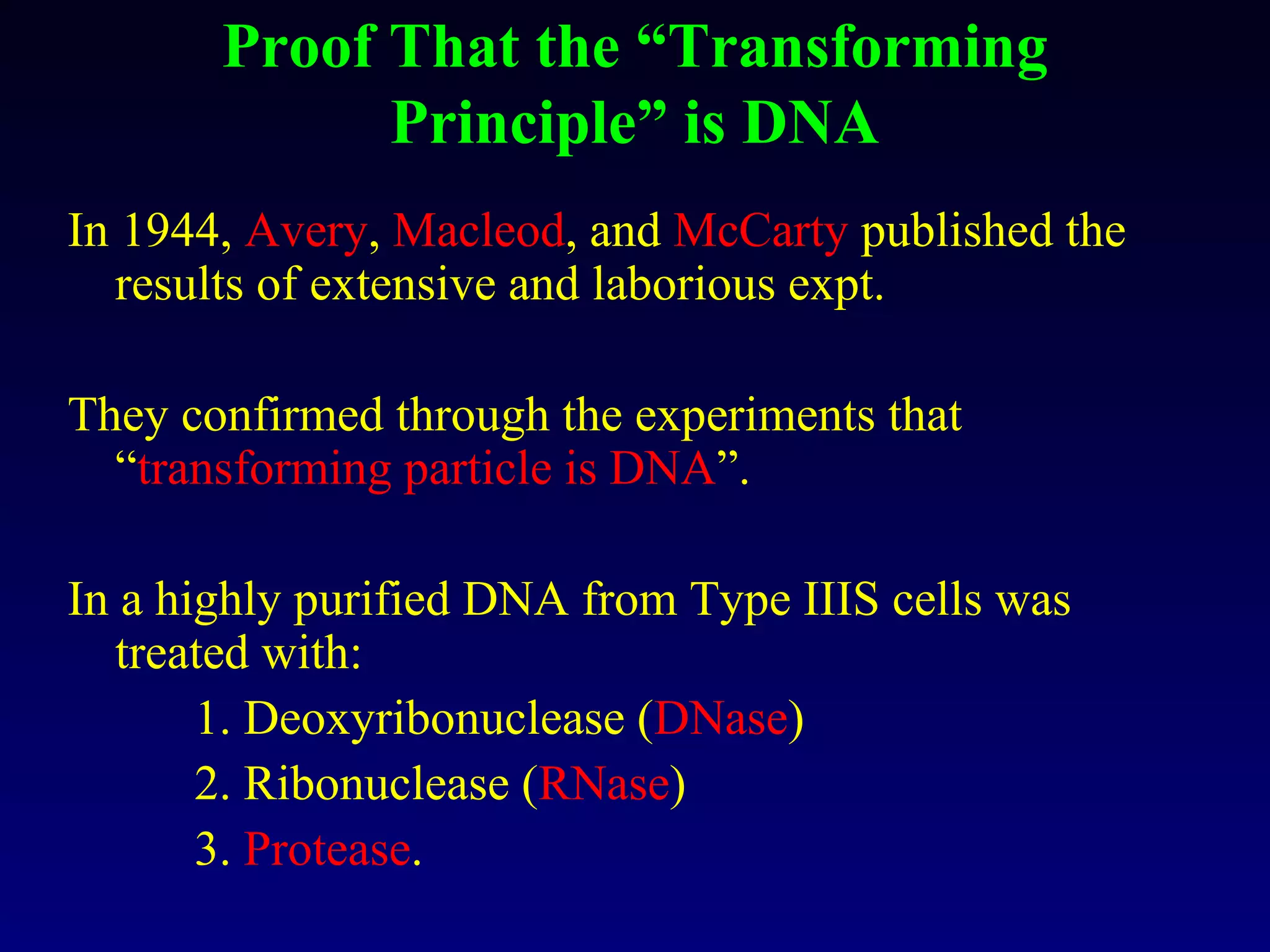
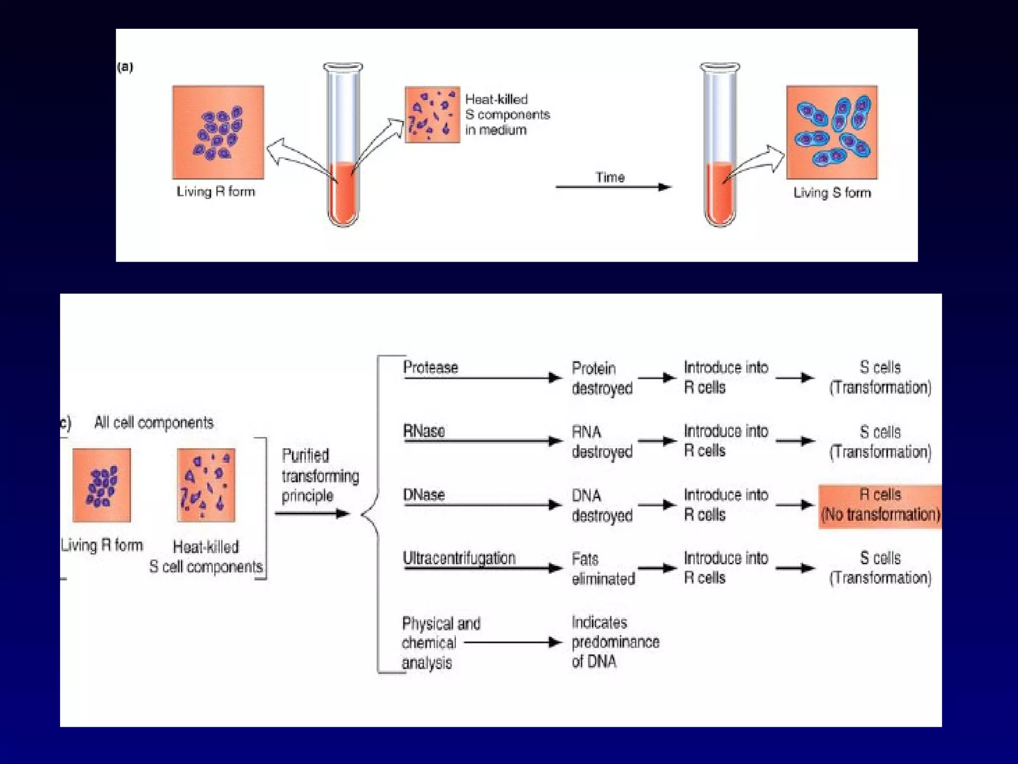
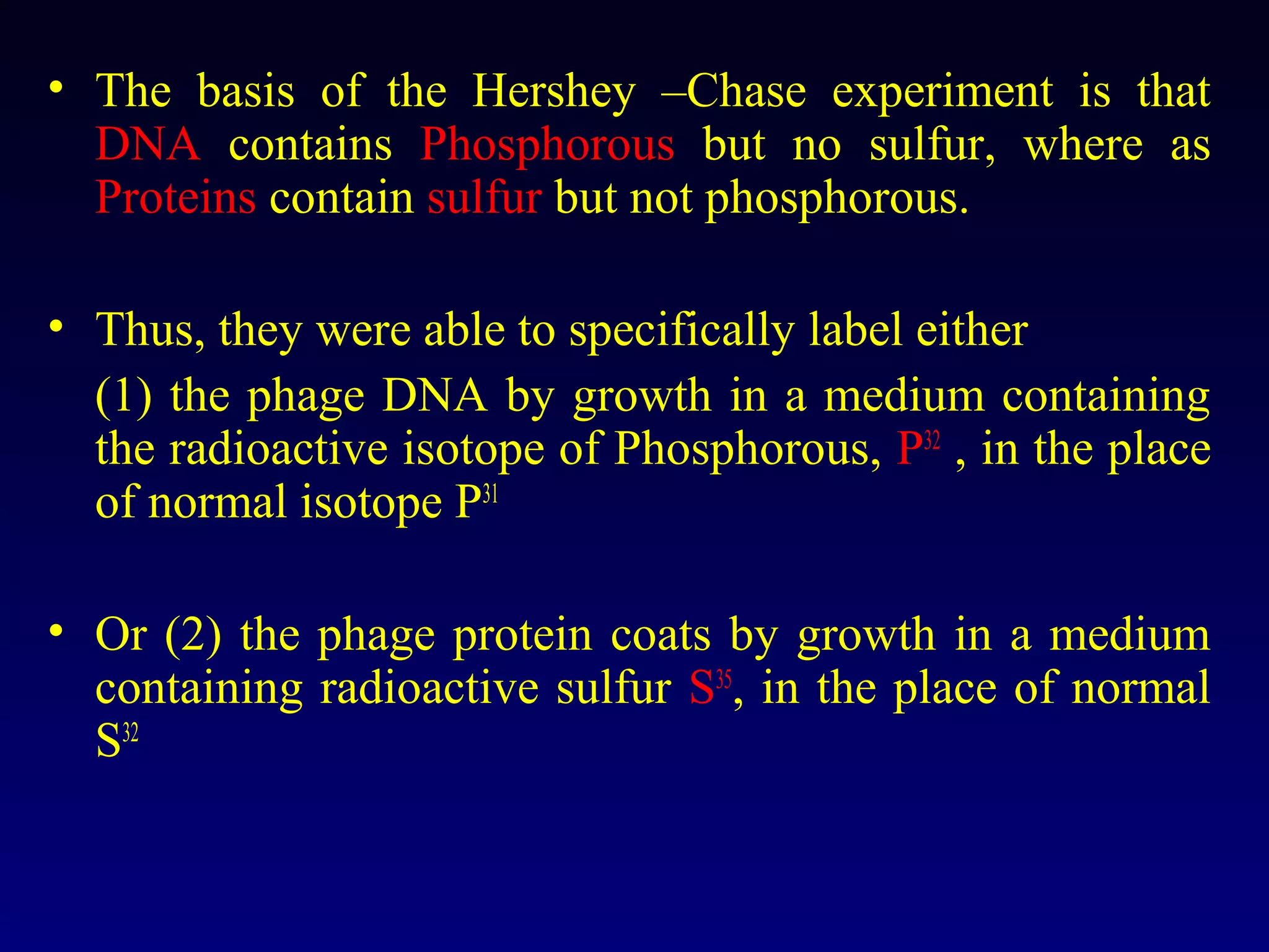
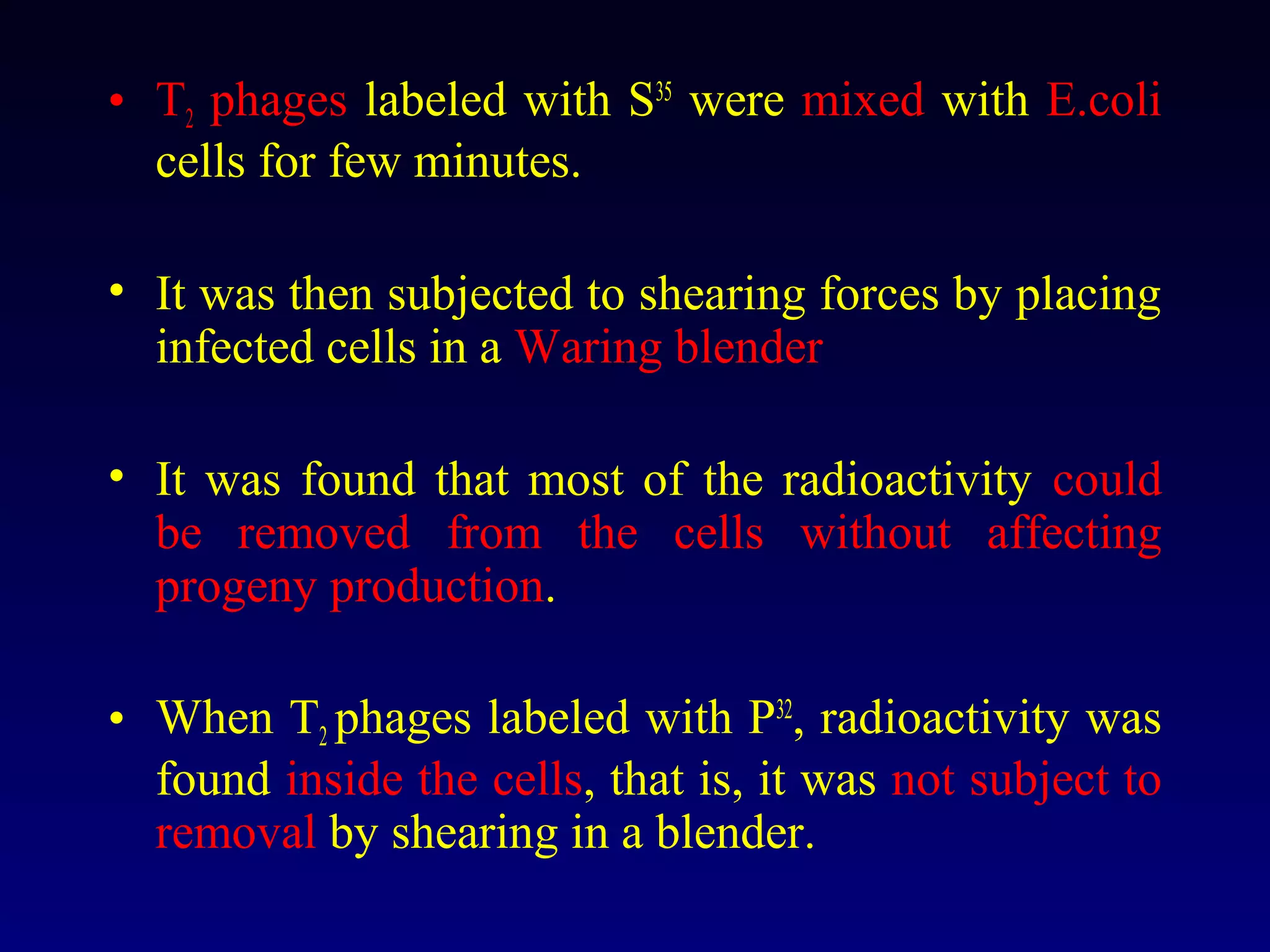
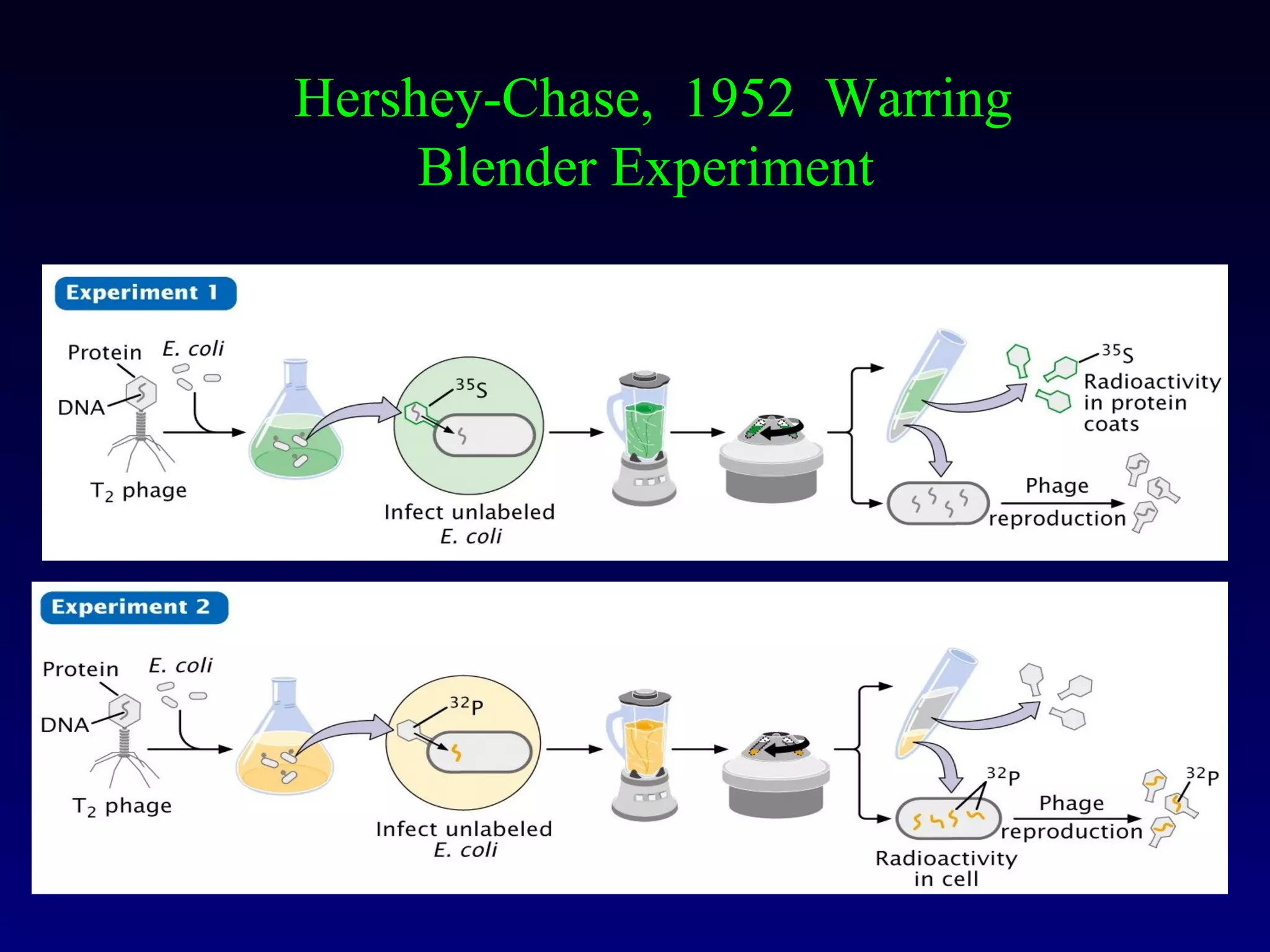
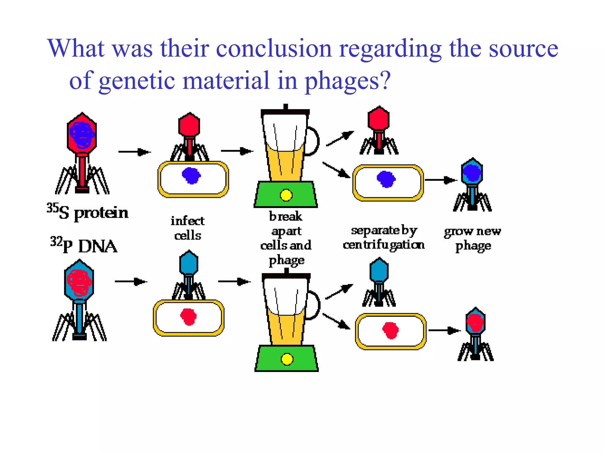
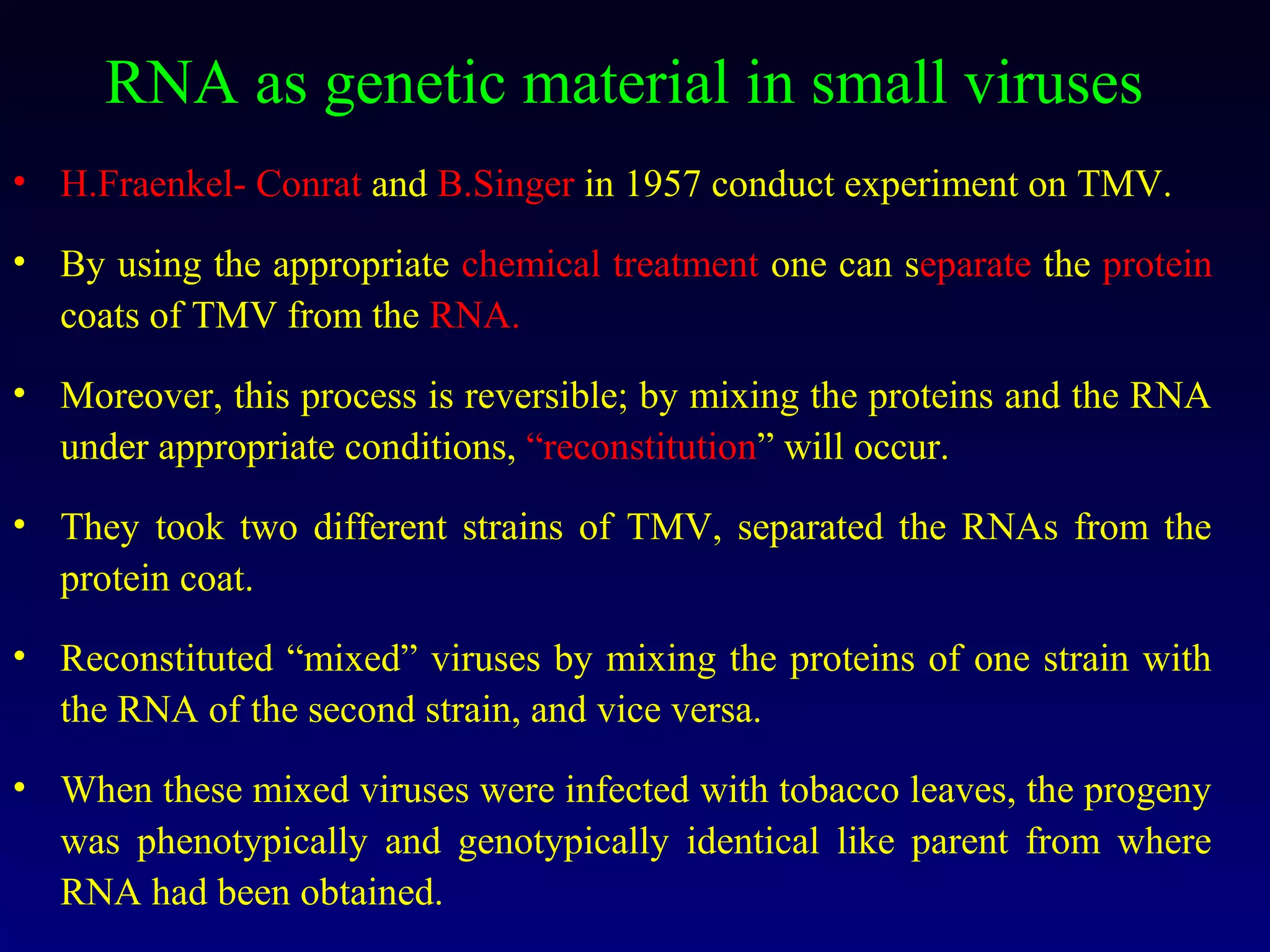
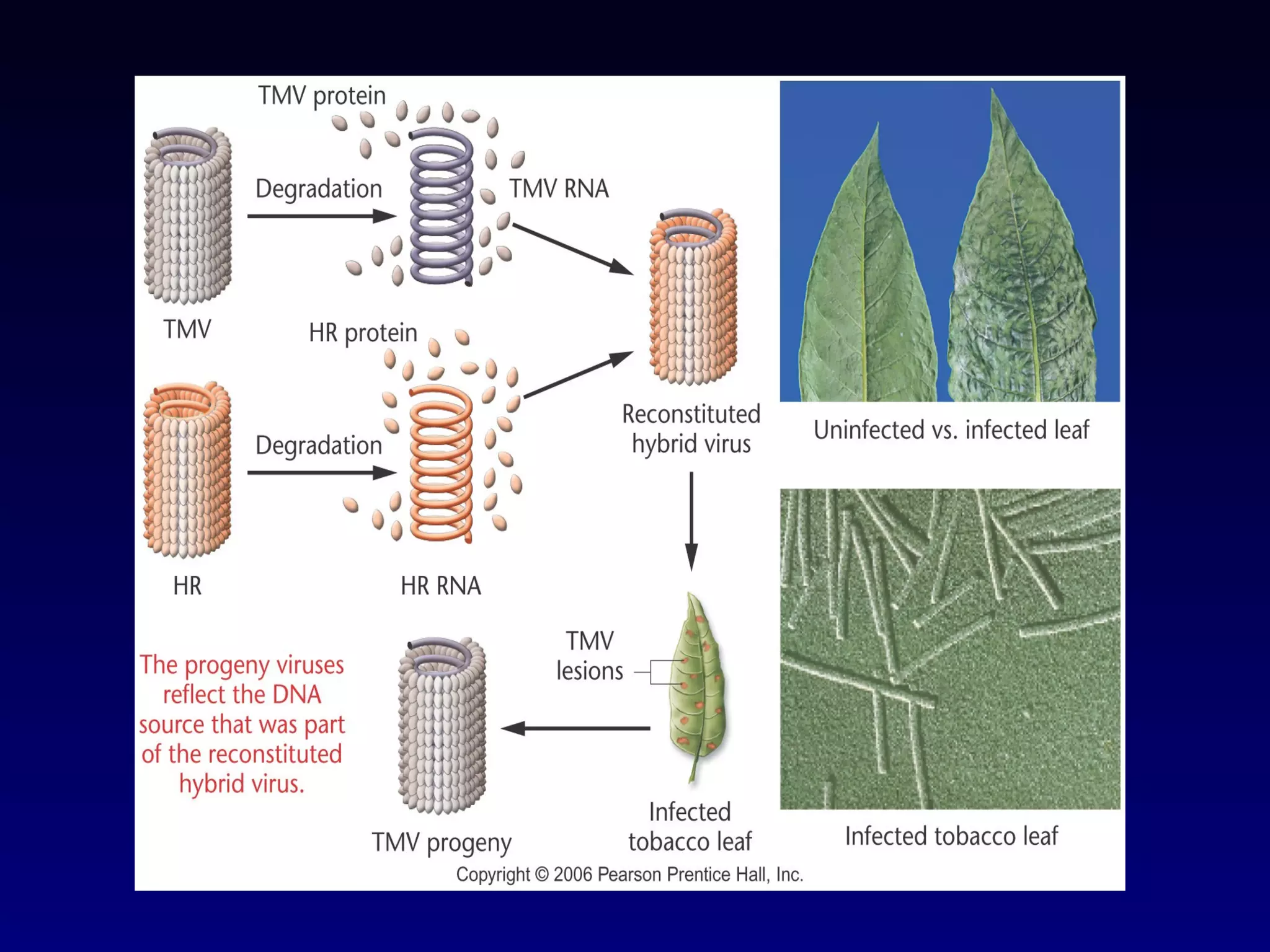
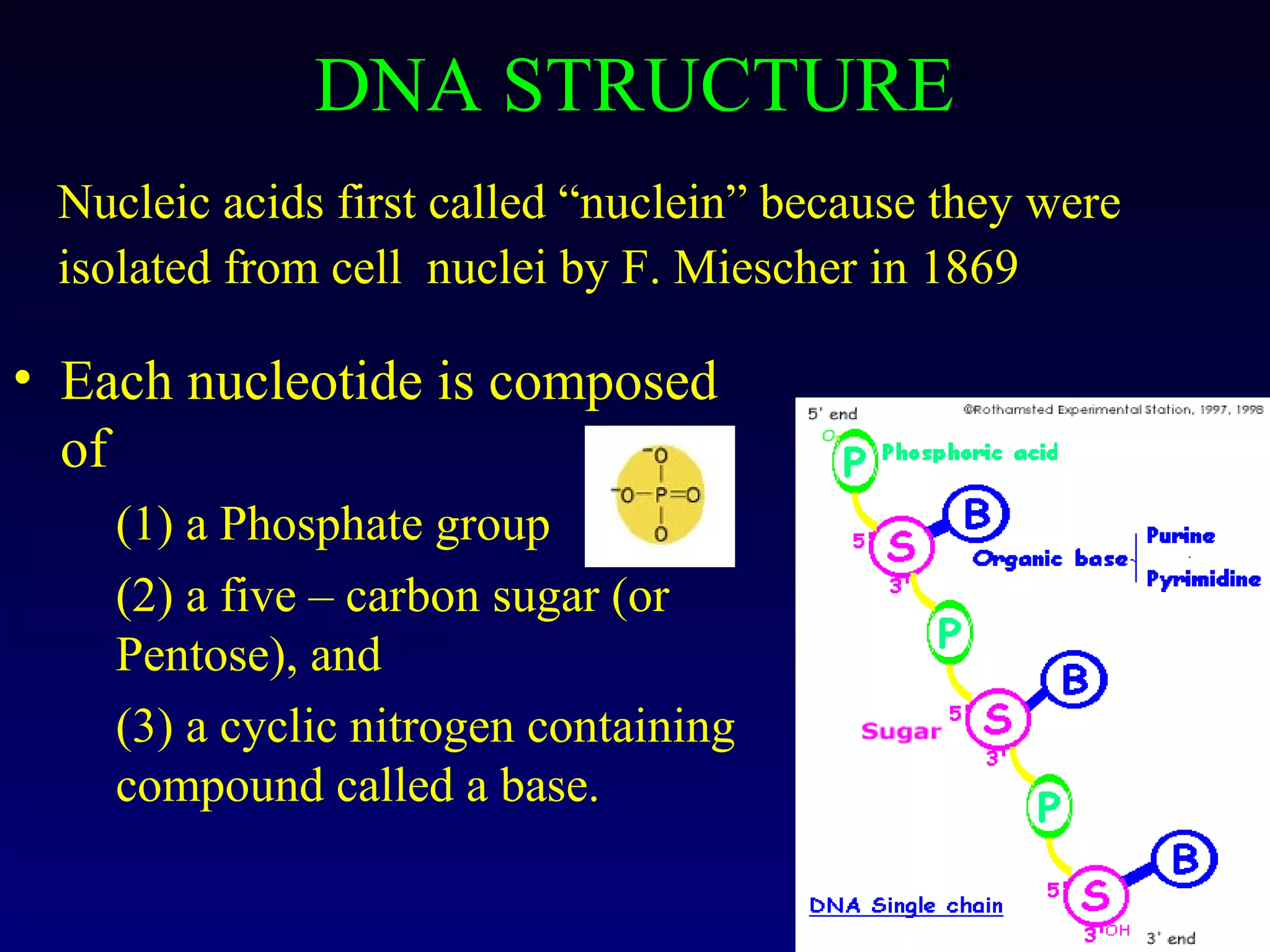
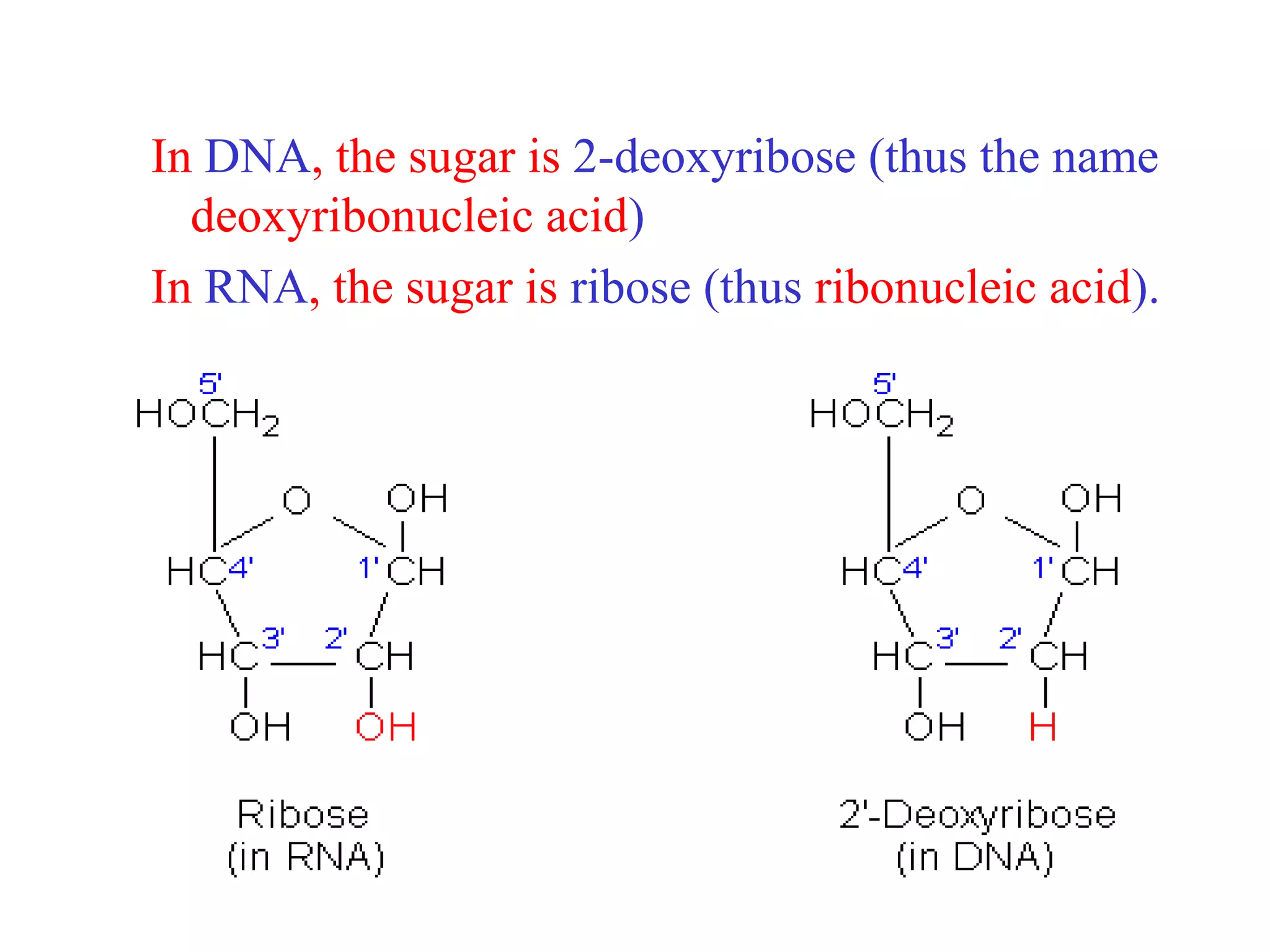
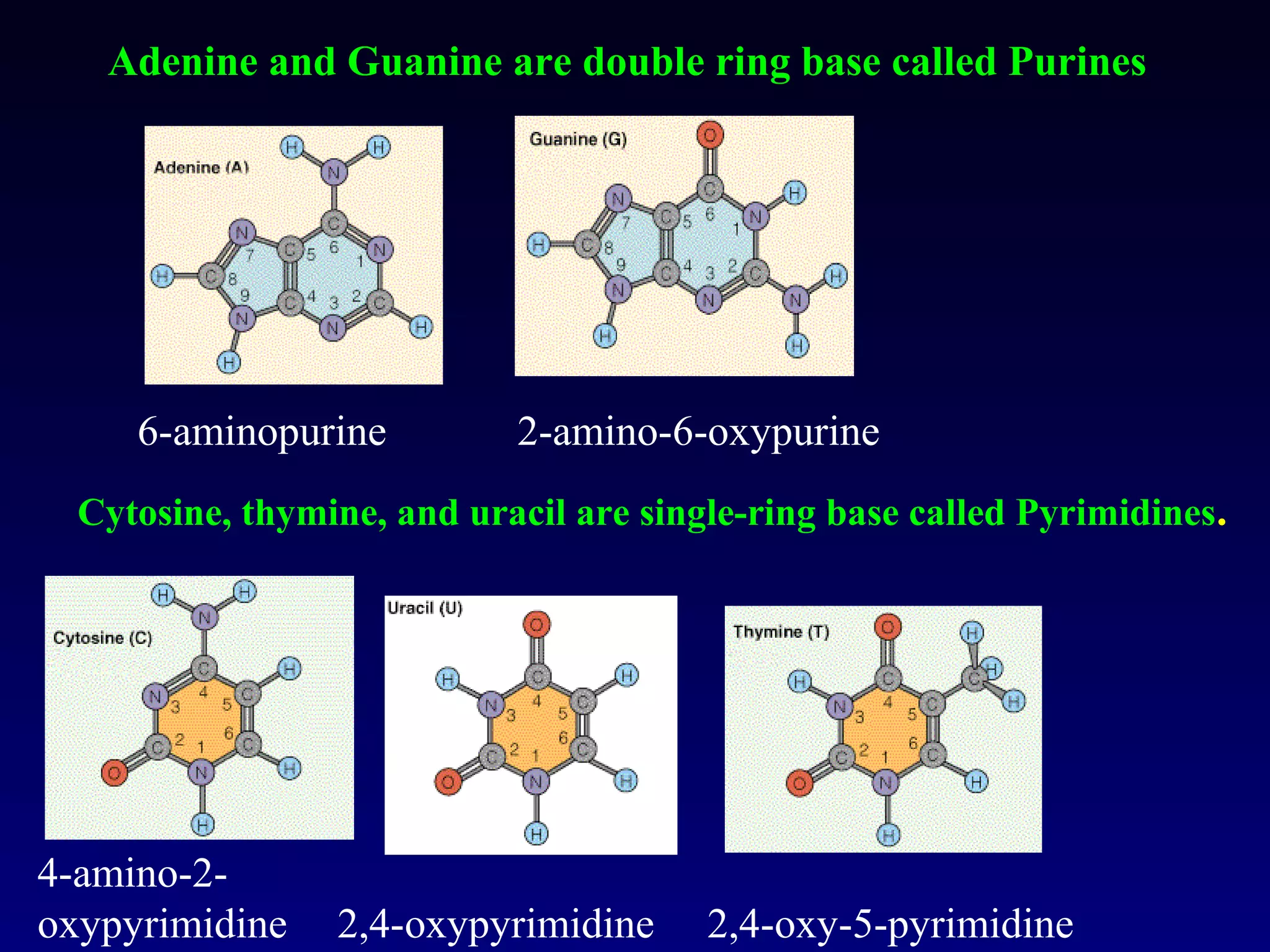
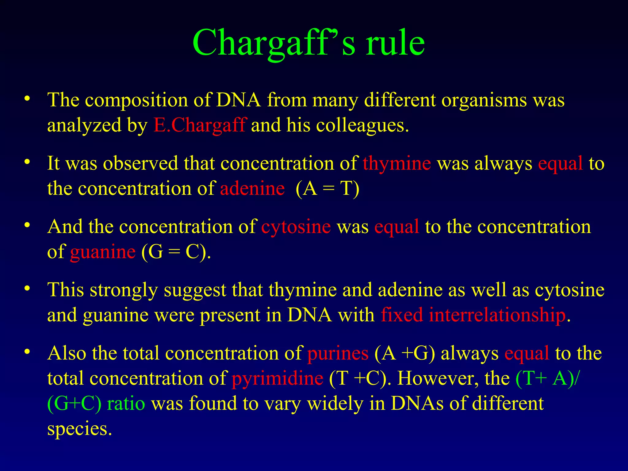
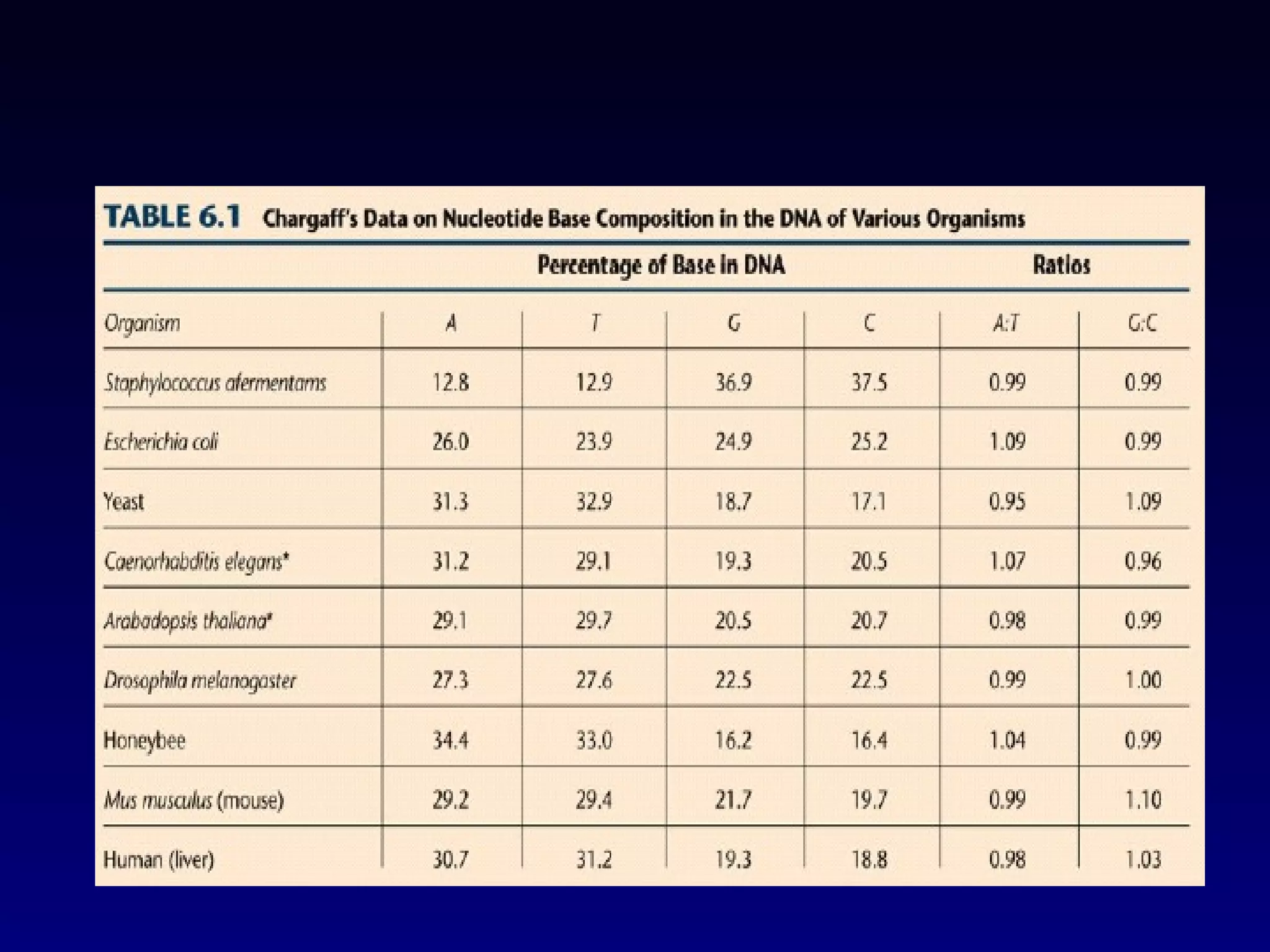
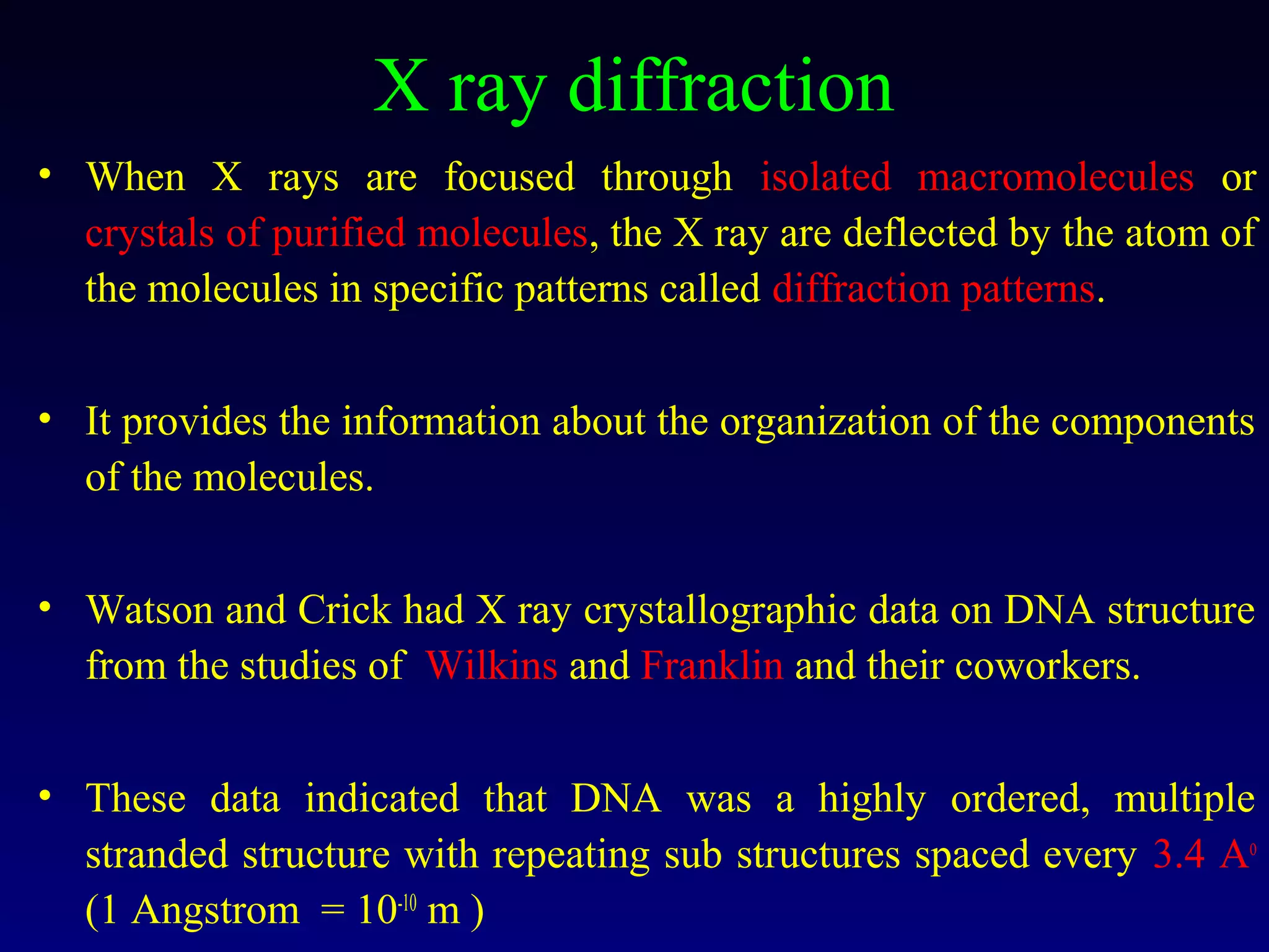
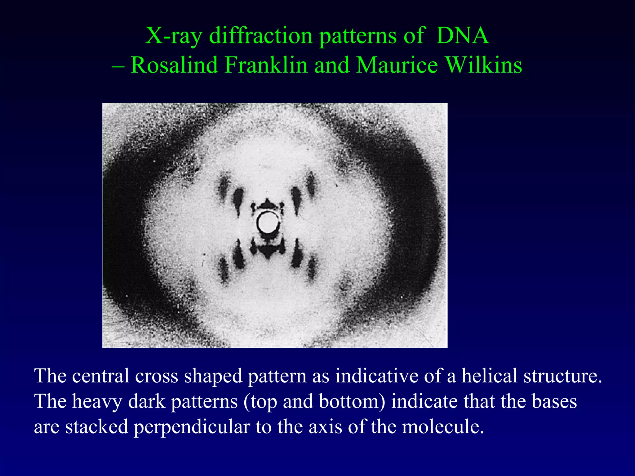
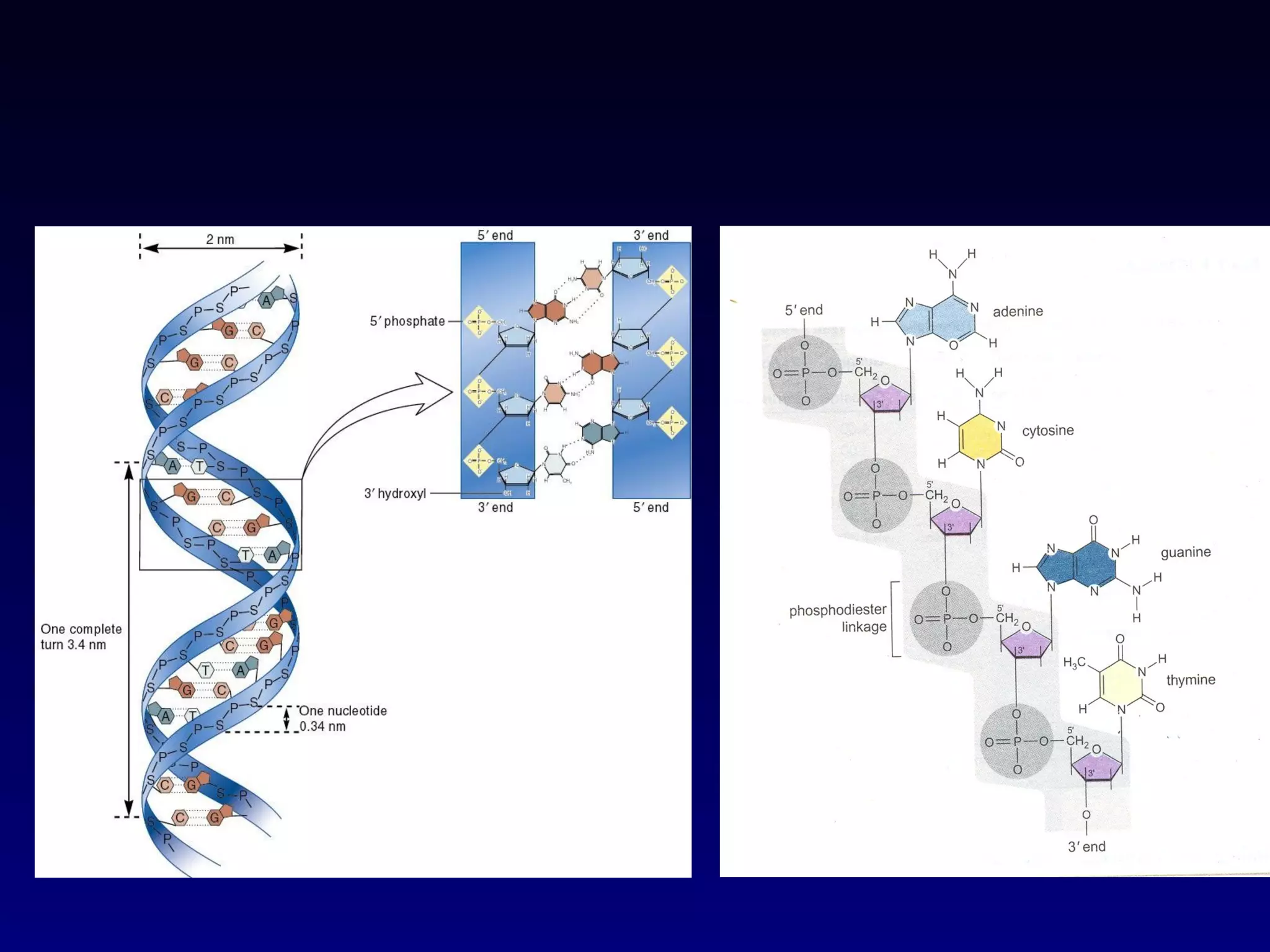
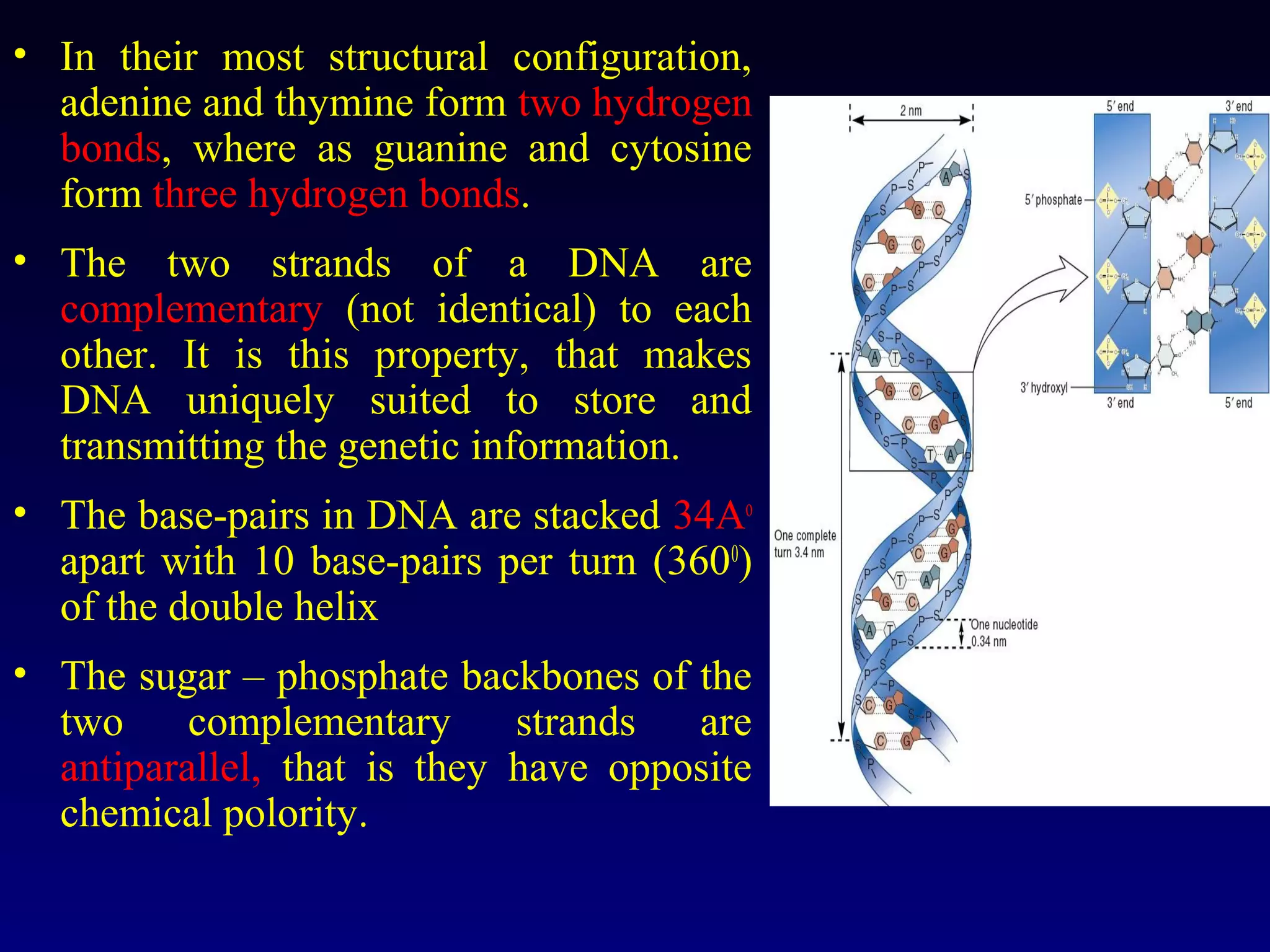
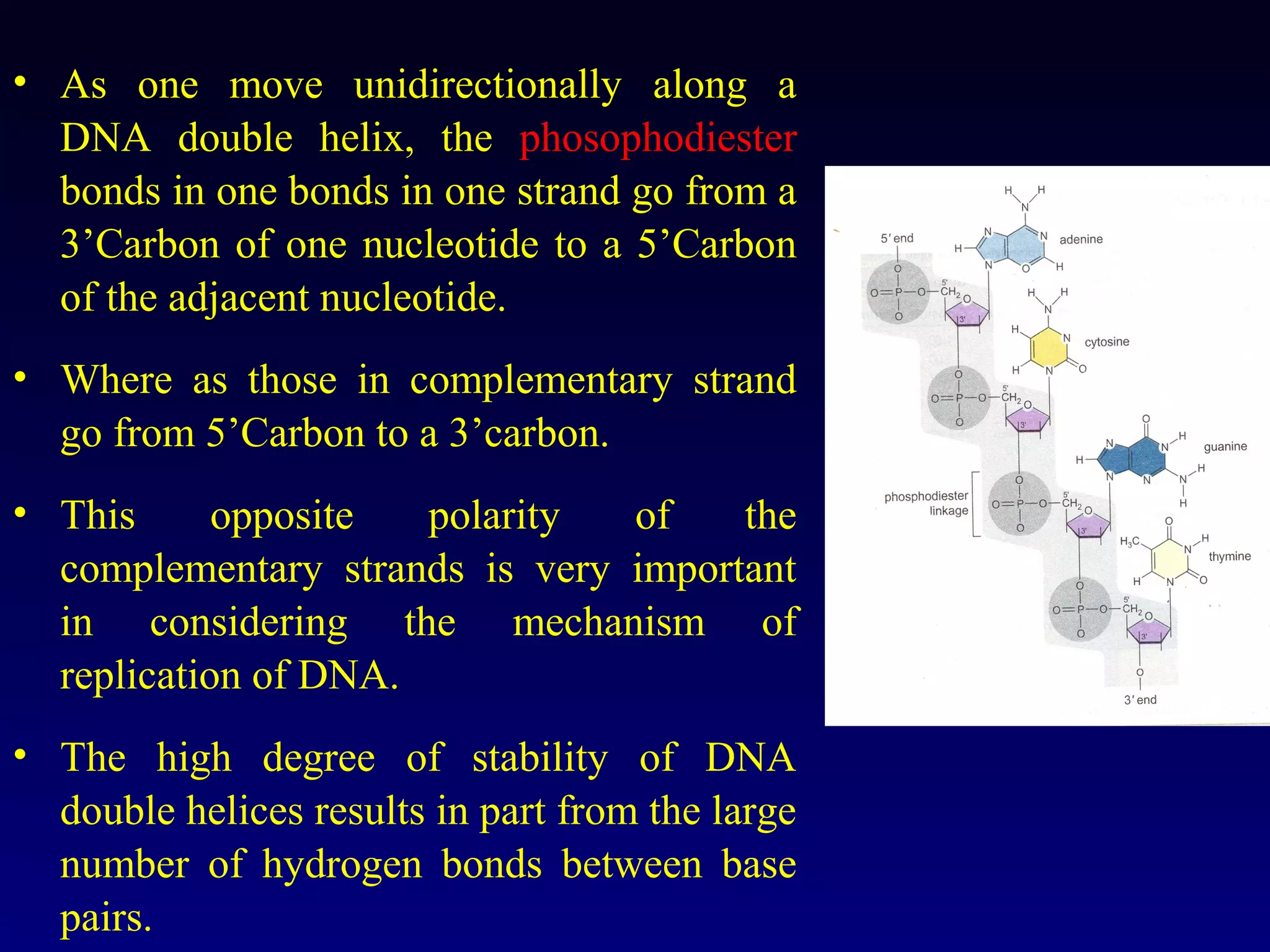
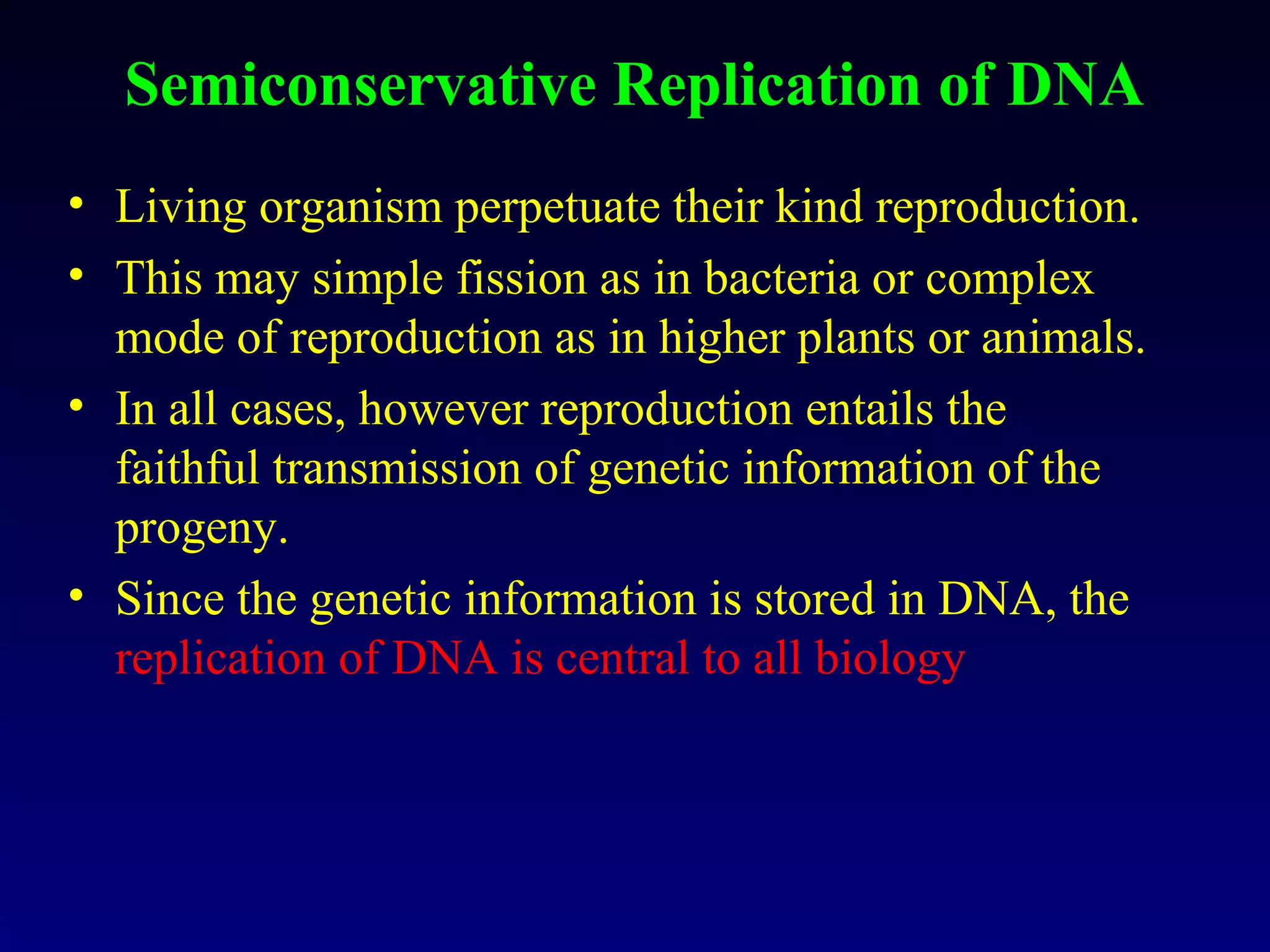
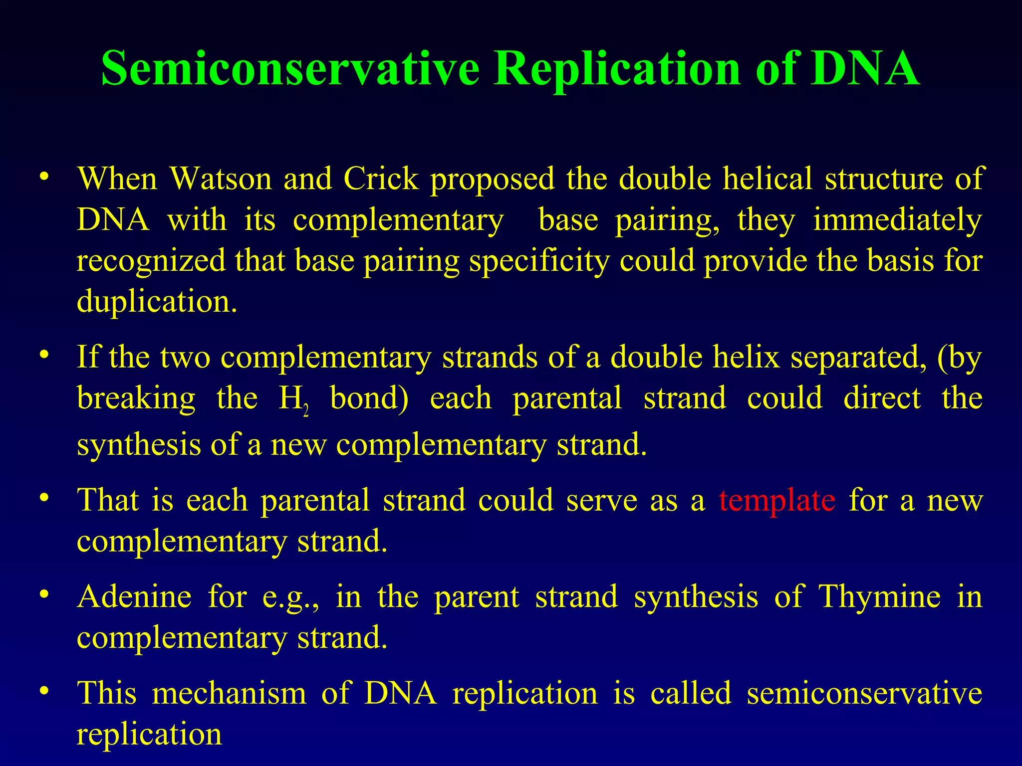
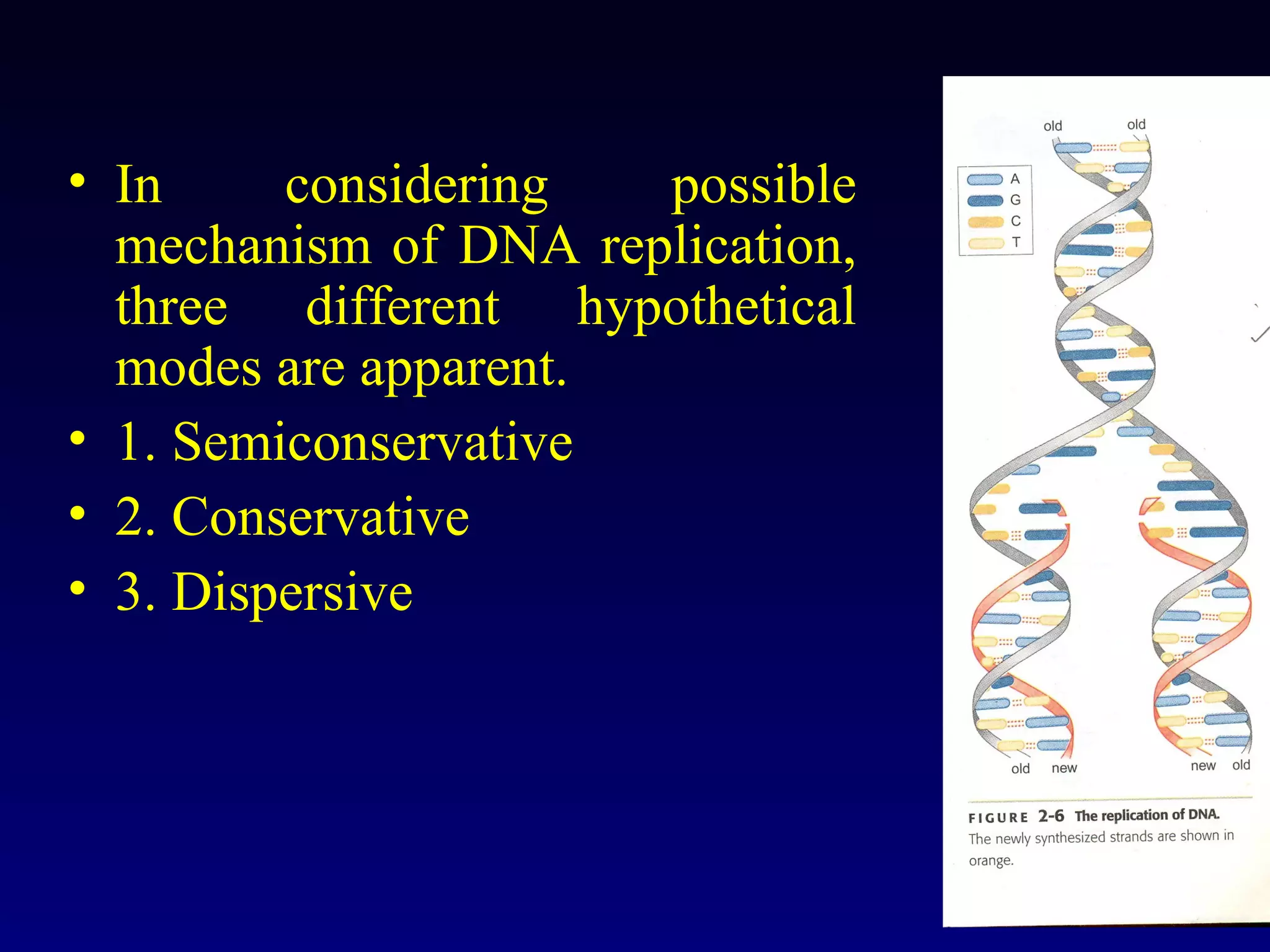
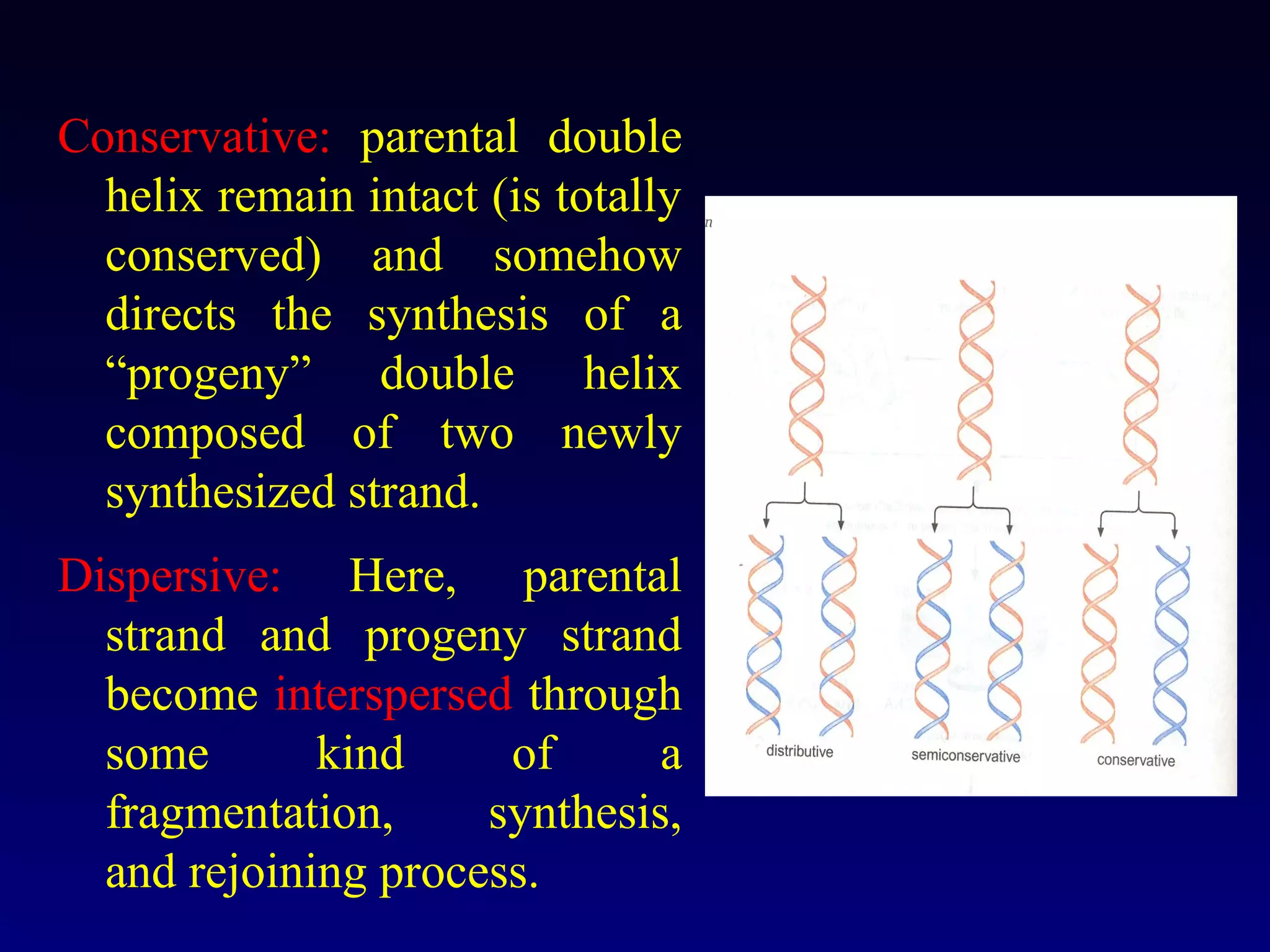
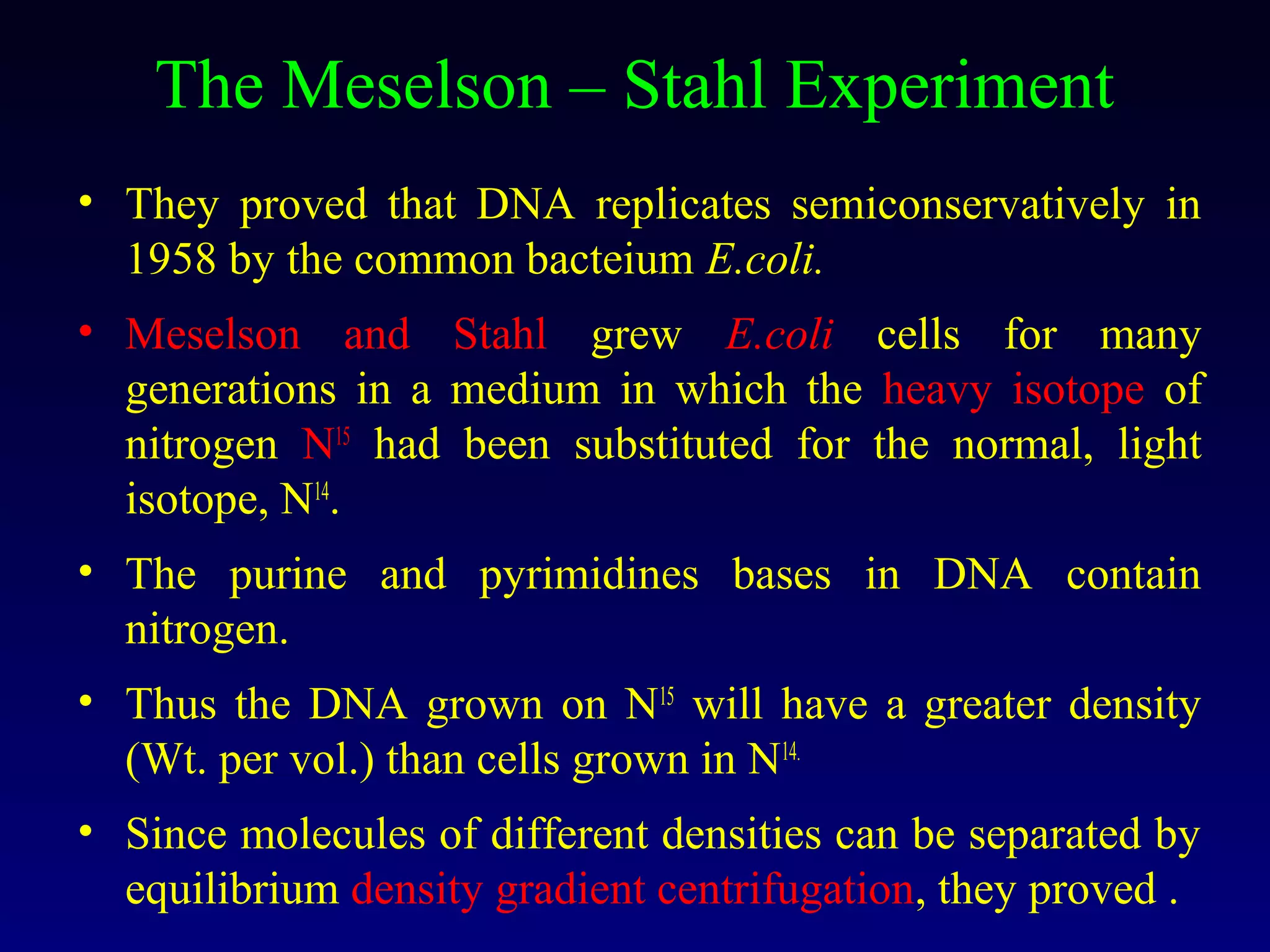
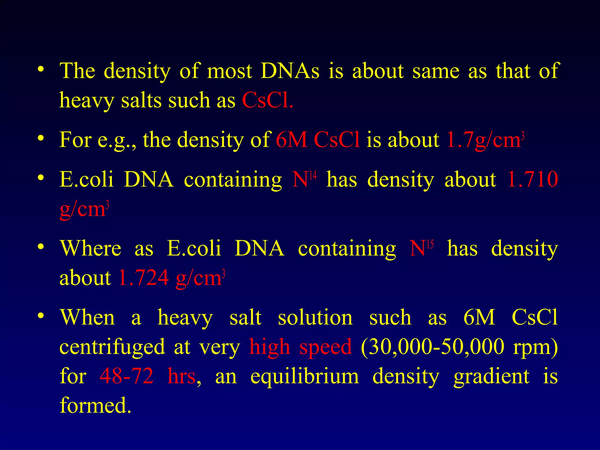
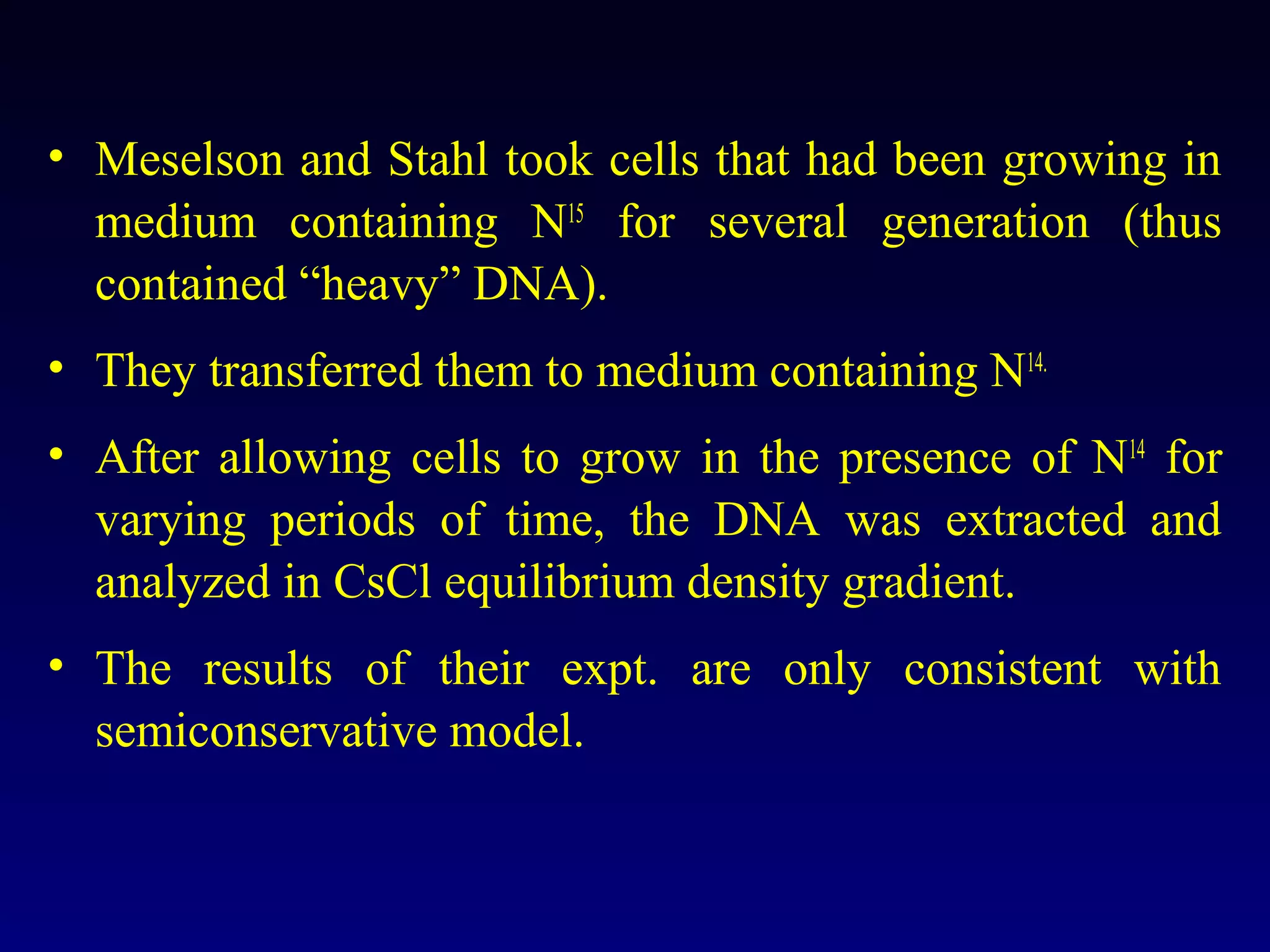
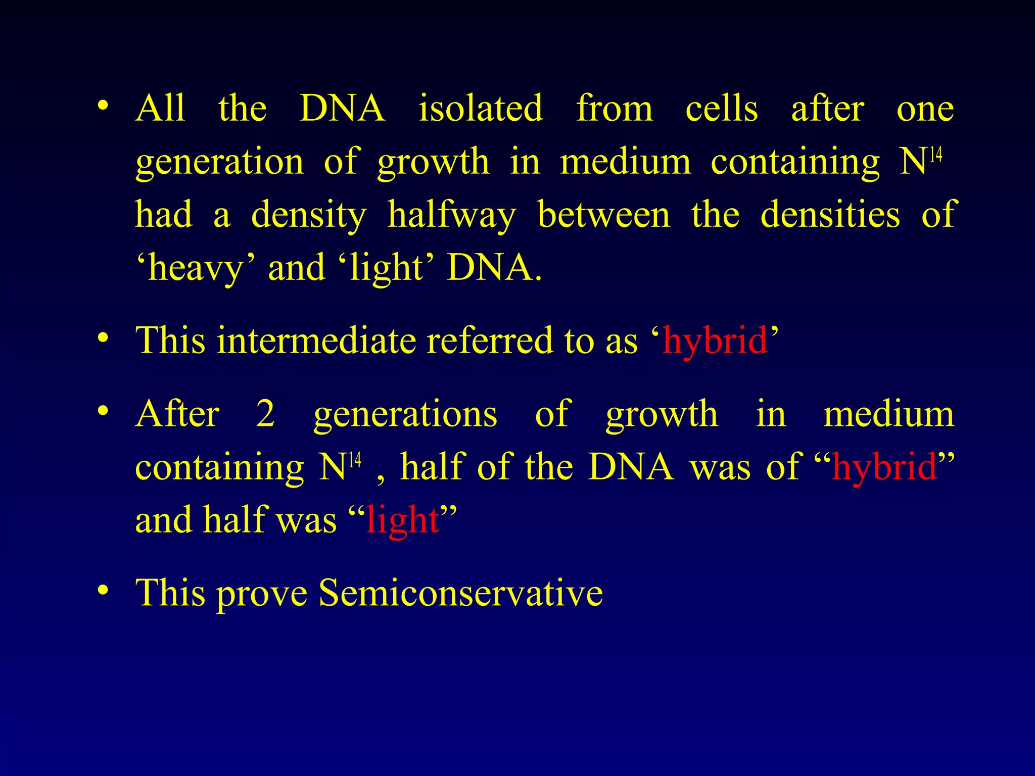
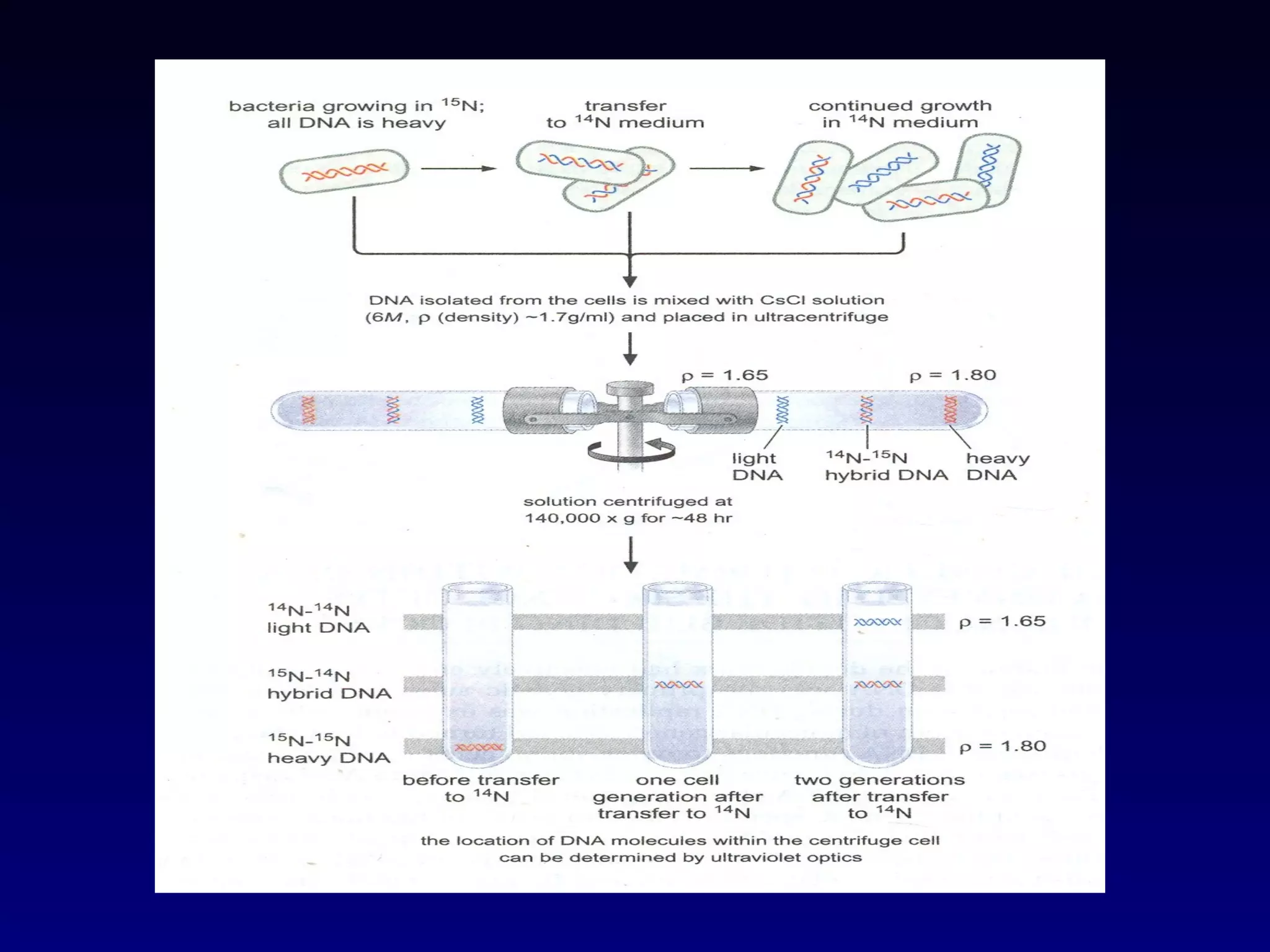
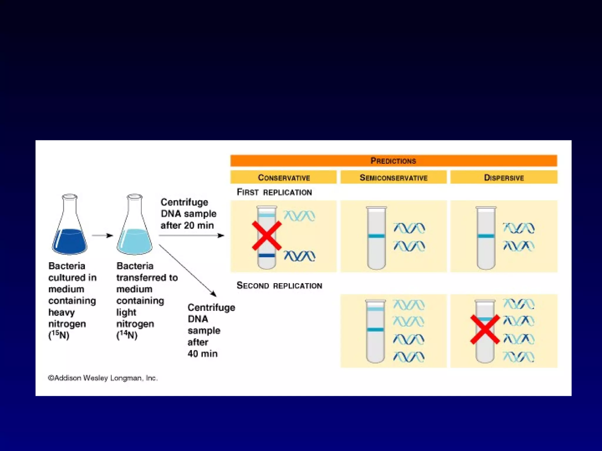
This document provides an overview of DNA and genetics. It discusses how DNA was established as the genetic material through experiments in the 1940s-1950s, including Griffith's transformation experiments, Avery et al.'s work demonstrating the transforming principle was DNA, and Hershey and Chase's experiments with bacterial viruses. It also summarizes the discovery of the DNA double helix structure by Watson and Crick in 1953, based on Chargaff's rules and X-ray crystallography data. The key properties of DNA structure, including specific base pairing and semiconservative replication, are briefly outlined.

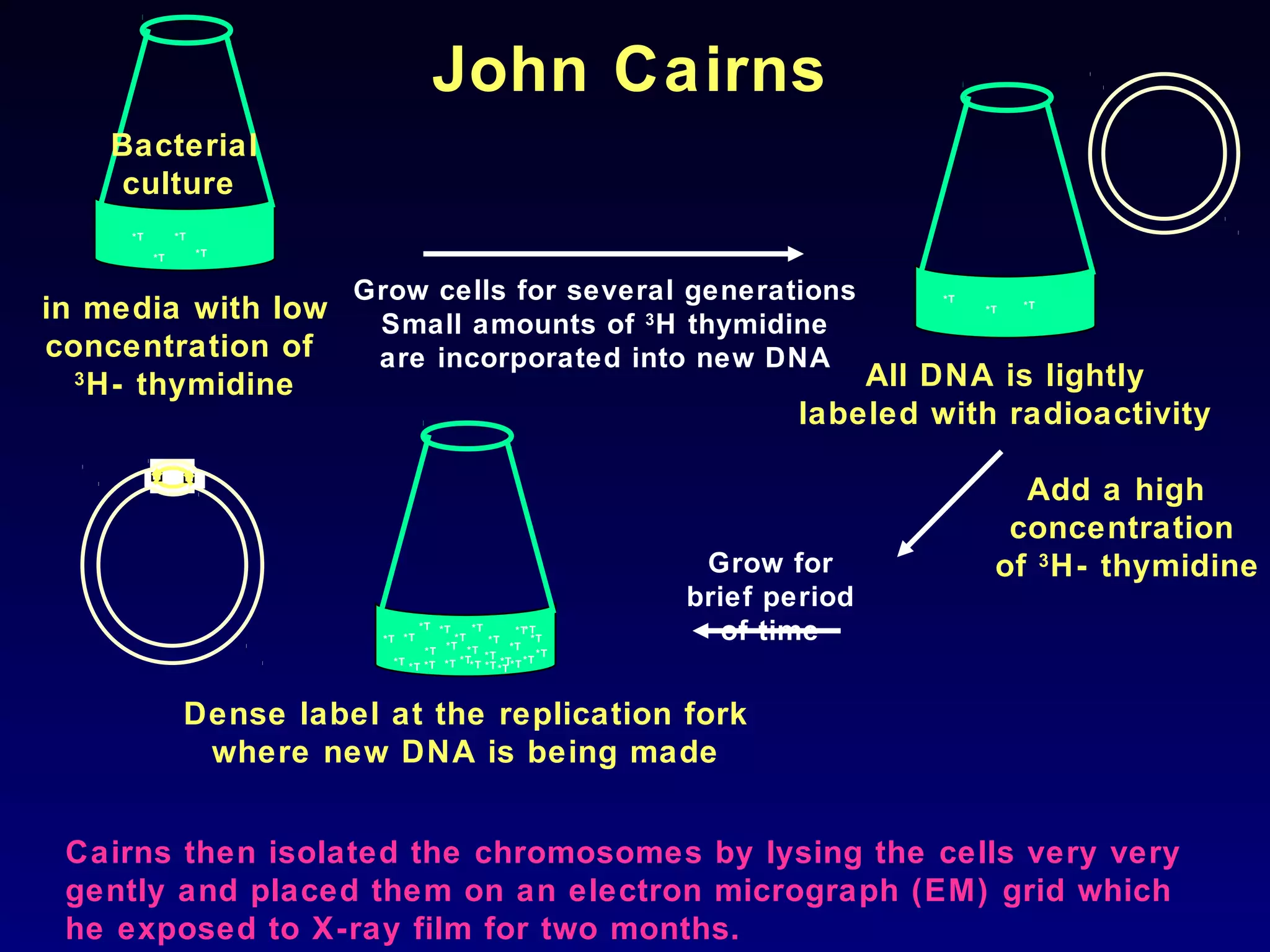
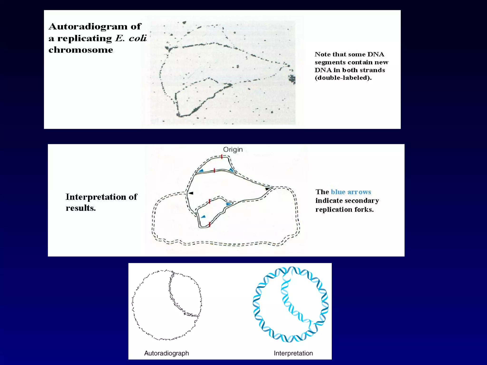
![Cairn’s Experiment
• The visualization of replicating chromosome was first
accomplished by J. Cairns in 1963 using the technique called
autoradiography.
• Autoradiography is a method of detecting and localizing
radioactive isotopes in macromolecules by exposure to
photographic emulsion that is sensitive to low energy radiation.
• Autoradiography is particularly useful in studying DNA
metabolism because DNA can be specifically labeled by growing
cells on [H3]thymidine, the tritiated deoxyribonucleoside of
thymidine.
• Thymidine is incorporated exclusively into DNA; it is not present
in any other major component of the cell.](https://image.slidesharecdn.com/dna-anoverview-130211024022-phpapp02/75/Dna-an-overview-53-2048.jpg)
![• Cairns grew E.coli cells in medium containing [H3]thymidine for
varying period of time.
• He lysed the cell very gently so as not to break the chromosomes
and he carefully collected the chromsomes on membrane filter.
• These filters are affixed to glass slides, coated with emulsion
sensitive to β – particles (the low energy electrons emitted
during decay of tritium) and store in dark for radioactive decays.
• The autoradiograph observed when the films were developed.
• It showed that the chromosomes of E.coli are circular
structures that exist as θ shaped intermediates during
replication.](https://image.slidesharecdn.com/dna-anoverview-130211024022-phpapp02/75/Dna-an-overview-54-2048.jpg)